Published online Feb 27, 2019. doi: 10.4240/wjgs.v11.i2.85
Peer-review started: November 2, 2018
First decision: January 5, 2019
Revised: February 23, 2019
Accepted: February 24, 2019
Article in press: February 25, 2019
Published online: February 27, 2019
Processing time: 117 Days and 3.7 Hours
Formation of intraperitoneal adhesions is one of the major complications after abdominal surgery, which may lead to bowel obstruction. Thrombospondin 1 (TSP-1) is an extracellular matrix modulating glycoprotein during tissue regeneration and collagen deposition.
To evaluated the therapeutic potential of overexpressed TSP-1 in suppressing pelvic adhesion formations in rat models.
Pelvic adhesion was induced in anesthetized rats by laparotomy cecal abrasion. The animals were randomly assigned to treatment of local application with Seprafilm (an antiadhesive bioresorbable membrane) or adenoviral vectors encoding mouse TSP-1 (AdTSP-1) on the surfaces of the injured cecum. The severity of the peritoneal adhesions was evaluated by blinded observers 14 d later.
Compared with control (no treatment) group, the application of Sperafilm significantly reduced the formation of adhesion band, and local administration of AdTSP-1 on the injured cecum the also attenuated the severity of peritoneal adhesion score. However, systemic delivery of AdTSP-1 did not affect the formation of adhesion.
We conclude that therapeutic approaches in inducing regional overexpression of TSP-1 may serve as alternative treatment strategies for preventing postoperative peritoneal adhesion.
Core tip: Formation of intraperitoneal adhesions is one of the major complications after abdominal surgery, which may lead to bowel obstruction. Thrombospondin-1 (TSP-1) is an extracellular matrix modulating glycoprotein during tissue regeneration and collagen deposition. This study evaluated the therapeutic potential of overexpressed TSP-1 in suppressing pelvic adhesion formations in rat models. We found the application of Sperafilm reduced the formation of adhesion band, and local administration of adenoviral vectors encoding TSP-1 on the injured cecum the also attenuated the severity of peritoneal adhesion score. Therefore, therapeutic approaches in inducing regional overexpression of TSP-1 may serve as alternative treatment strategies for preventing postoperative peritoneal adhesion.
- Citation: Tai YS, Jou IM, Jung YC, Wu CL, Shiau AL, Chen CY. In vivo expression of thrombospondin-1 suppresses the formation of peritoneal adhesion in rats. World J Gastrointest Surg 2019; 11(2): 85-92
- URL: https://www.wjgnet.com/1948-9366/full/v11/i2/85.htm
- DOI: https://dx.doi.org/10.4240/wjgs.v11.i2.85
Peritoneal adhesion formation is a major complication of abdominal surgery and can lead to infertility in women, chronic adhesion bowel symptoms, and acute bowel obstruction needing urgent surgical re-exploration[1]. Abdominal and pelvic adhesions are defined as pathologic bands that are formed during the scaring process of peritoneal injury in the peritoneal and pelvic cavities and connect two or more organ surfaces[2]. The presentation of these bands can vary from thin simple films of connective tissue to thick fibrous bridges containing blood vessels[3]. It has been estimated that up to 95% of abdominal-pelvic surgery patients developed varying levels of intra-abdominal adhesion[4]. Thick fibrous peritoneal adhesions can complicate with abdominal re-exploration surgery and increases the risk of inadvertent enterostomy[5].
Several anti-adhesive agents designed to treat peritoneal adhesions have been developed and tested in both pre-clinical and clinical settings. However, none of these agents are widely used clinically due to a lack of long-term efficacy. Thrombospondins (TSPs) are multi-domain, calcium-binding extracellular glycoproteins that mediate interactions with other extracellular matrix components[6]. Thrombospondin-1 (TSP-1) is a prototypical thrombospondin and an important regulator of platelet aggregation, inflammatory responses, and angiogenesis in wound healing[6]. Our previous study demonstrated that direct intra-articular administration of adenoviral vectors encoding TSP-1 (AdTSP-1) significantly ameliorated the progress of collagen-induced arthritis by reducing both synovial hypertrophy and angiogenesis[7]. The present study aimed to examine the therapeutic potential of overexpressed TSP-1 in suppressing pelvic adhesion formations in rat models.
A recombinant adenoviral vector encoding with mouse TSP-137 complementary DNA was prepared as described in a previous article[7].
Forty male albino Wistar rats weighing 180 to 220 g were anesthetized with intraperitoneal injections of 1.5 mL/kg of 1% (w/v) sodium amobarbital (Amytal Sodium; Eli Lilly and Company, Indianapolis, IN). Each rat had its abdomen shaved and a midline laparotomy performed. The cecum was identified and a surface defect area (1 × 2 cm2) was created on the ventral surface of the cecum by gently rubbing the cecal surface against an electrocautery tip cleaner[8]. A trans-peritoneal defect similar in size to the one created on the cecum was then excised on the corresponding retroperitoneal wall. The rats were then randomly assigned to four treatment groups. Group I (controls, n = 10) received the standard operation that excised the surface defects. Group II (Seprafilm group, n = 10) received the standard operation and the cecal surface defects were covered with an anti-adhesive bioresorbable membrane (Seprafilm; Genzyme Corporation, Cambridge, MA). Group III (n = 10) received the standard operation and the cecal surface defects were coated with 5 × 10⁷ plaque forming units of AdTSP-1. Group IV (n = 10) received the standard operation and AdTSP-1 (5 × 10⁷) was administered intra-peritoneally on postoperative days (POD) 1, 3, and 5. At the end of each operation, the abdominal incision was closed in two layers and the animals were allowed to recover from the anesthesia.
All rats were sacrificed on POD 14 with an overdose of ethyl ether. The abdomen of each rat was re-explored to evaluate adhesion formation through histological examinations and immunohistochemical staining. The severity of the peritoneal adhesions was graded by a blinded observer according to the criteria listed on Table 1[9].
| Grade | Observation |
| 0 | No adhesions |
| 1 | One single adhesion |
| 2 | Mild adhesions |
| 3 | Moderate adhesions |
| 4 | Severe adhesion that adhere to abdominal wall |
Peritoneal adhesions that were attached to the cecum were excised in their full thickness en-block. The tissues were then fixed in 10% phosphate-buffered formalin and processed for hematoxylin and eosin staining. The frozen tissue sections (5 μm in thickness) were stained against anti-TSP-1 (N-20) or anti-TGFβ (sc-146-G) antibodies (Santa Cruz Biotechnology, Santa Cruz, CA). A pathologist, who was blinded to the treatment groups, independently assessed the histological sections under a light microscope.
All data are expressed as means ± SD. Statistical comparisons between the four groups were analyzed using the Kruskal-Wallis test. Statistical significance was accepted at P < 0.05.
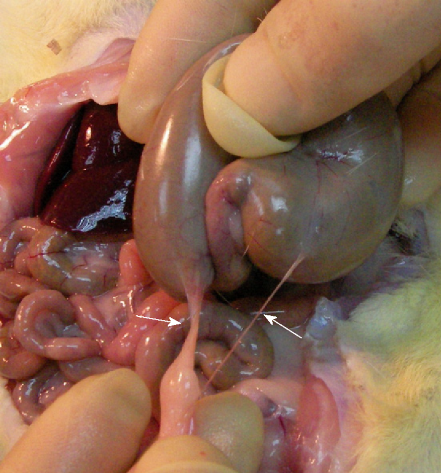
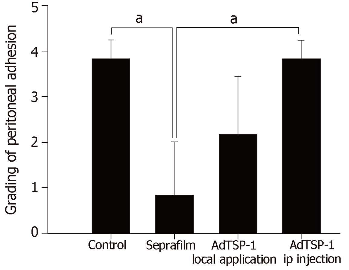
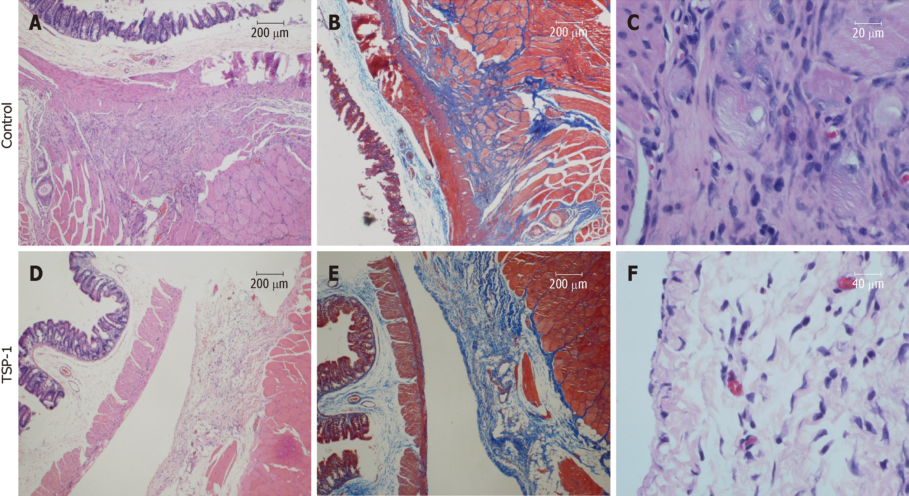
It was found that peritoneal adhesions mainly formed on the apical region of the cecum with the adjacent omemtum, as opposed to other organs or the abdominal wall (Figure 1). Group I (controls) was found to have developed peritoneal bands that were more tightly adhered to the cecum. One rat in Group I developed bowel perforation due to an adhesion band restricting the cecum. In comparison, rats that received AdTSP-1 (Groups III, IV) developed loose, detachable connective tissue bands around the cecum. Rats that were treated with Seprafilm coverings (Group II) were found to have developed significantly less peritoneal adhesion formations than the controls (Group I) (Figure 2). Peritoneal adhesion development in Group III was found to be attenuated to a similar degree as in Group II. Group IV showed no effect in peritoneal adhesion formation reduction (Figure 2). Histological examination of the adhesion bands resected from the muscularis propria and abdominal skeletal muscle of the controls showed increased deposition of collagen fibers and infiltration of mononuclear cells (Figure 3).
In the present study, we aimed to examine the therapeutic effects of overexpressed TSP-1 on peritoneal adhesion formation suppression. It was found that Seprafilm application showed the most pronounced preventative effects against peritoneal adhesion formation at 14 d after lesioning; local transfection with TSP-1 showed similar preventative effects to Seprafilm application. Seprafilm is an adhesion barrier and is used clinically in the prevention of peritoneal adhesion formation. Seprafilm application is unlikely to produce any adverse effects on surgical sites, but its anti-adhesive effects only extend to areas that the film covers[10]. Therefore, the placement of this adhesion barrier requires an accurate assessment of all intraperitoneal regions that may potentially form adhesion bands after an operation. However, there are certain regions in the peritoneal cavity where it is impossible to perfectly apply Seprafilm; and as a result, up to 50% of patients who had Seprafilm application during restorative proctolectomy or ileostomy still developed postoperative adhesion[11]. The use of these commercially available adhesion barriers significantly reduced the incidence of adhesion formation. It should be noted, however, that the formulation of adhesion barriers (i.e., gel, solid, solution) can affect their efficacy and associated adverse effects[12]. A deeper understanding of the molecular mechanisms underlying adhesion may provide insight to alternative molecule-targeting approaches to adhesion prevention without the potential adverse effects from biomaterials and their degraded matter.
TSP-1 is a matricellular glycoprotein that was first isolated in the 1970s as a 190000-Da thrombin-sensitive protein[13]. TSP-1 has been recognized as a cell adhesion molecule as well as a major component of platelet α-granules that are released upon platelet activation. TSP-1 mediates diverse pleiotropic biological functions, including the anti-angiogenic properties and modulation of the interactions between cells and matrices[14]. To the best of our knowledge, this is the first experimental study investigating the efficacy of regional delivery of TSP-1 as an anti-adhesive strategy in the prevention of peritoneal adhesion following cecal abrasion injury.
TSP-1 plays an important role in both wound healing and adhesion formation. The normal process of wound healing involves an initial period of hemostasis mediated by platelets, which is followed an inflammatory phase. Angiogenesis and fibroplasia, mediated by endothelial cells and fibroblasts respectively, occur during the late stages of the inflammatory phase. TSP-1 is an important molecule that mediates cell-cell interactions. Importantly, TSP-1 modulates the procession of all three stages of wound healing via the TGF-β-mediated pathway; TSP-1 is involved in the stimulation of cell adhesion, cell migration, anoikis resistance, collagen expression, and matrix deposition[15,16]. However, overexpression of TSP-1 can lead to decreased wound healing due to reductions in fibroblast migration, granulation tissue formation, and angiogenesis[17].
In this study, we tested a novel concept to prevent the formation of peritoneal adhesion, namely the regional overexpression of TSP-1 in a solution delivery form. In our opinion, direct application of TSP-1 onto the injured sites of the serosa may provide two major advantages over other routes of administration: (1) the constant expression of TSP-1 mediates anti-inflammatory and anti-adhesive effects locally at the lesion sites; and (2) ease and efficacy in the delivery of the soluble form of an anti-adhesive agent onto the targeting surfaces in the pelvic cavity that are anatomically unapproachable. Our study also demonstrated that systemic delivery of TSP-1 failed to prevent the formation of peritoneal adhesion, suggesting that tissue adhesion is a regional tissue reaction and local application is a more appropriate treatment approach.
There are a number of limitations in this study. Firstly, we did not test for a concentration-dependent response. Nevertheless, our results showed that direct application of TSP-1 is not inferior to Seprafilm. Secondly, this is a proof-of-concept study and there was a lack of mechanical investigations. Thirdly, gene delivery system is not a clinical appreciable method of treatment; other TSP-1 targeting delivery system is under investigation in our laboratory.
In conclusion, this experimental, proof-of-concept study suggests that therapeutic approaches in inducing regional overexpression of TSP-1 may serve as alternative treatment strategies for preventing peritoneal adhesion after intraabdominal surgery or other interventional procedures.
Formation of intraperitoneal adhesions is one of the major complications after abdominal surgery, and up to 95% of abdominal-pelvic surgery patients developed varying levels of intra-abdominal adhesion.
Thrombospondin-1 (TSP-1) is a prototypical thrombospondin and an important regulator of platelet aggregation, inflammatory responses, and angiogenesis in wound healing.
The present study aimed to evaluate the therapeutic potential of overexpressed TSP-1 in suppressing pelvic adhesion formations in rat models.
Laparoscopic cecal abrasion caused by laparoscopic adhesion in anesthetized rats. Animals were randomized to topical application of Seprafilm (an adhesive bioabsorbable membrane) or an adenoviral vectors encoding mouse TSP-1 (AdTSP-1) to treat damaged cecal surfaces. The severity of peritoneal adhesions was observed blindly after 14 d.
Compared with the control group (no treatment group), the application of Sperafilm significantly reduced the formation of adhesion bands, and local administration of the injured cecal AdTSP-1 also reduced the severity of the peritoneal adhesion score. However, systemic administration of AdTSP-1 did not affect the formation of adhesions.
The treatment of local overexpression of TSP-1 can be used as an alternative treatment strategy for prevention of postoperative peritoneal adhesions.
We did not test the concentration-related response. However, our results indicate that the direct application of TSP-1 is not inferior to Seprafilm. Second, this is a proof-of-concept study that lacks a mechanical survey. Third, the gene delivery system is not a clinically evaluable treatment; our laboratory is also investigating other TSP-1 targeted delivery systems.
| 1. | Aarons CB, Cohen PA, Gower A, Reed KL, Leeman SE, Stucchi AF, Becker JM. Statins (HMG-CoA reductase inhibitors) decrease postoperative adhesions by increasing peritoneal fibrinolytic activity. Ann Surg. 2007;245:176-184. [RCA] [PubMed] [DOI] [Full Text] [Cited by in Crossref: 103] [Cited by in RCA: 114] [Article Influence: 6.0] [Reference Citation Analysis (0)] |
| 2. | Brüggmann D, Tchartchian G, Wallwiener M, Münstedt K, Tinneberg HR, Hackethal A. Intra-abdominal adhesions: definition, origin, significance in surgical practice, and treatment options. Dtsch Arztebl Int. 2010;107:769-775. [RCA] [PubMed] [DOI] [Full Text] [Cited by in Crossref: 30] [Cited by in RCA: 91] [Article Influence: 5.7] [Reference Citation Analysis (0)] |
| 3. | Hellebrekers BW, Trimbos-Kemper TC, Trimbos JB, Emeis JJ, Kooistra T. Use of fibrinolytic agents in the prevention of postoperative adhesion formation. Fertil Steril. 2000;74:203-212. [RCA] [PubMed] [DOI] [Full Text] [Cited by in Crossref: 148] [Cited by in RCA: 152] [Article Influence: 5.8] [Reference Citation Analysis (0)] |
| 4. | Ellis H, Moran BJ, Thompson JN, Parker MC, Wilson MS, Menzies D, McGuire A, Lower AM, Hawthorn RJ, O'Brien F, Buchan S, Crowe AM. Adhesion-related hospital readmissions after abdominal and pelvic surgery: a retrospective cohort study. Lancet. 1999;353:1476-1480. [RCA] [PubMed] [DOI] [Full Text] [Cited by in Crossref: 651] [Cited by in RCA: 650] [Article Influence: 24.1] [Reference Citation Analysis (0)] |
| 5. | Liu HJ, Wu CT, Duan HF, Wu B, Lu ZZ, Wang L. Adenoviral-mediated gene expression of hepatocyte growth factor prevents postoperative peritoneal adhesion in a rat model. Surgery. 2006;140:441-447. [RCA] [PubMed] [DOI] [Full Text] [Cited by in Crossref: 18] [Cited by in RCA: 26] [Article Influence: 1.3] [Reference Citation Analysis (0)] |
| 6. | Adams JC, Lawler J. The thrombospondins. Int J Biochem Cell Biol. 2004;36:961-968. [RCA] [PubMed] [DOI] [Full Text] [Cited by in Crossref: 312] [Cited by in RCA: 333] [Article Influence: 15.1] [Reference Citation Analysis (0)] |
| 7. | Jou IM, Shiau AL, Chen SY, Wang CR, Shieh DB, Tsai CS, Wu CL. Thrombospondin 1 as an effective gene therapeutic strategy in collagen-induced arthritis. Arthritis Rheum. 2005;52:339-344. [RCA] [PubMed] [DOI] [Full Text] [Cited by in Crossref: 42] [Cited by in RCA: 43] [Article Influence: 2.0] [Reference Citation Analysis (0)] |
| 8. | Buckenmaier CC, Pusateri AE, Harris RA, Hetz SP. Comparison of antiadhesive treatments using an objective rat model. Am Surg. 1999;65:274-282. [PubMed] |
| 9. | Nair SK, Bhat IK, Aurora AL. Role of proteolytic enzyme in the prevention of postoperative intraperitoneal adhesions. Arch Surg. 1974;108:849-853. [PubMed] |
| 10. | Mohri Y, Uchida K, Araki T, Inoue Y, Tonouchi H, Miki C, Kusunoki M. Hyaluronic acid-carboxycellulose membrane (Seprafilm) reduces early postoperative small bowel obstruction in gastrointestinal surgery. Am Surg. 2005;71:861-863. [PubMed] |
| 11. | Beck DE. The role of Seprafilm bioresorbable membrane in adhesion prevention. Eur J Surg Suppl. 1997;49-55. [PubMed] |
| 12. | Ten Broek RPG, Stommel MWJ, Strik C, van Laarhoven CJHM, Keus F, van Goor H. Benefits and harms of adhesion barriers for abdominal surgery: a systematic review and meta-analysis. Lancet. 2014;383:48-59. [RCA] [PubMed] [DOI] [Full Text] [Cited by in Crossref: 204] [Cited by in RCA: 234] [Article Influence: 19.5] [Reference Citation Analysis (0)] |
| 13. | Baenziger NL, Brodie GN, Majerus PW. A thrombin-sensitive protein of human platelet membranes. Proc Natl Acad Sci U S A. 1971;68:240-243. [RCA] [PubMed] [DOI] [Full Text] [Cited by in Crossref: 212] [Cited by in RCA: 240] [Article Influence: 4.4] [Reference Citation Analysis (0)] |
| 14. | Bornstein P, Agah A, Kyriakides TR. The role of thrombospondins 1 and 2 in the regulation of cell-matrix interactions, collagen fibril formation, and the response to injury. Int J Biochem Cell Biol. 2004;36:1115-1125. [RCA] [PubMed] [DOI] [Full Text] [Cited by in Crossref: 155] [Cited by in RCA: 175] [Article Influence: 8.0] [Reference Citation Analysis (0)] |
| 15. | Sweetwyne MT, Murphy-Ullrich JE. Thrombospondin1 in tissue repair and fibrosis: TGF-β-dependent and independent mechanisms. Matrix Biol. 2012;31:178-186. [RCA] [PubMed] [DOI] [Full Text] [Cited by in Crossref: 153] [Cited by in RCA: 196] [Article Influence: 14.0] [Reference Citation Analysis (0)] |
| 16. | Esemuede N, Lee T, Pierre-Paul D, Sumpio BE, Gahtan V. The role of thrombospondin-1 in human disease. J Surg Res. 2004;122:135-142. [RCA] [PubMed] [DOI] [Full Text] [Cited by in Crossref: 92] [Cited by in RCA: 96] [Article Influence: 4.4] [Reference Citation Analysis (0)] |
| 17. | Streit M, Velasco P, Brown LF, Skobe M, Richard L, Riccardi L, Lawler J, Detmar M. Overexpression of thrombospondin-1 decreases angiogenesis and inhibits the growth of human cutaneous squamous cell carcinomas. Am J Pathol. 1999;155:441-452. [RCA] [PubMed] [DOI] [Full Text] [Cited by in Crossref: 206] [Cited by in RCA: 216] [Article Influence: 8.0] [Reference Citation Analysis (0)] |
Open-Access: This article is an open-access article which was selected by an in-house editor and fully peer-reviewed by external reviewers. It is distributed in accordance with the Creative Commons Attribution Non Commercial (CC BY-NC 4.0) license, which permits others to distribute, remix, adapt, build upon this work non-commercially, and license their derivative works on different terms, provided the original work is properly cited and the use is non-commercial. See:
Manuscript source: Unsolicited manuscript
Specialty type: Gastroenterology and hepatology
Country of origin: Taiwan
Peer-review report classification
Grade A (Excellent): A
Grade B (Very good): 0
Grade C (Good): 0
Grade D (Fair): 0
Grade E (Poor): E
P- Reviewer: Lee JI, McMillin MA S- Editor: Dou Y L- Editor: A E- Editor: Wu YXJ


