©The Author(s) 2016.
World J Gastrointest Surg. Jun 27, 2016; 8(6): 472-475
Published online Jun 27, 2016. doi: 10.4240/wjgs.v8.i6.472
Published online Jun 27, 2016. doi: 10.4240/wjgs.v8.i6.472
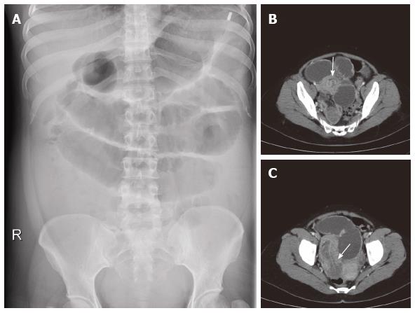
Figure 1 Plain abdominal X-ray revealed dilated small bowel loops.
A: Plain abdominal radiograph showing dilated small bowel; B: Abdominal computed tomography showing grossly dilated small bowel loops with “doughnut”; and C: “sausage” signs of small bowel intussusception.
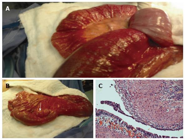
Figure 2 The leading point of intussusception was a cystic lesion measuring 4 cm in diameter which was located adjacent to the mesenteric side of the ileum.
Intraoperative photography revealing the (A) ileoileal intussusception (B) enteric cyst in the mesenteric side of the ileal loop which was the lead point of the intussusception and (C) Histopathology of enteric duplication cyst lined by columnar intestinal mucosa with underling muscular layer. The outer layer of the cyst wall was fused with normal intestine (H/E stain × 200).
- Citation: Al-Qahtani HH. Enteric duplication cyst as a leading point for ileoileal intussusception in an adult: A rare cause of complete small intestinal obstruction. World J Gastrointest Surg 2016; 8(6): 472-475
- URL: https://www.wjgnet.com/1948-9366/full/v8/i6/472.htm
- DOI: https://dx.doi.org/10.4240/wjgs.v8.i6.472


