Copyright
©The Author(s) 2016.
World J Gastrointest Surg. Feb 27, 2016; 8(2): 124-133
Published online Feb 27, 2016. doi: 10.4240/wjgs.v8.i2.124
Published online Feb 27, 2016. doi: 10.4240/wjgs.v8.i2.124
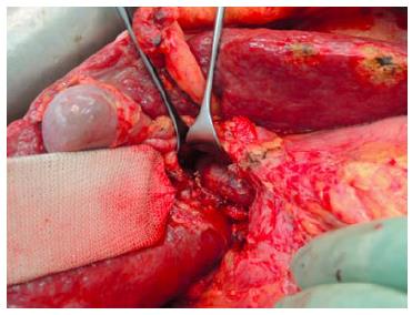
Figure 1 Exposure of the portal vein by lifting the common bile duct and right hepatic artery using a lid retractor.
Here the right portal vein branches were transected.
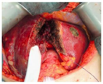
Figure 2 Liver parenchyma transection along the falciform ligament.
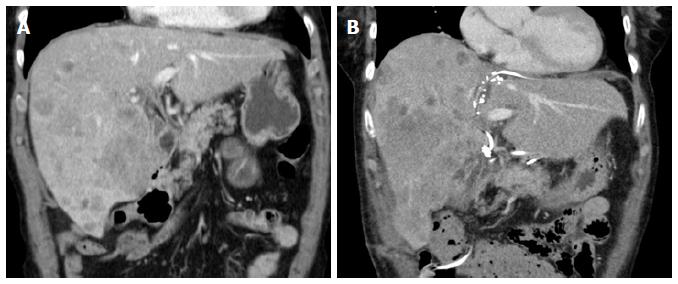
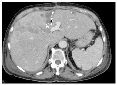
Figure 3 Computed tomography scan before associating liver partition and portal vein ligation for staged hepatectomy in a patient with intrahepatic cholangiocarcinoma and on day 10 after liver partition.
A: The future liver remnant consisted of segment 2 and 3 with volume of 347 mL (23% of the standardized total liver volume); B: Showing the hypertrophy of the segment 2 and 3 with volume of 610 mL (41% of the standardized total liver volume).
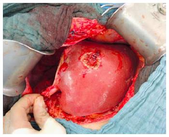
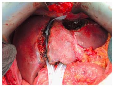
Figure 4 Completion of right trisectionectomy.
Figure 5 Partial associating liver partition and portal vein ligation for staged hepatectomy.
The non-total liver parenchymal transection is indicated by the clips, left along the liver split area in a computed tomography scan performed on day 10 after the liver partition.
Figure 6 The future liver remnant was separated by two Penrose drains from the right liver lobe in a patient with bilobar colorectal liver metastases during the first stage operation.
Three lesions at the left hemi-liver were resected.
- Citation: Li J, Ewald F, Gulati A, Nashan B. Associating liver partition and portal vein ligation for staged hepatectomy: From technical evolution to oncological benefit. World J Gastrointest Surg 2016; 8(2): 124-133
- URL: https://www.wjgnet.com/1948-9366/full/v8/i2/124.htm
- DOI: https://dx.doi.org/10.4240/wjgs.v8.i2.124


