©2012 Baishideng.
World J Gastrointest Surg. May 27, 2012; 4(5): 131-134
Published online May 27, 2012. doi: 10.4240/wjgs.v4.i5.131
Published online May 27, 2012. doi: 10.4240/wjgs.v4.i5.131
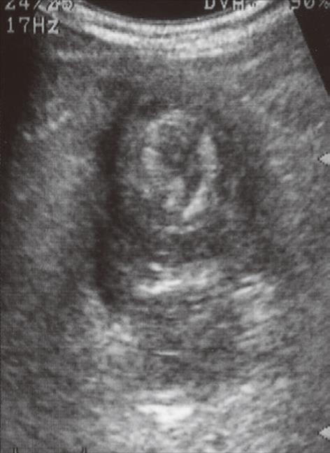
Figure 1 Abdominal ultrasonography of the right hypochondrium showing a target-like mass appearing as multiple concentric rings, which is suggestive of intussusception.
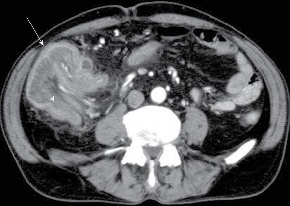
Figure 2 Abdominal computed tomography showing the sausage-shaped appearance that is characteristic of intussusception, comprising edematous bowel wall (arrow) with accompanying mesenteric fat and mesenteric blood vessels (arrowhead) within the lumen.
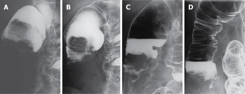
Figure 3 A water-soluble contrast enema showed a cup-shaped filling defect caused by the intussusception in the right upper quadrant (A); the cecum (intussusceptum) with the tumor was gradually pushed outward from the ascending colon (intussuscipiens) by the enema pressure (B and C); and finally, the intussusception was reduced, and contrast material could flow into the terminal ileum (D).
- Citation: Namikawa T, Okamoto K, Okabayashi T, Kumon M, Kobayashi M, Hanazaki K. Adult intussusception with cecal adenocarcinoma: Successful treatment by laparoscopy-assisted surgery following preoperative reduction. World J Gastrointest Surg 2012; 4(5): 131-134
- URL: https://www.wjgnet.com/1948-9366/full/v4/i5/131.htm
- DOI: https://dx.doi.org/10.4240/wjgs.v4.i5.131


