
















Copyright: ©Author(s) 2026.
World J Gastrointest Surg. May 27, 2026; 18(5): 118473
Published online May 27, 2026. doi: 10.4240/wjgs.v18.i5.118473
Published online May 27, 2026. doi: 10.4240/wjgs.v18.i5.118473
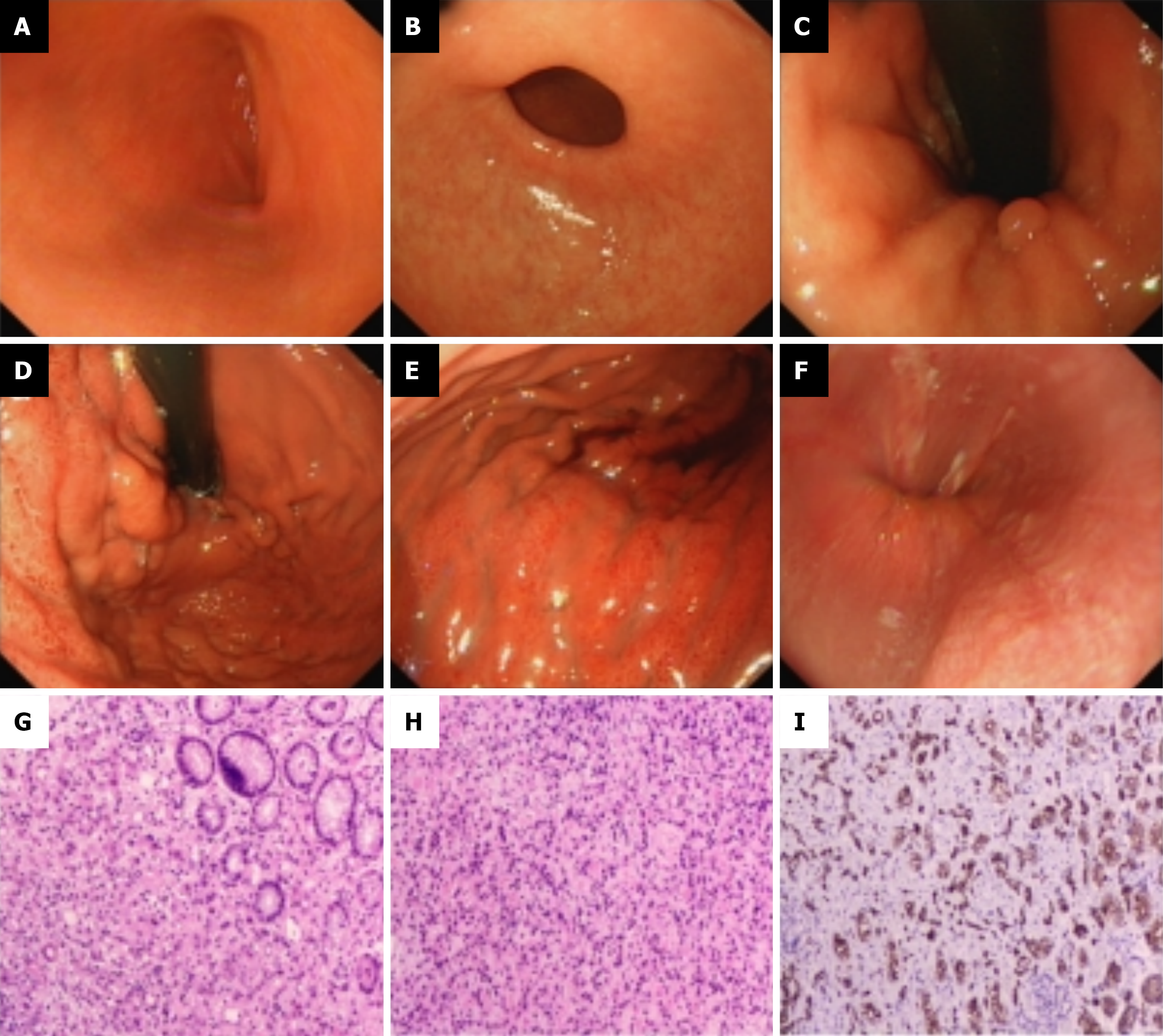
Figure 1 Electronic gastroscopic and routine biopsy pathology findings of the gastric mucosa.
A: Duodenal bulb; B: Antrum; C: Gastric angle; D: Fundus; E: Body; F: Esophagus; G and H: Hematoxylin and eosin staining of gastric body mucosal tissue; I: Immunohistochemical staining of biopsy specimen.
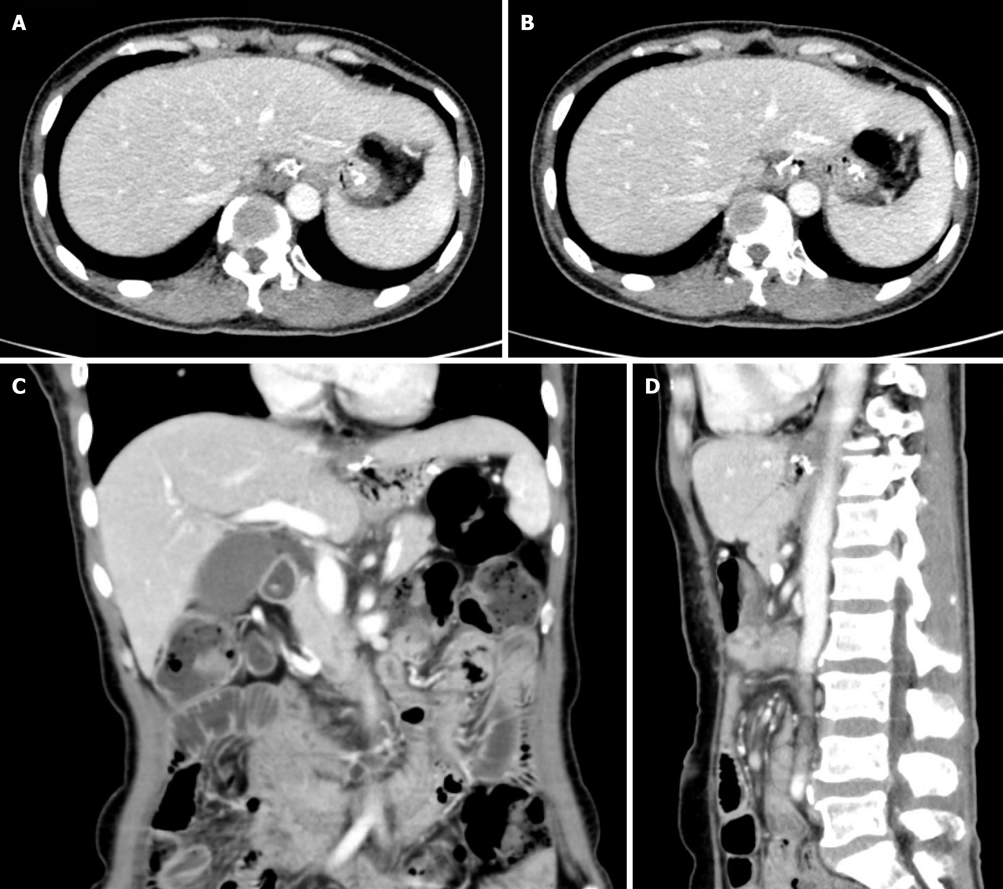
Figure 2 Contrast-enhanced computed tomography scans of the upper abdomen.
A and B: Axial; C: Coronal; D: Sagittal.
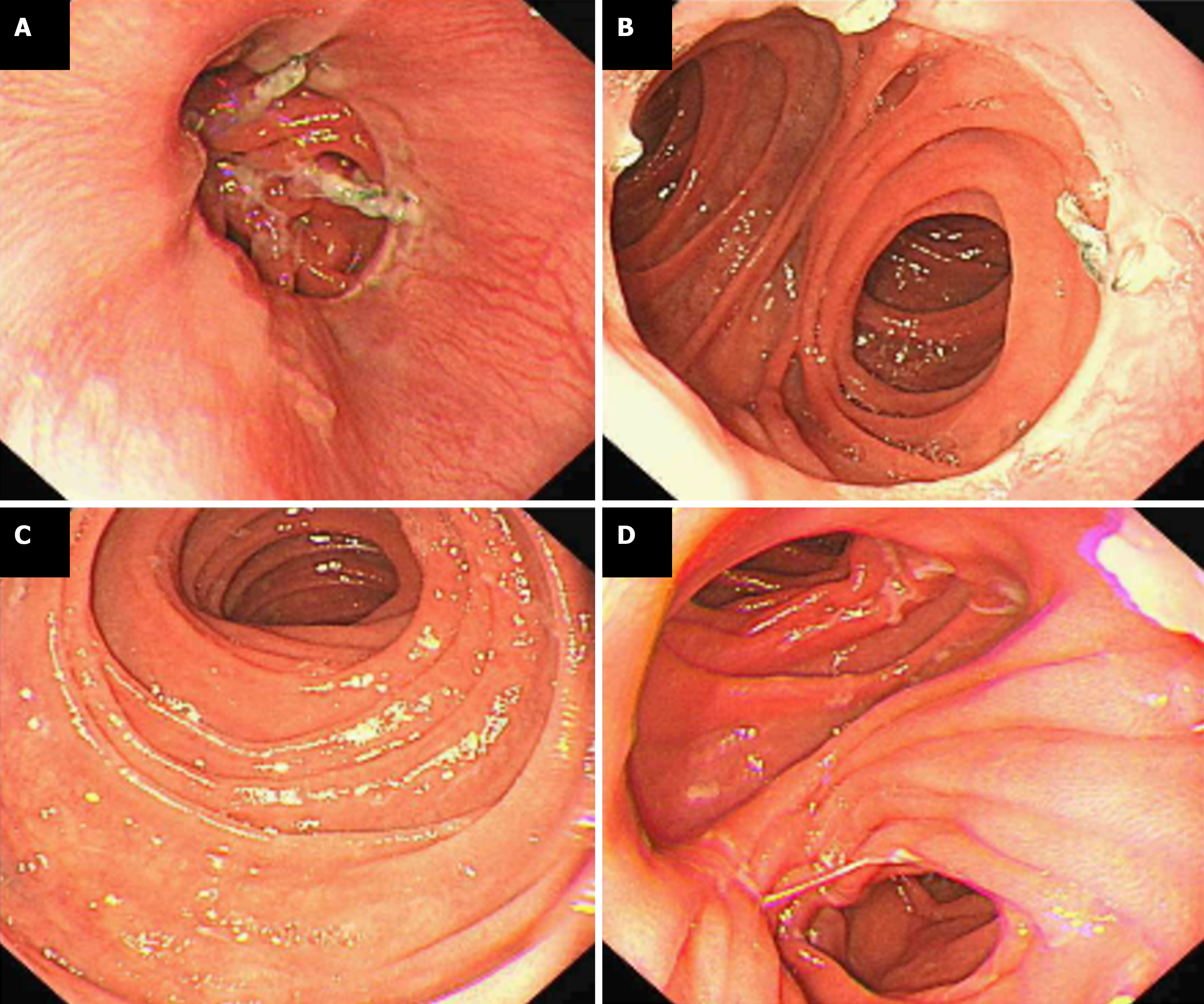
Figure 3 Electronic gastroscopy findings.
A: Lower esophagus; B: Anastomosis; C: Efferent loop; D: Saddle region.
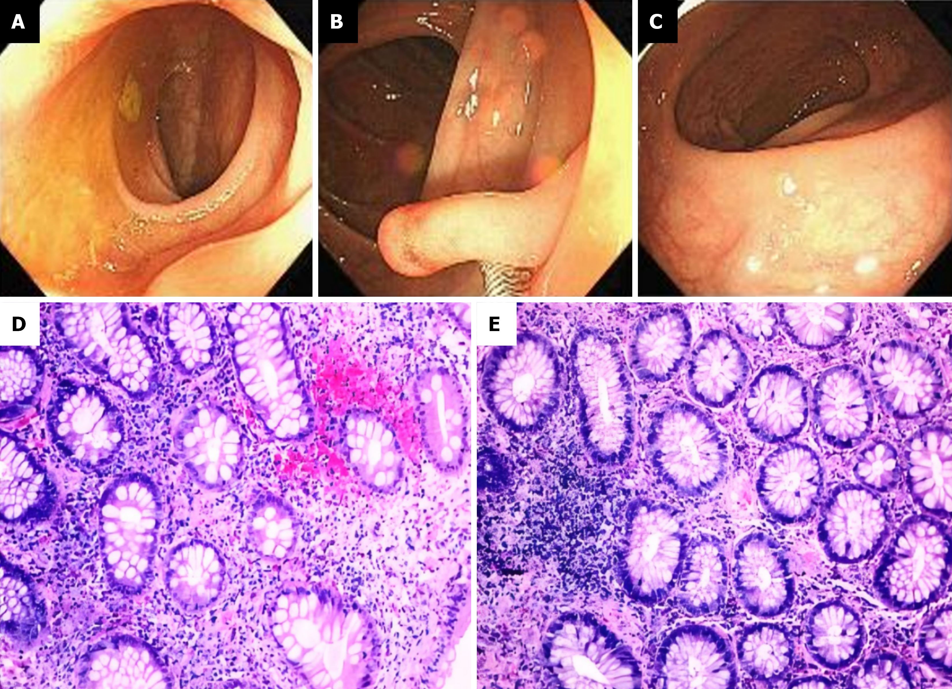
Figure 4 Electronic colonoscopy and pathological biopsy findings.
A: Terminal ileum; B: Ascending colon; C: Rectum; D: Immunohistochemical staining of biopsy specimen; E: Hematoxylin and eosin staining of biopsy tissue.
- Citation: Lu B, Wen K. Cancer-related fatigue and functional gastrointestinal disorders after total gastrectomy: A case report. World J Gastrointest Surg 2026; 18(5): 118473
- URL: https://www.wjgnet.com/1948-9366/full/v18/i5/118473.htm
- DOI: https://dx.doi.org/10.4240/wjgs.v18.i5.118473