
















Copyright: ©Author(s) 2026.
World J Gastrointest Surg. May 27, 2026; 18(5): 117998
Published online May 27, 2026. doi: 10.4240/wjgs.v18.i5.117998
Published online May 27, 2026. doi: 10.4240/wjgs.v18.i5.117998
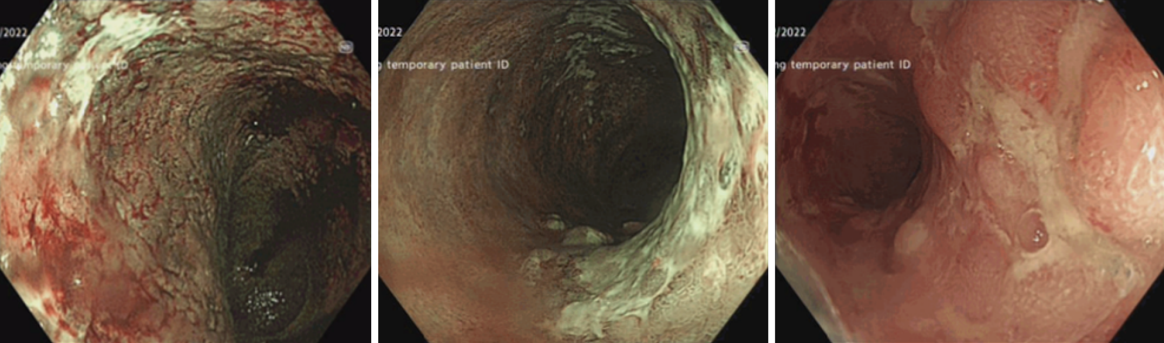
Figure 1 Baseline colonoscopic findings.
Initial colonoscopic evaluation revealing severe active pancolitis (Mayo Endoscopic Score 3) with diffuse erythema, friable mucosa, and deep ulcerations prior to biologic therapy. The terminal ileum was endoscopically normal with no evidence of inflammation, and random biopsies from the ileum and colon showed features consistent with ulcerative colitis without granulomas or other findings suggestive of Crohn’s disease.
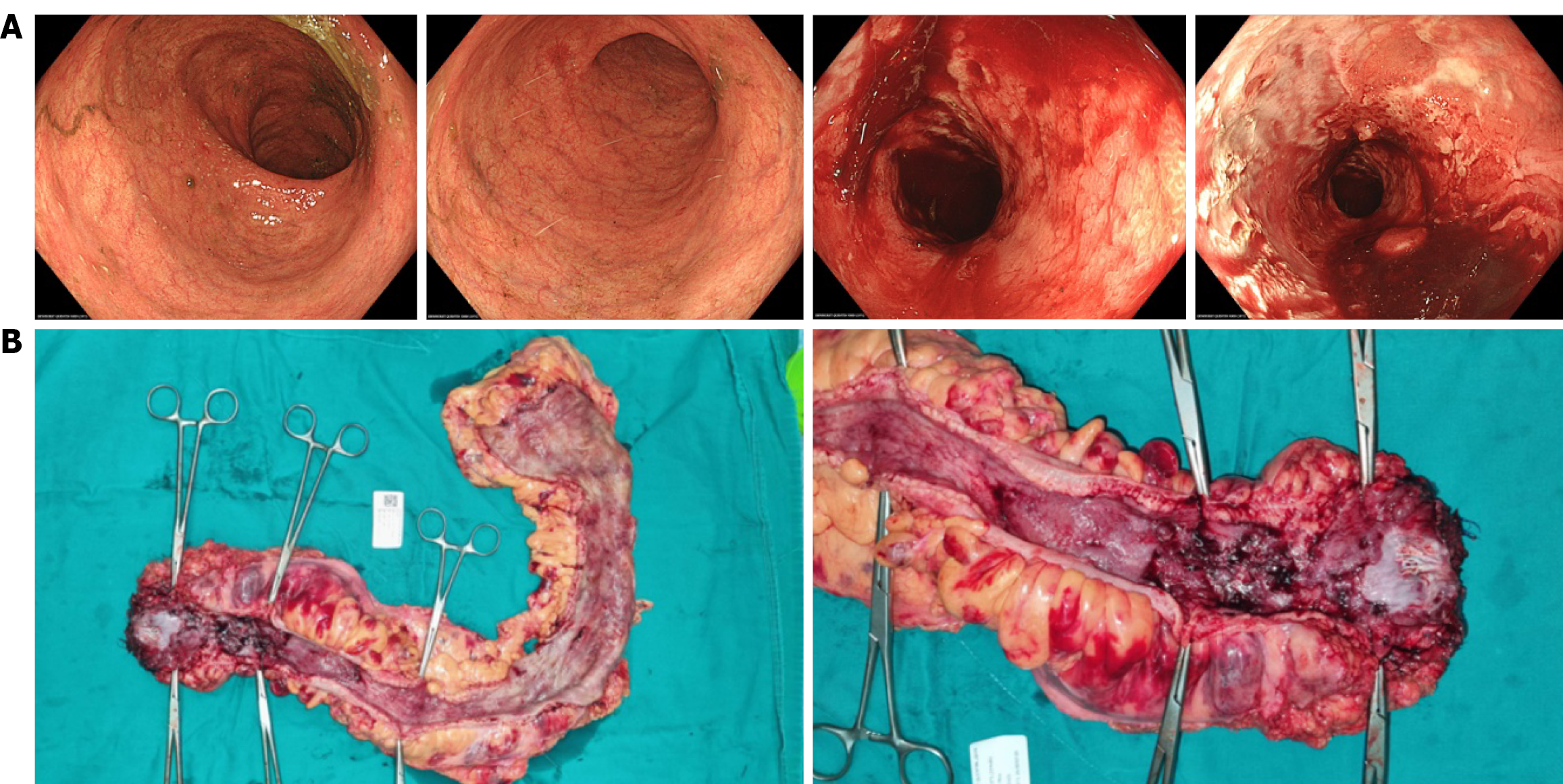
Figure 2 Endoscopic and gross pathological evidence of differential mucosal healing post-tofacitinib therapy.
A: Follow-up colonoscopy illustrating a heterogeneous healing pattern: Mucosal remission in the right colon (Mayo Endoscopic Score 0-1) contrasted with persistent severe inflammation in the distal segments, including the rectum and sigmoid colon (Mayo Endoscopic Score 3); B: Gross specimen of the resected colorectum demonstrating severe inflammation, mural thickening, and extensive ulceration in the sigmoid colon and rectum, confirming the extent of medically refractory disease.
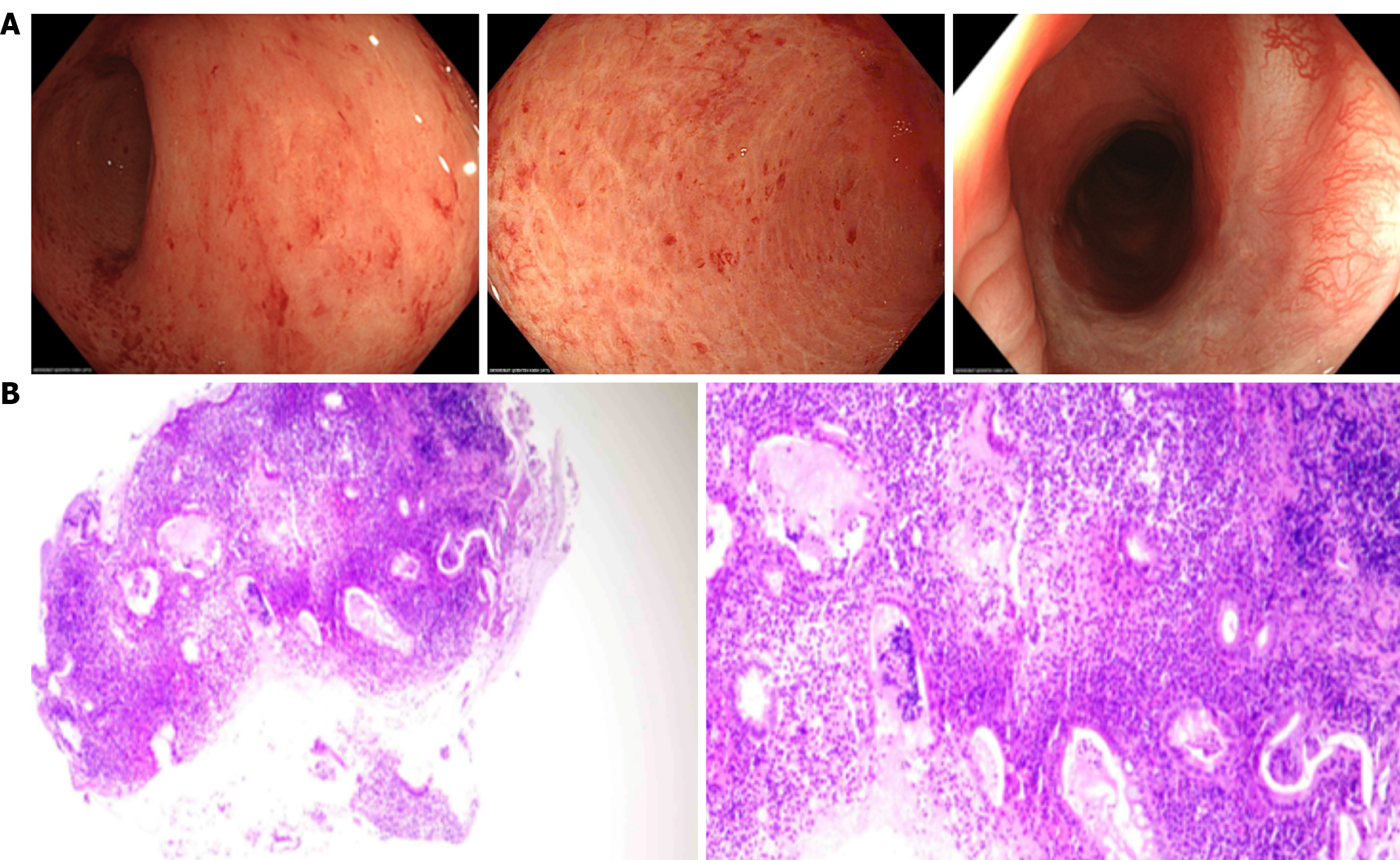
Figure 3 Postoperative endoscopic and histopathological findings at six months following subtotal colectomy.
A: Colonoscopy of the remaining colon shows a patent colo-anal anastomosis and only mild, diffuse erythema; B: Corresponding histopathology (hematoxylin and eosin stain) of biopsies reveals features consistent with diversion colitis, including chronic inflammation and glandular atrophy, with a complete absence of neutrophils, indicating no evidence of active ulcerative colitis.
- Citation: Nguyen CD, Dang LM, Trinh TTT, Tran HD, Phan AT, Vo DTN, Le HDM, Vo TD, Bui HH. Subtotal colectomy with Deloyers colo-anal anastomosis for refractory ulcerative colitis: A case report. World J Gastrointest Surg 2026; 18(5): 117998
- URL: https://www.wjgnet.com/1948-9366/full/v18/i5/117998.htm
- DOI: https://dx.doi.org/10.4240/wjgs.v18.i5.117998