Copyright
©The Author(s) 2025.
World J Gastrointest Surg. Jul 27, 2025; 17(7): 107804
Published online Jul 27, 2025. doi: 10.4240/wjgs.v17.i7.107804
Published online Jul 27, 2025. doi: 10.4240/wjgs.v17.i7.107804
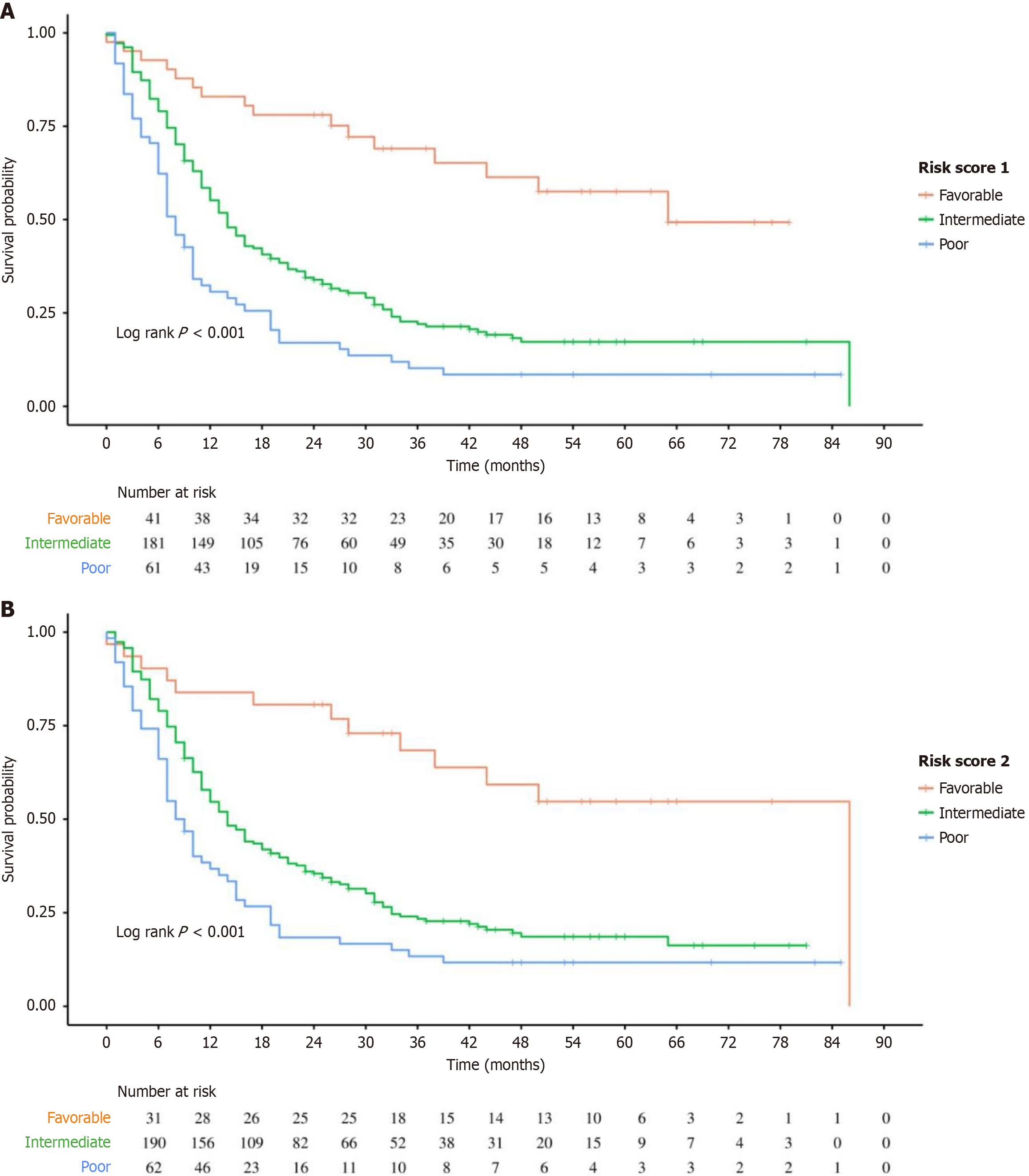
Figure 1 Kaplan Meier survival curves for patients stratified by risk group.
The low-risk group showed significantly better survival outcomes than the moderate and high-risk groups, whereas the high-risk group exhibited the poorest prognosis. Differences in survival among the groups were statistically significant (log-rank test, P < 0.001), indicating the predictive value of the risk score for patient prognosis. A: Risk score 1 from Reader 1; B: Risk score 2 from Reader 2.
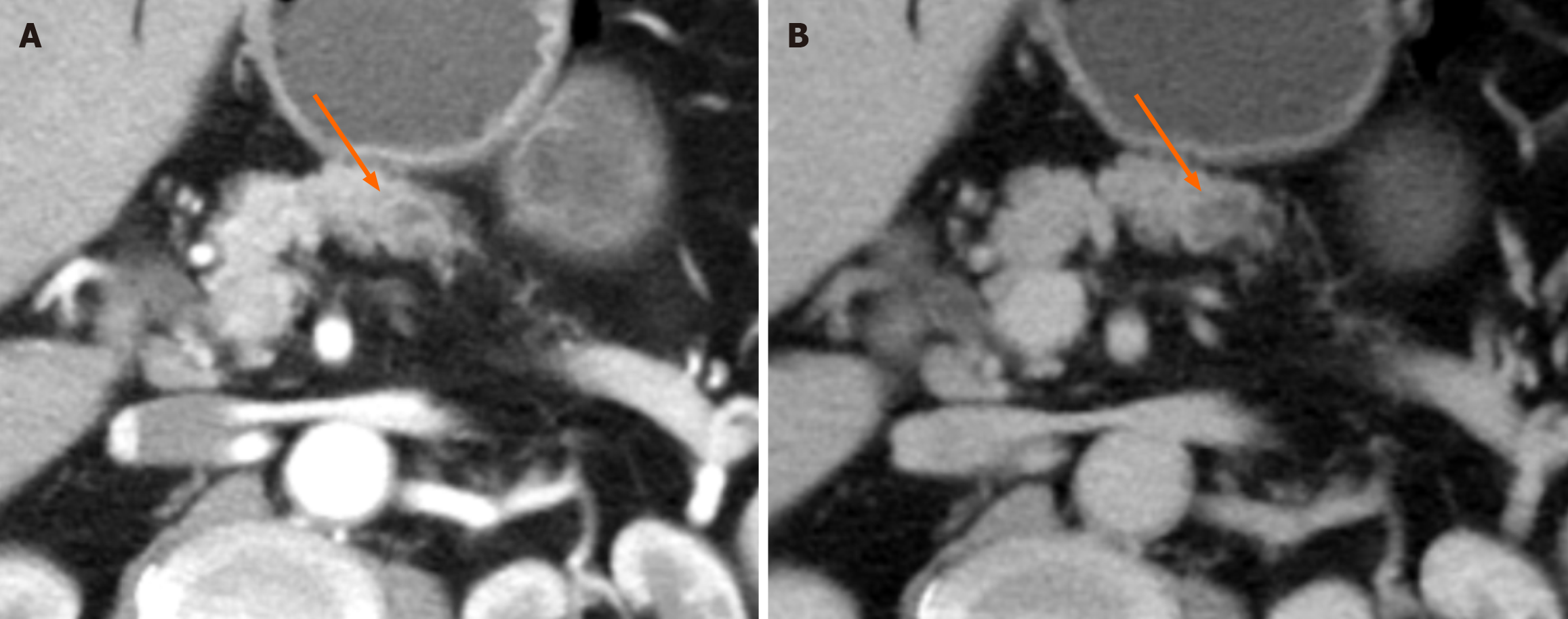
Figure 2 A 61-year-old male with resectable pancreatic ductal adenocarcinoma.
Axial contrast-enhanced computed tomography images revealed a 1.7 cm mass in the body of the pancreas (indicated by the arrow), which was hypodense in both. The image scale was approximately 0.55 mm per pixel. A: Arterial phase; B: Portal venous phase. There was no evidence of tumor necrosis, peritumoral invasion, or suspicious metastatic lymph nodes. The patient was given a risk score of 1, placing him in the good prognosis group. Following a standard pancreaticoduodenectomy, the patient survived for 65 months without any tumor recurrence.
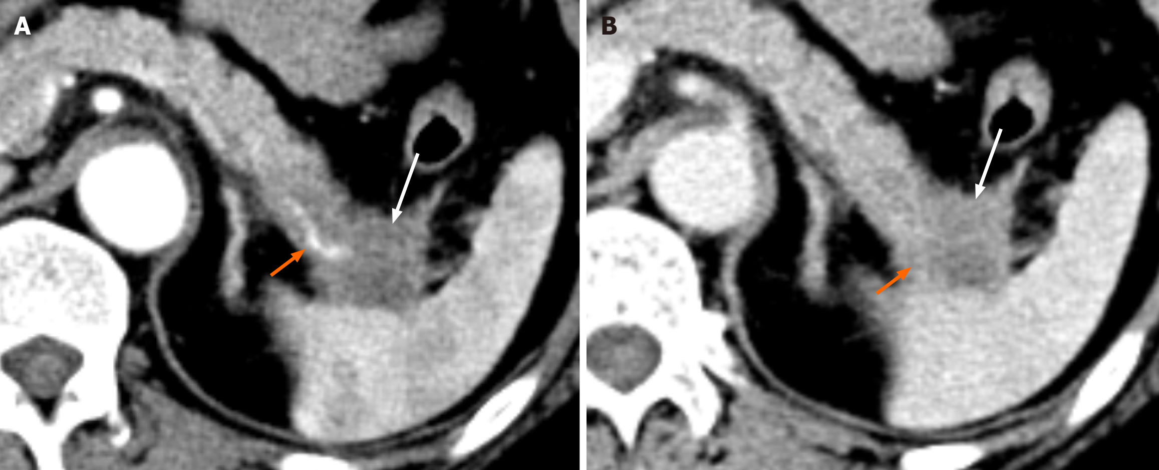
Figure 3 Computed tomography images of a 61-year-old female with resectable pancreatic ductal adenocarcinoma.
A: Arterial phase; B: Portal venous phase axial contrast-enhanced computed tomography images revealed a 3.8 cm hypodense mass in the tail of the pancreas (indicated by a white arrow) with invasion of the splenic artery (indicated by a orange arrow). The image scale was approximately 0.31 mm per pixel. There was no evidence of tumor necrosis or suspicious metastatic lymph nodes. With a risk score of 3, the patient was placed in the moderate prognosis group. A standard distal pancreatectomy was performed, and the tumor recurred 18 months postoperatively.
Figure 4 Computed tomography images of a 64-year-old male with resectable pancreatic ductal adenocarcinoma.
A: Arterial phase; B and C: Portal venous phase computed tomography axial contrast enhancement showed a 3.4 cm hypodense mass in the head of the pancreas with adjacent vascular invasion (white arrow in A), and a suspicious metastatic lymph node adjacent to the pancreatic head with central necrosis (orange arrow in C). The image scale was approximately 0.41 mm per pixel. The patient’s risk score was 5, placing him in the poor prognosis group. A standard pancreaticoduodenectomy was performed, and pathological examination confirmed the diagnosis of poorly differentiated pancreatic ductal adenocarcinoma. The tumor recurred 9 months postoperatively.
- Citation: Liu XH, Xie JH, Zhu XS, Liu LH. Preoperative computed tomography-based risk stratification model validation for postoperative pancreatic ductal adenocarcinoma recurrence. World J Gastrointest Surg 2025; 17(7): 107804
- URL: https://www.wjgnet.com/1948-9366/full/v17/i7/107804.htm
- DOI: https://dx.doi.org/10.4240/wjgs.v17.i7.107804


