Copyright
©The Author(s) 2025.
World J Gastrointest Surg. Jul 27, 2025; 17(7): 105956
Published online Jul 27, 2025. doi: 10.4240/wjgs.v17.i7.105956
Published online Jul 27, 2025. doi: 10.4240/wjgs.v17.i7.105956
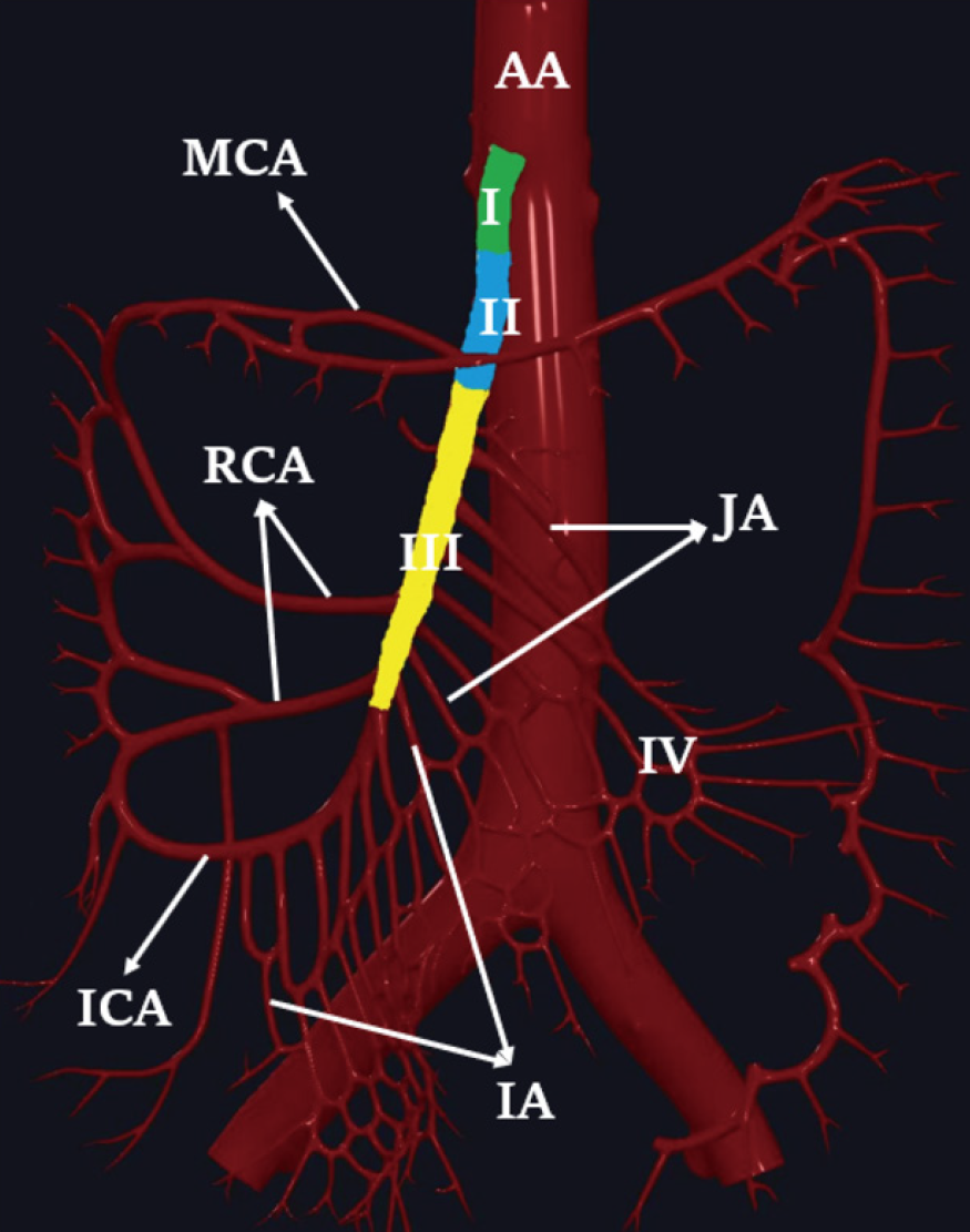
Figure 1 Schematic diagram of the superior mesenteric artery.
AA: Abdominal aorta; MCA: Middle colon artery; RCA: Right colon artery; JA: Jejunal artery; ICA: Ileocolic artery; IA: Ileal artery.
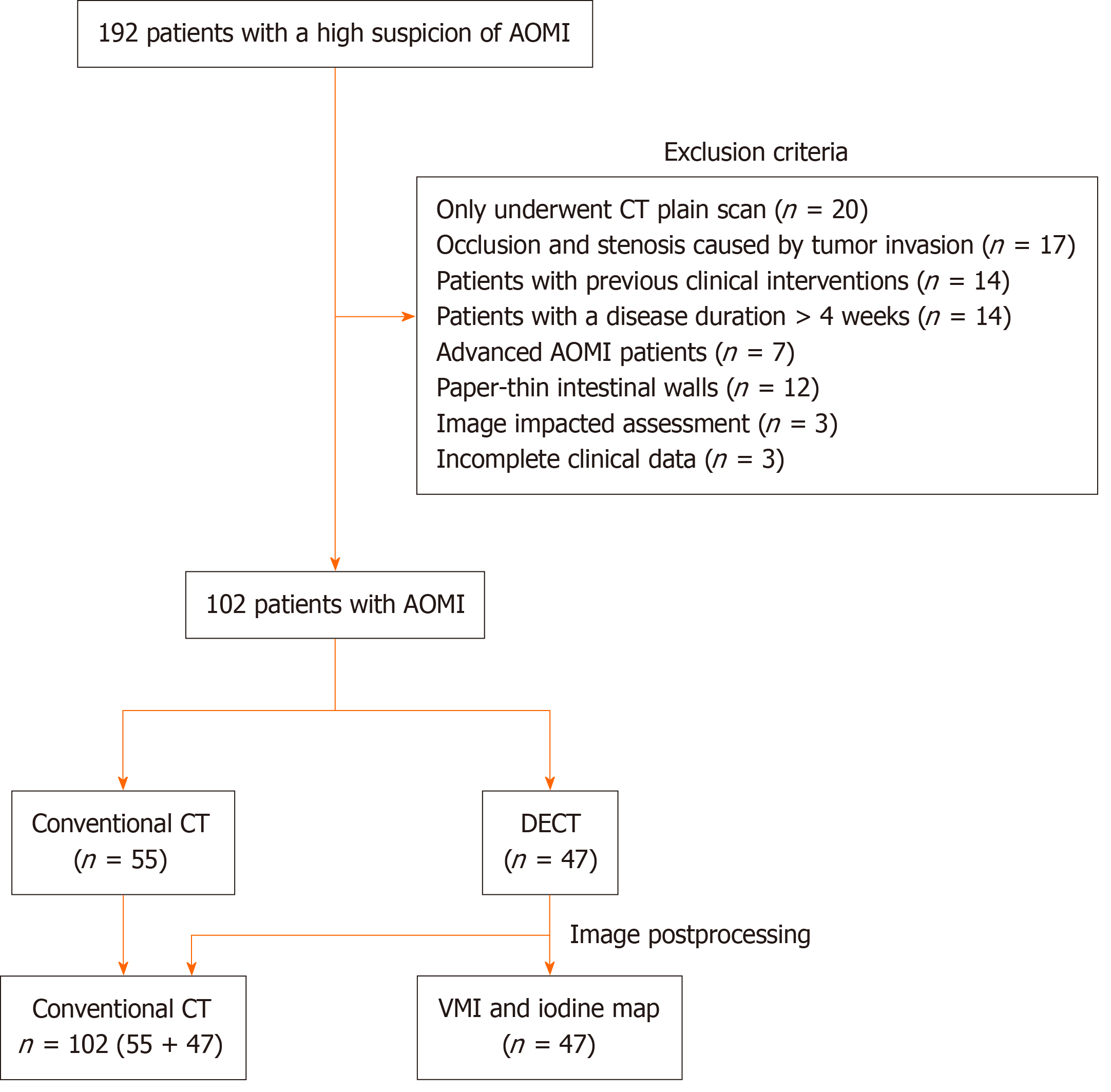
Figure 2 Flow diagram of the study population.
AOMI: Acute occlusive mesenteric ischemia; DECT: Dual-energy computed tomography; VMI: Virtual monoenergetic image; CT: Computed tomography.
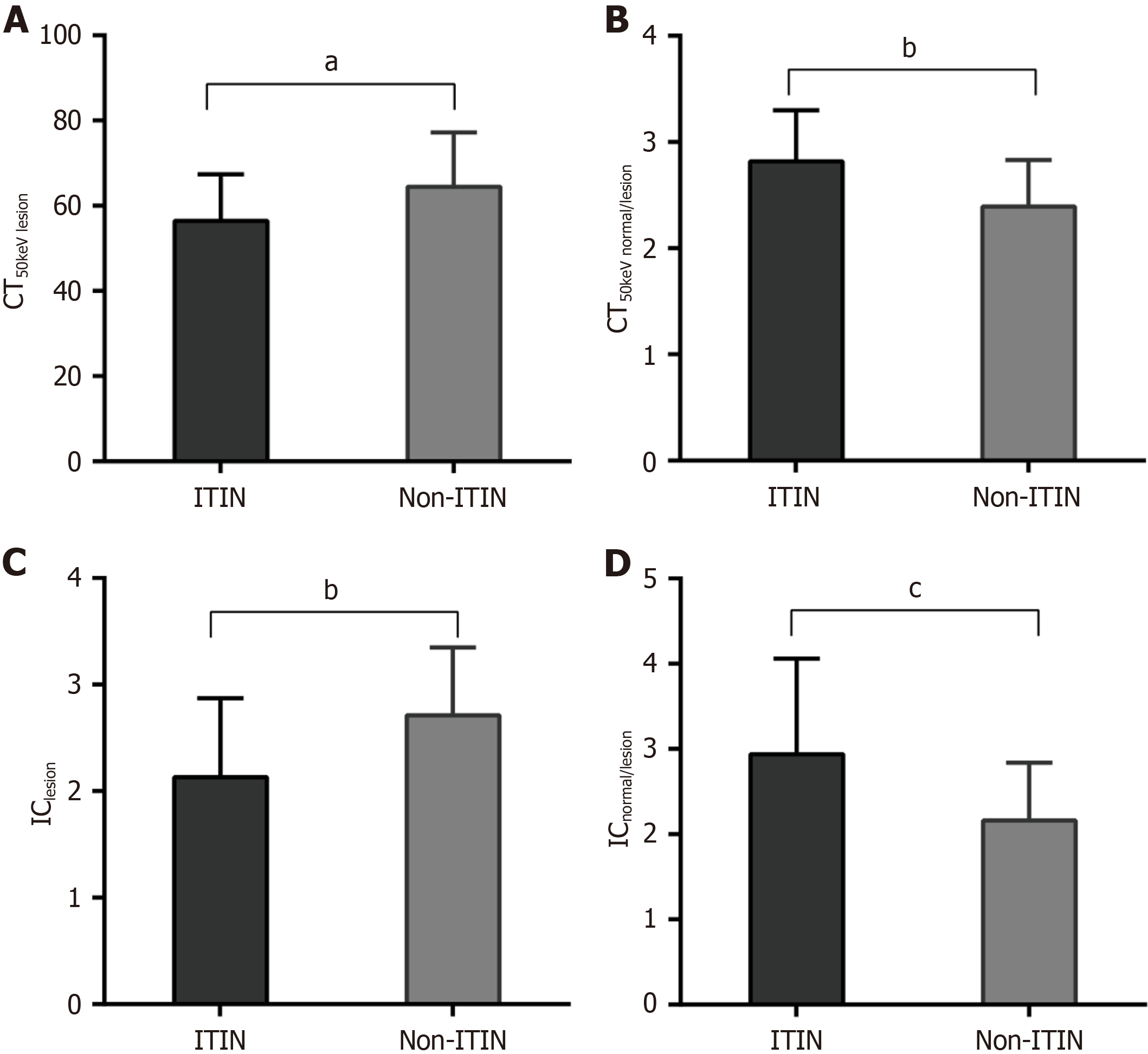
Figure 3 Bar graph shows the differences in the computed tomography 50 keV and iodine concentration values between irreversible transmural intestinal necrosis and non-irreversible transmural intestinal necrosis groups.
A: Computed tomography, 50 keV lesion; B: Computed tomography, 50 keV normal/lesion; C: Iodine concentration, lesion; D: Iodine concentration, normal/lesion. aP < 0.05, bP < 0.01, cP < 0.001. CT: Computed tomography; IC: Iodine concentration; ITIN: Irreversible transmural intestinal necrosis.
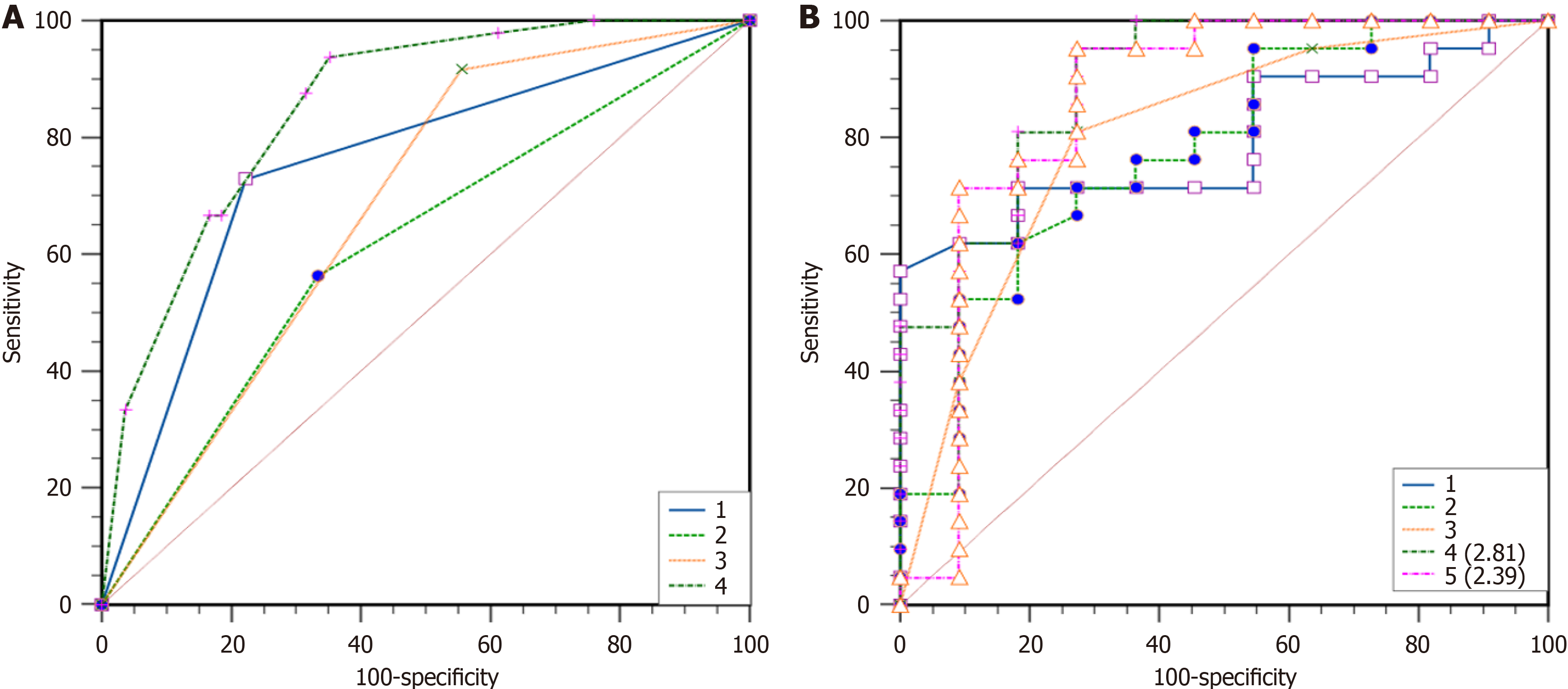
Figure 4 Receiver operating characteristic curves of dual energy computed tomography parameters, computed tomography subjective signs, and combined model for predicting irreversible transmural intestinal necrosis.
A: Comparison of computed tomography (CT) subjective signs in predicting irreversible transmural intestinal necrosis among 102 patients. (1) Multidetector CT combined signs; (2) Decreased or absent intestinal wall reinforcement; (3) Intestinal dilatation; and (4) Substantial organ infarction; B: Comparison of the efficacy of different CT indicators in predicting irreversible transmural intestinal necrosis among 47 patients with dual energy computed tomography scans. (1) Iodine concentration, normal/lesion (ICnormal/lesion); (2) CT50 keV normal/lesion; (3) Combined CT signs; (4) CT50 keV normal/lesion combined with intestinal dilatation and substantial organ infarction (the cut-off value of CT50 keV normal/lesion is 2.81); and (5) ICnormal/lesion combined with intestinal dilatation and substantial organ infarction (the cut-off value of ICnormal/lesion is 2.39).
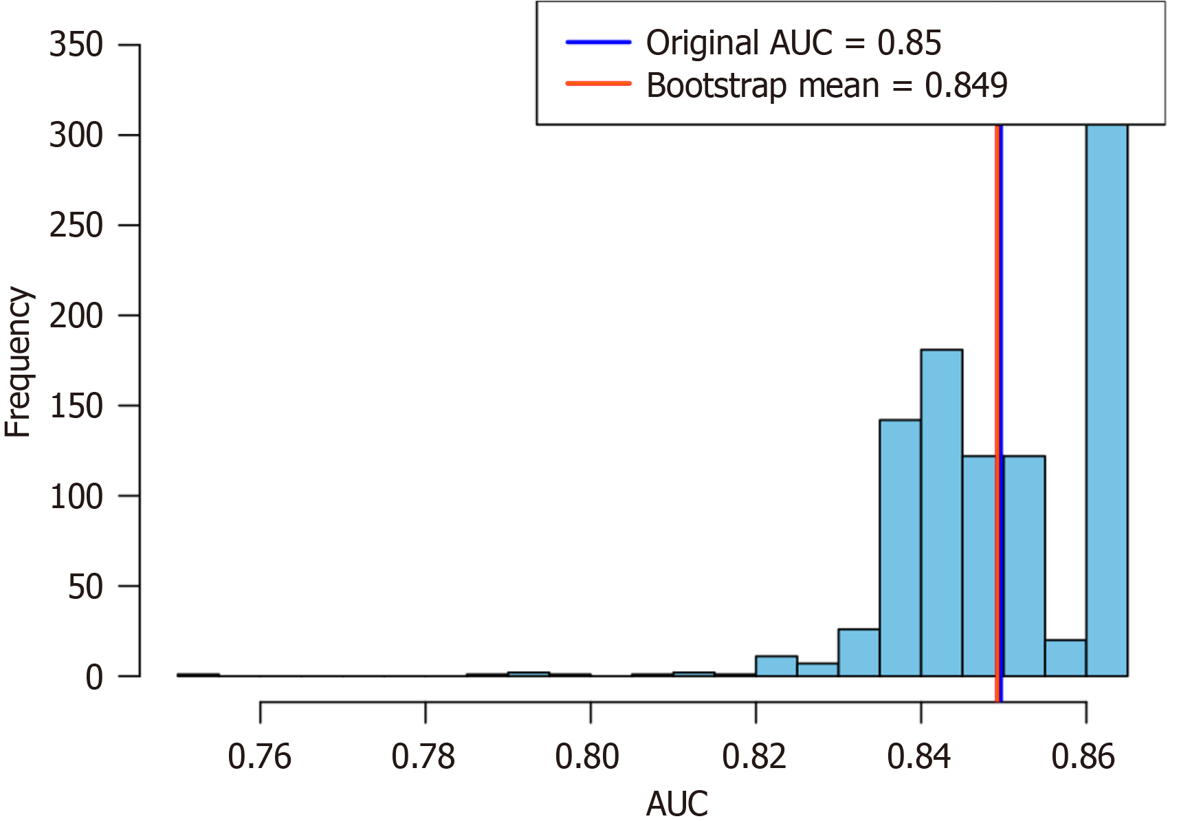
Figure 5 A histogram depicting the area under the curve distribution from the bootstrap analysis (1000 iterations) revealed an original area under the curve of 0.
894 and a mean area under the curve of 0.888. AUC: Area under the curve.
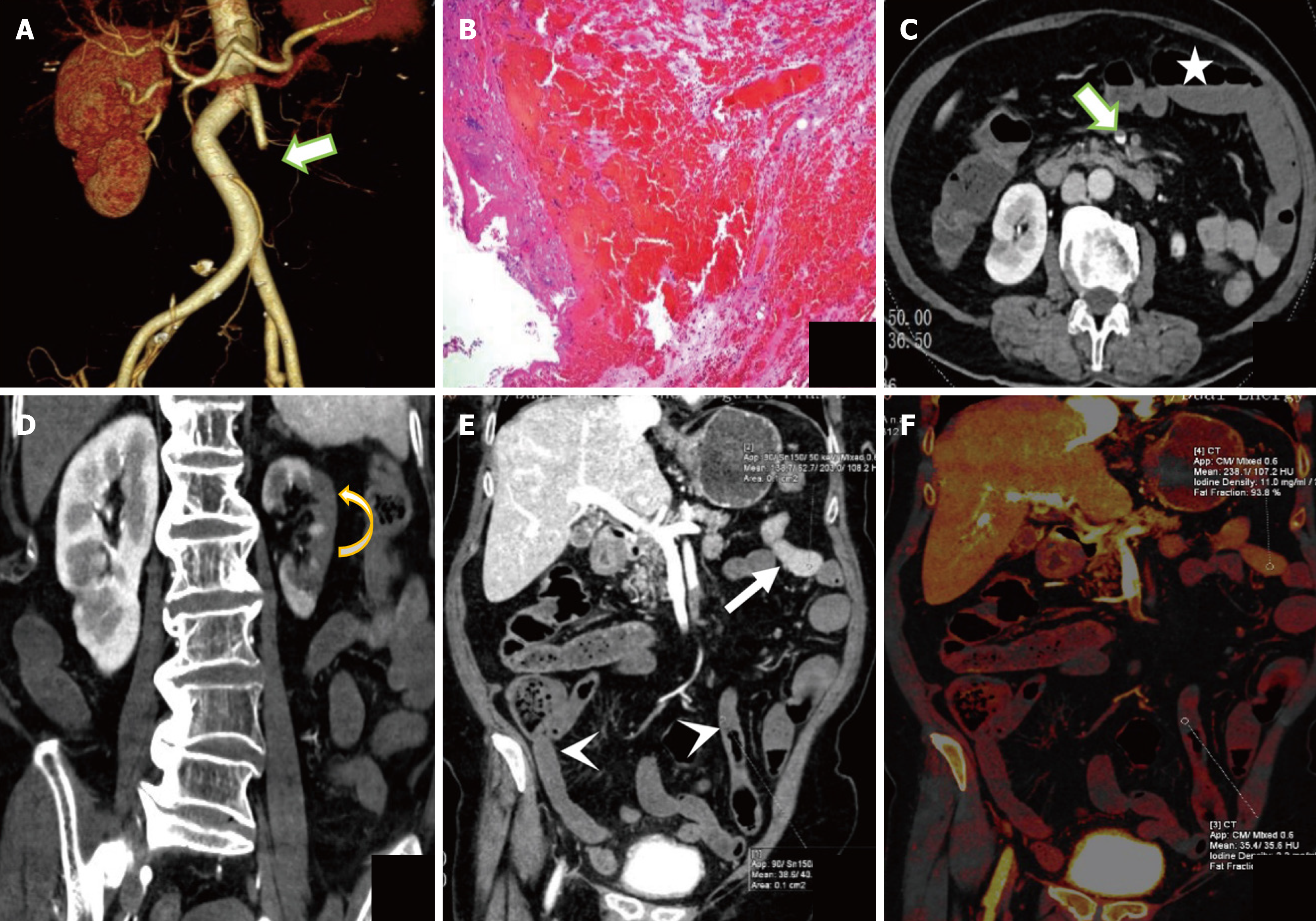
Figure 6 The representative dual energy computed tomography image of a 76-year-old woman with irreversible transmural intestinal necrosis.
A: Volume rendering technique image; B: Hematoxylin and eosin staining images; C and D: Transverse and coronal images of the portal vein phase at 120 kVp; E: Portal vein phase at 50 keV image; F: Iodine density. Dual energy computed tomography examination reveals filling defect in superior mesenteric artery (area II + III + IV) and superior mesenteric vein (thick arrow in A and C), decreased and absent diffuse intestinal wall enhancement (arrowhead in E), normal intestinal duct (thin arrow in E), intestinal dilatation (star in C), and left renal infarction (curved arrow in D). Computed tomography 50 keV normal/lesion = 4.33, iodine concentration normal/lesion = 5.00.
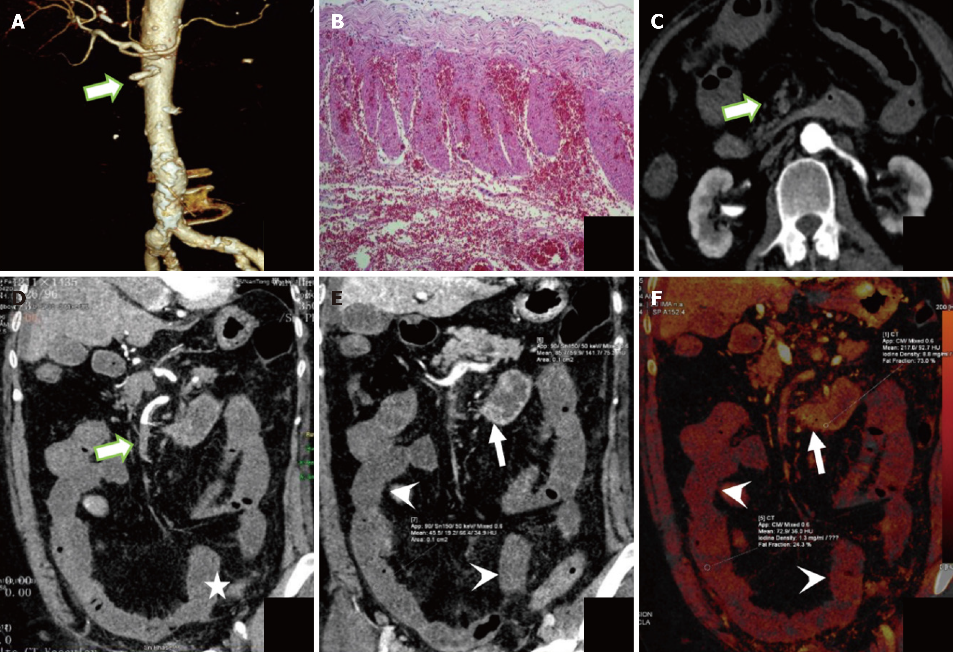
Figure 7 The representative dual energy computed tomography image of a 75-year-old woman with irreversible transmural intestinal necrosis.
A: Volume rendering technique image; B: Hematoxylin and eosin staining images; C and D: Transverse and coronal images of the portal vein phase at 120 kVp; E: Portal vein phase at 50 keV image; F: Iodine density. Dual energy computed tomography examination reveals filling defect in superior mesenteric artery (area I+ II + III + IV) (thick arrow in A, C, and D), decreased and absent diffuse intestinal wall enhancement (arrowhead in E and F), normal intestinal duct (thin arrow in E and F), intestinal dilatation (star in D). Computed tomography 50 keV normal/lesion = 2.13, iodine concentration normal/lesion = 6.77.
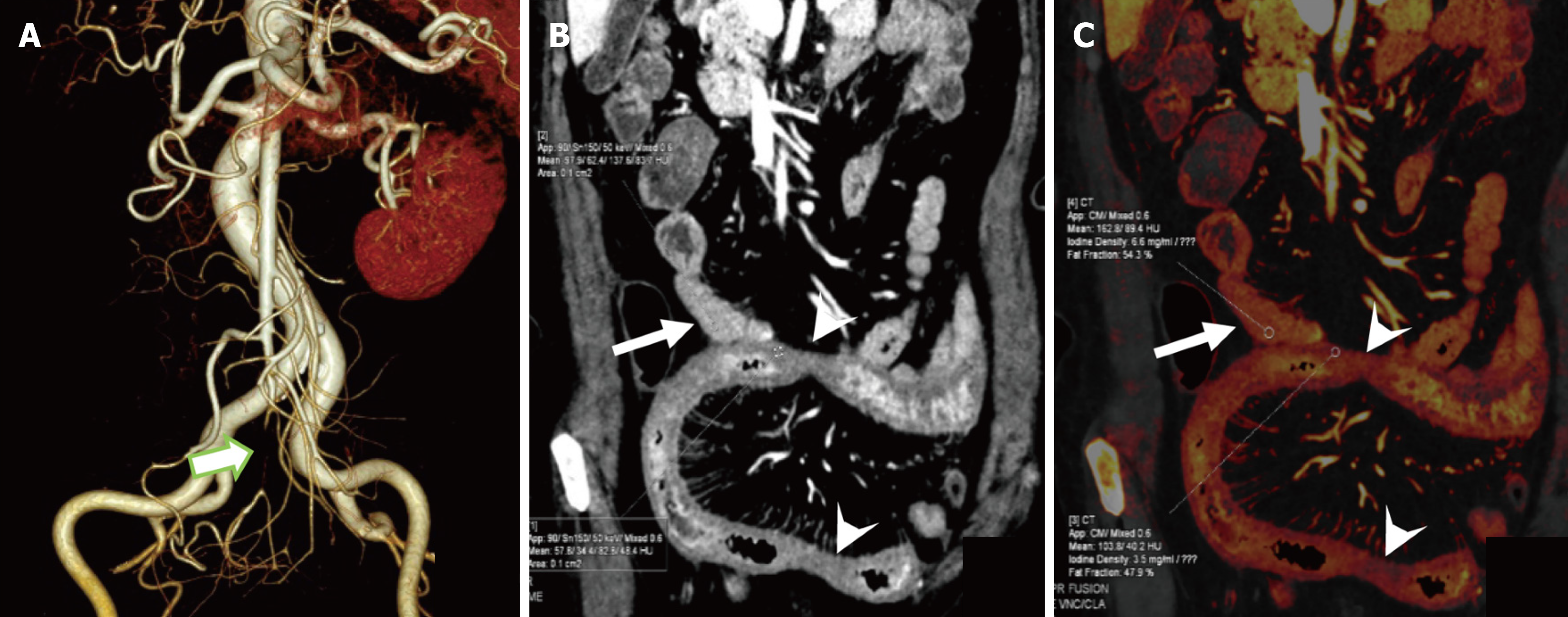
Figure 8 The representative dual energy computed tomography image of a 71-year-old woman without irreversible transmural intestinal necrosis.
A: Volume rendering technique image; B: Portal vein phase at 50 keV image; C: Iodine density. Dual energy computed tomography examination shows a filling defect in the superior mesenteric artery (area IV, thick arrow in A), diffuse thickening, and decreased enhancement of the ileal wall (arrowhead in B and C). Adjacent mesangial swelling, normal bowel (thin arrow in B and C), and no intestinal dilatation or other computed tomography signs of substantial organ infarction. Computed tomography 50 keV normal/lesion = 1.66; iodine concentration normal/lesion = 1.89.
- Citation: Yang JS, Xu ZY, Chen FX, Wang MR, Fan XL, He BS. Diagnostic value of dual-energy computed tomography in irreversible transmural intestinal necrosis in patients with acute occlusive mesenteric ischemia. World J Gastrointest Surg 2025; 17(7): 105956
- URL: https://www.wjgnet.com/1948-9366/full/v17/i7/105956.htm
- DOI: https://dx.doi.org/10.4240/wjgs.v17.i7.105956


