Copyright
©The Author(s) 2025.
World J Gastrointest Surg. Jul 27, 2025; 17(7): 104985
Published online Jul 27, 2025. doi: 10.4240/wjgs.v17.i7.104985
Published online Jul 27, 2025. doi: 10.4240/wjgs.v17.i7.104985
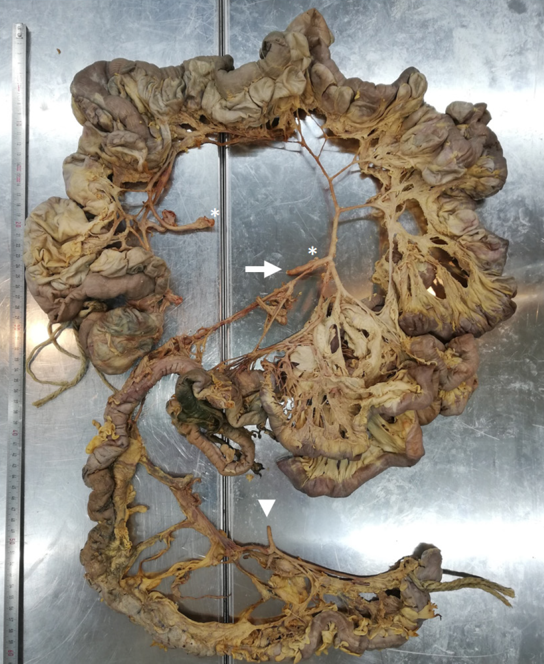
Figure 1 Overview of intestine harboring diverticula.
Intestinal tract including jejunum, ileum, and colon. Jejunum hosting diverticula over a length of 208 cm. Arrows mark superior mesenteric artery. Asterisks mark cut connection of artery branch due to preparation and visualization. Arrowheads mark inferior mesenteric artery.
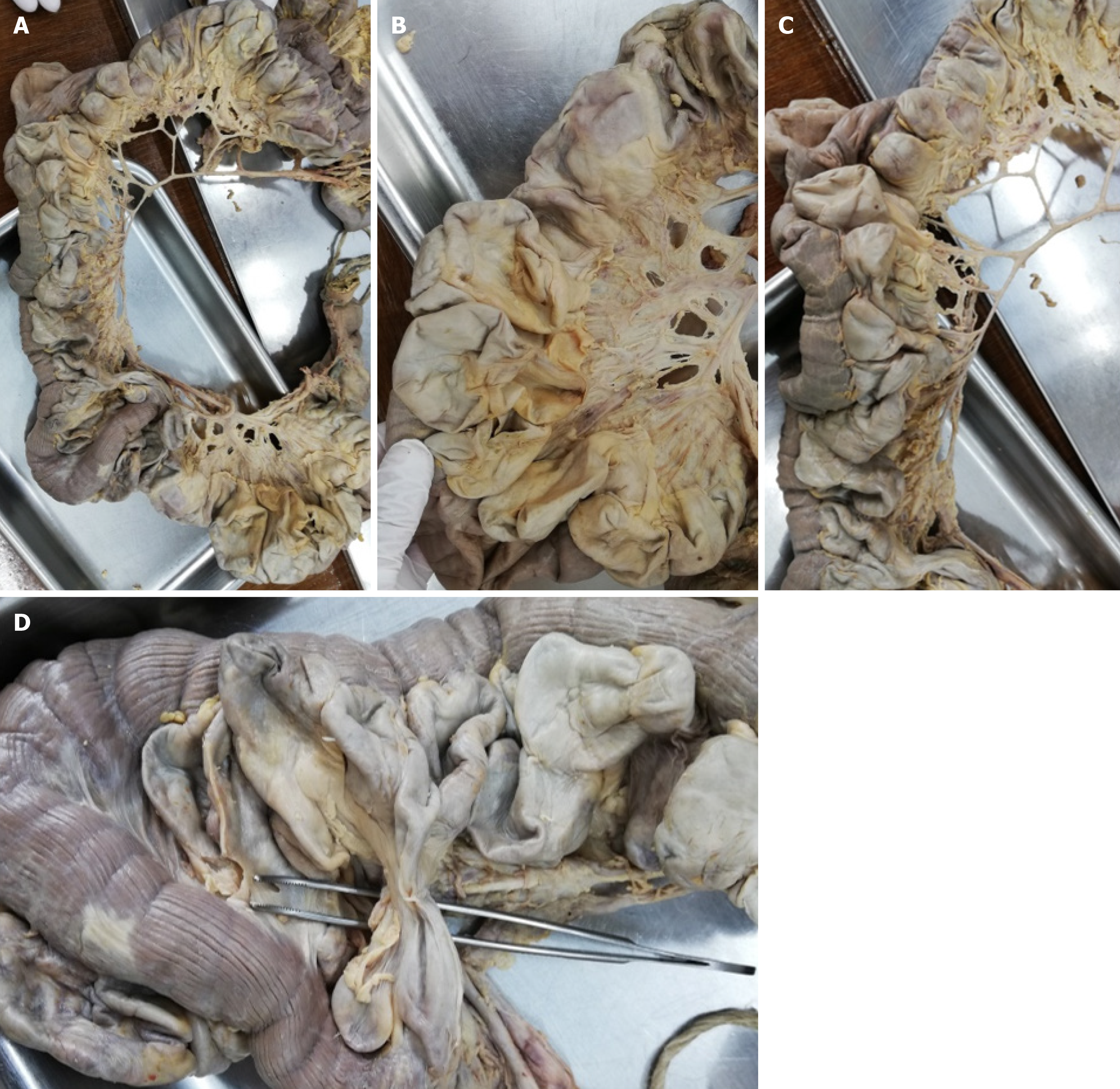
Figure 2 Diverticula.
A-C: Representative diverticula; D: Shortcut between two intestinal parts provided by diverticula.
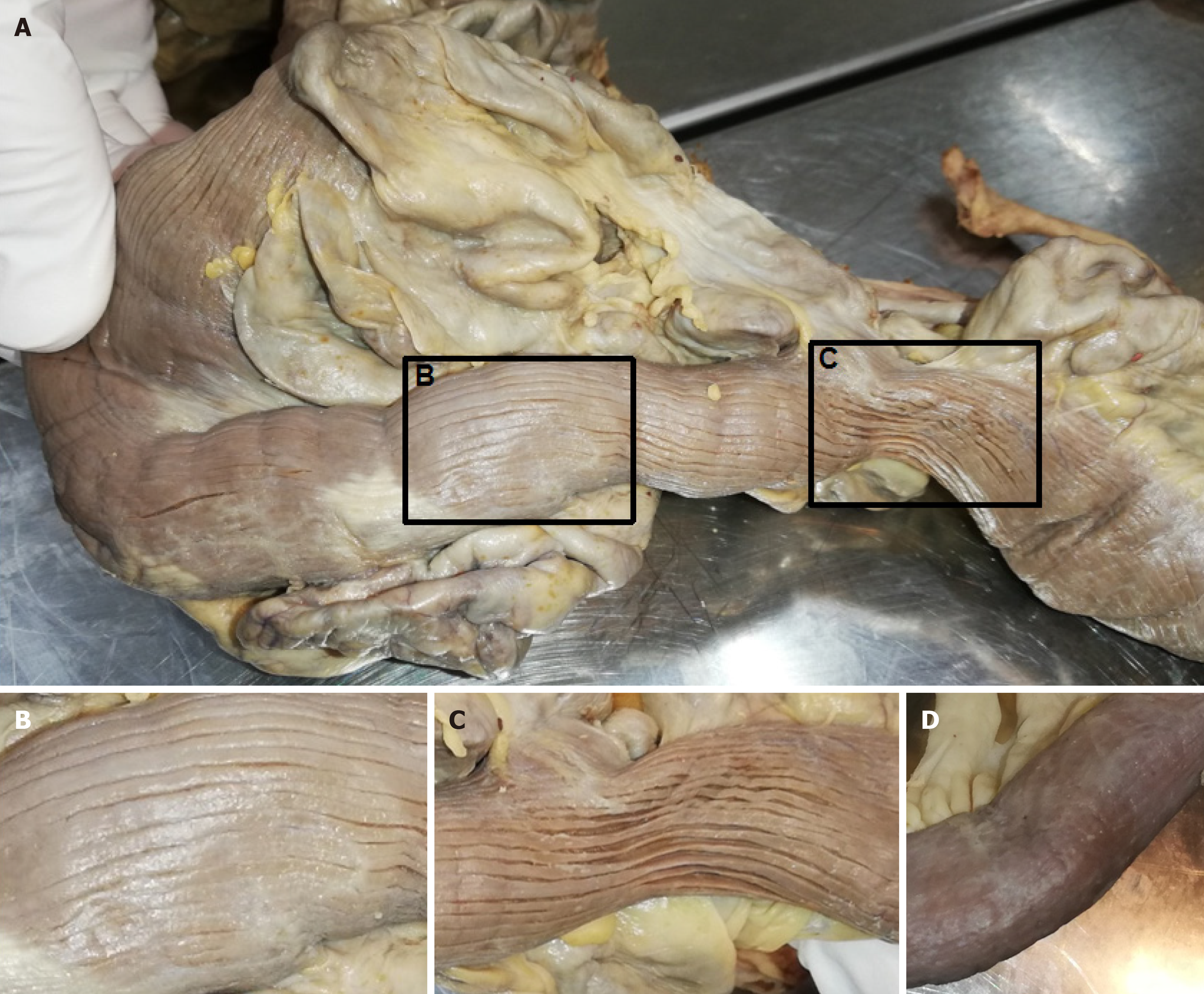
Figure 3 Longitudinal striation of jejunum with diverticula.
A: Intestinal part hosting diverticula showed remarkable longitudinal striation. Black box marks magnified part shown in B and C; B and C: Magnification of longitudinal striation; D: Intestinal part without diverticula.
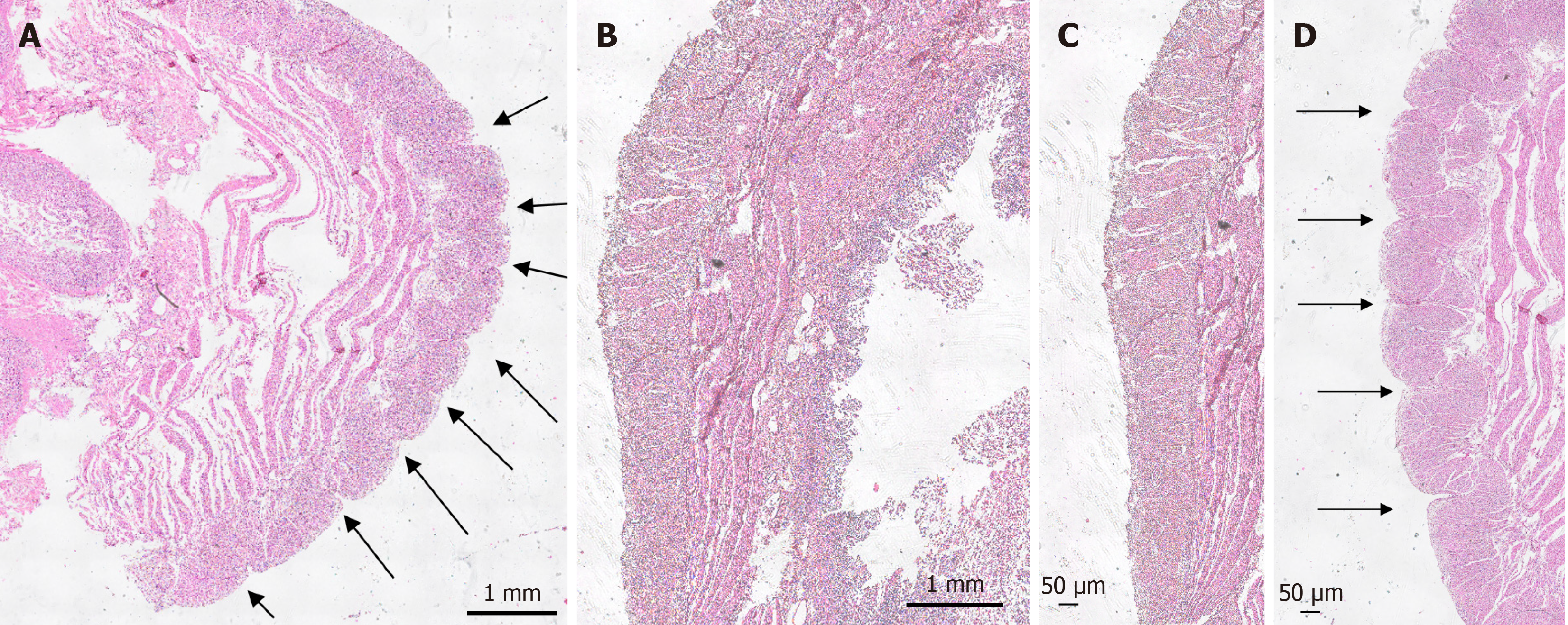
Figure 4 Microscopic investigation of longitudinal striation of jejunum with diverticula.
A and B: Hematoxylin and eosin (H&E)-stained diverticula and control tissue; C and D: Magnification of H&E-stained diverticula and control tissue. Arrows mark indentations responsible for longitudinal striation.
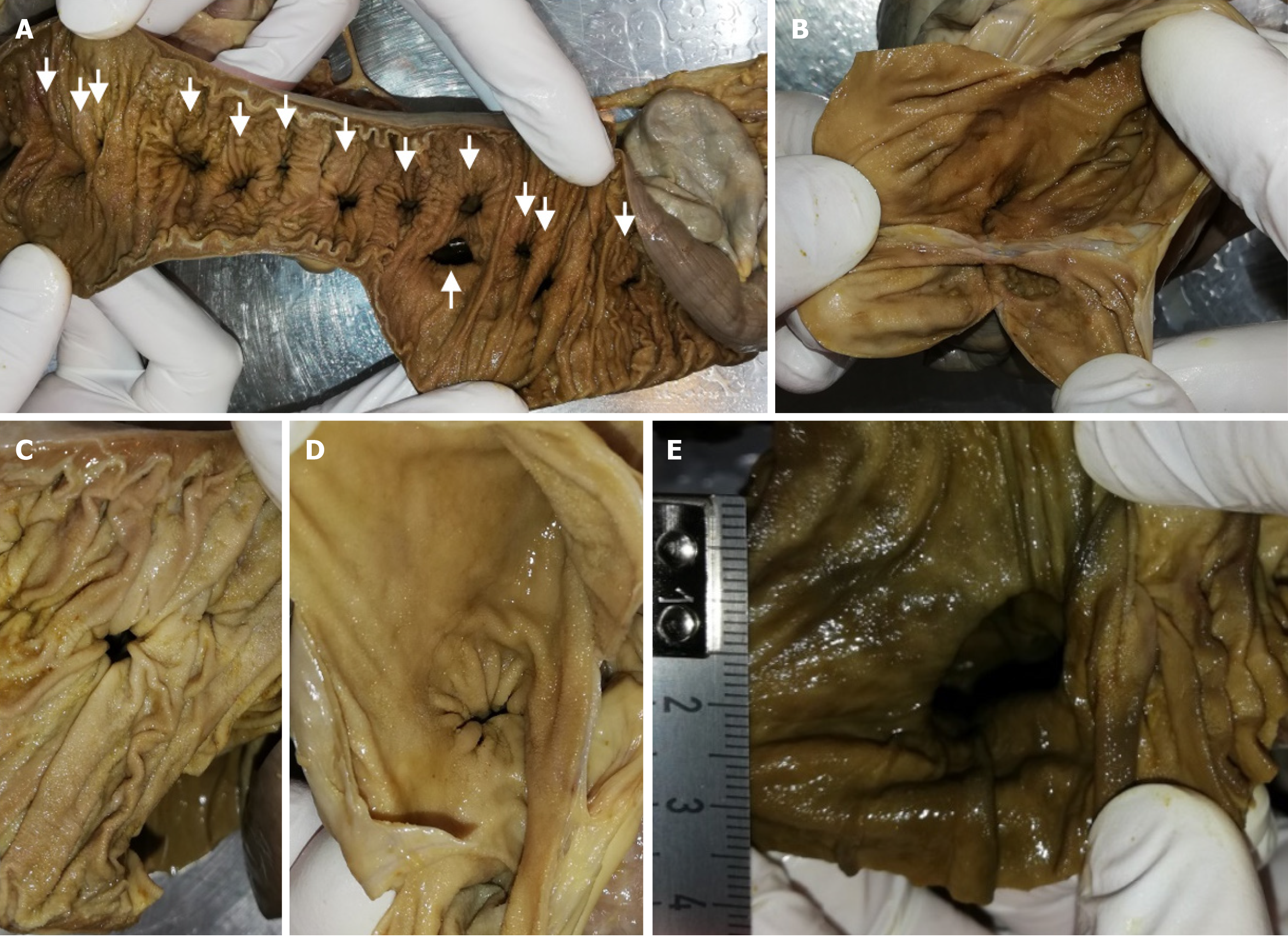
Figure 5 Lumen of intestine and diverticula.
A: Intestinal tract was cut longitudinally. Arrows mark diverticula entry points; B: Representative diverticula were opened; C and D: Diverticula entry points pictured from intestinal site (C) and from diverticulum site (D); E: Large diverticulum entry points with a diameter of 2 cm.
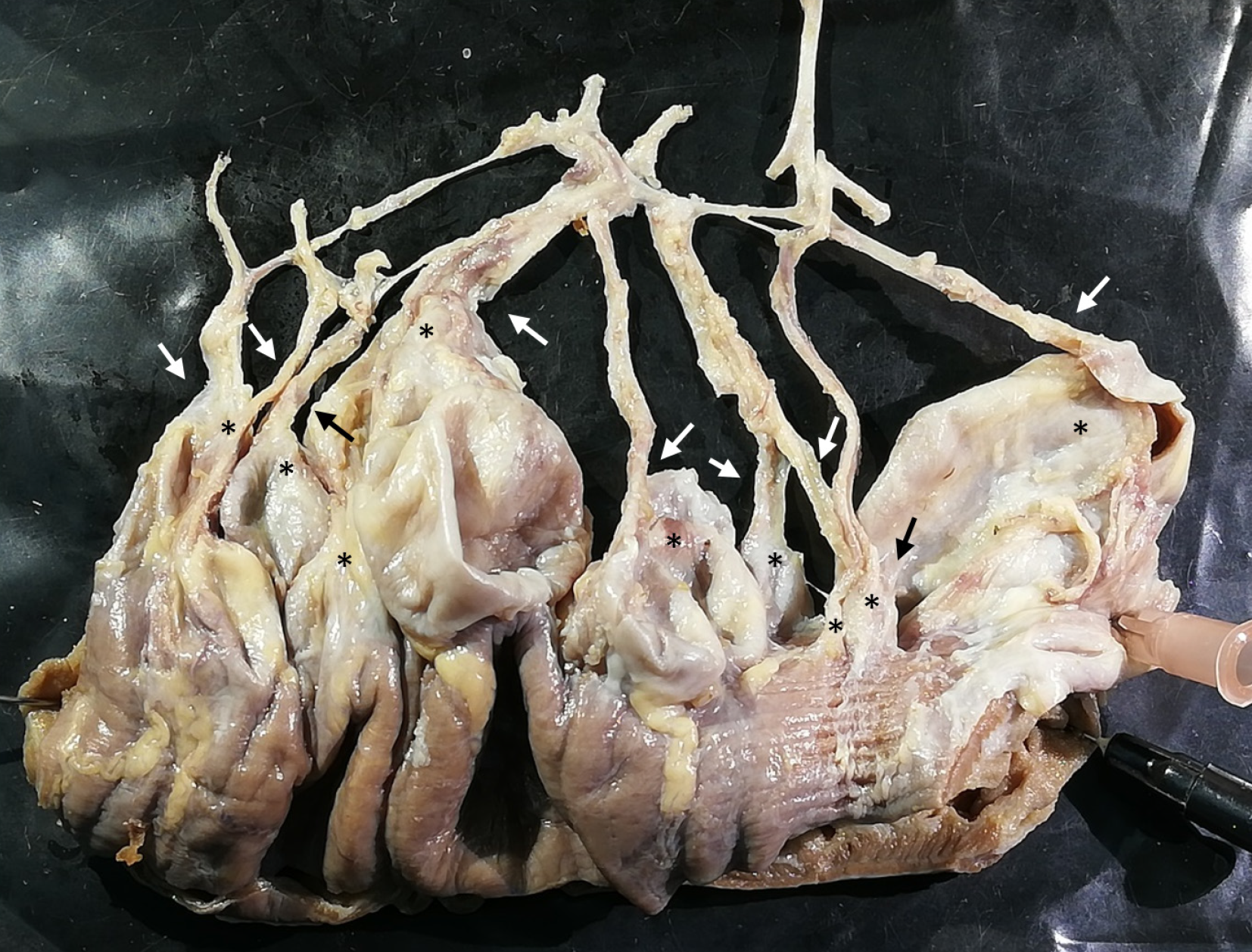
Figure 6 Relationship between vessels and diverticula.
Representative part of the jejunum (8 cm) with diverticula was cut out and dissected utilizing a dissecting microscope. All remaining fat tissue and intestine covering tunica serosa was removed, vessels were stretched out, and entry sites were exposed. Arrows mark arteria recta entering diverticula (marked with an asterisk). All investigated vessels end in diverticula, and all investigated diverticula were supplied by a vessel.
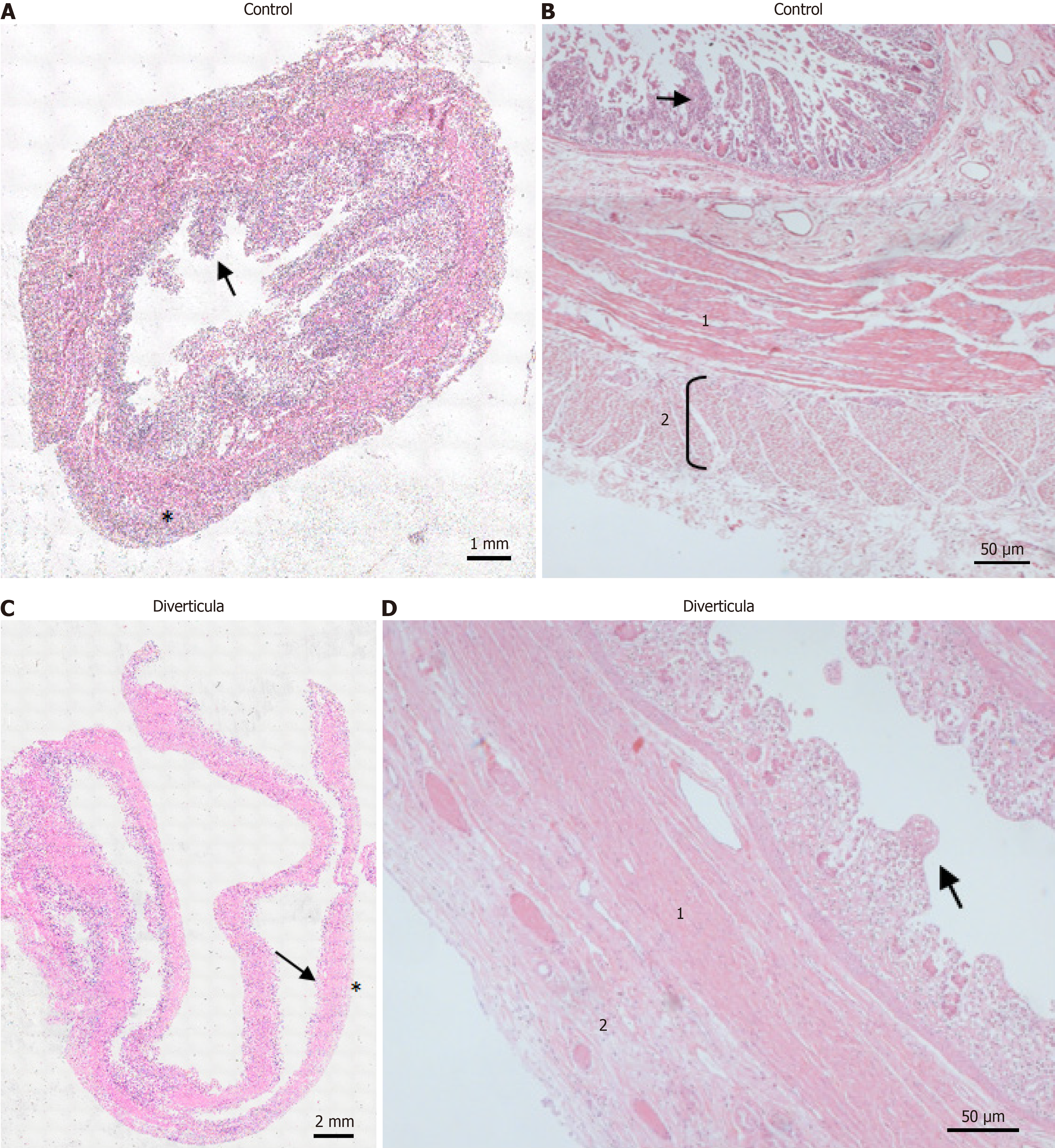
Figure 7 Histological investigation of diverticula.
A and C: Hematoxylin and eosin (H&E)-stained control tissue (A) and diverticula (C). Arrows mark intestinal villi. Asterisks mark stratum longitudinale; B and D: H&E-stained control tissue (B) and diverticula (D) in a higher magnification. 1: Stratum circular; 2: Stratum longitudinale. Arrows mark intestinal villi.
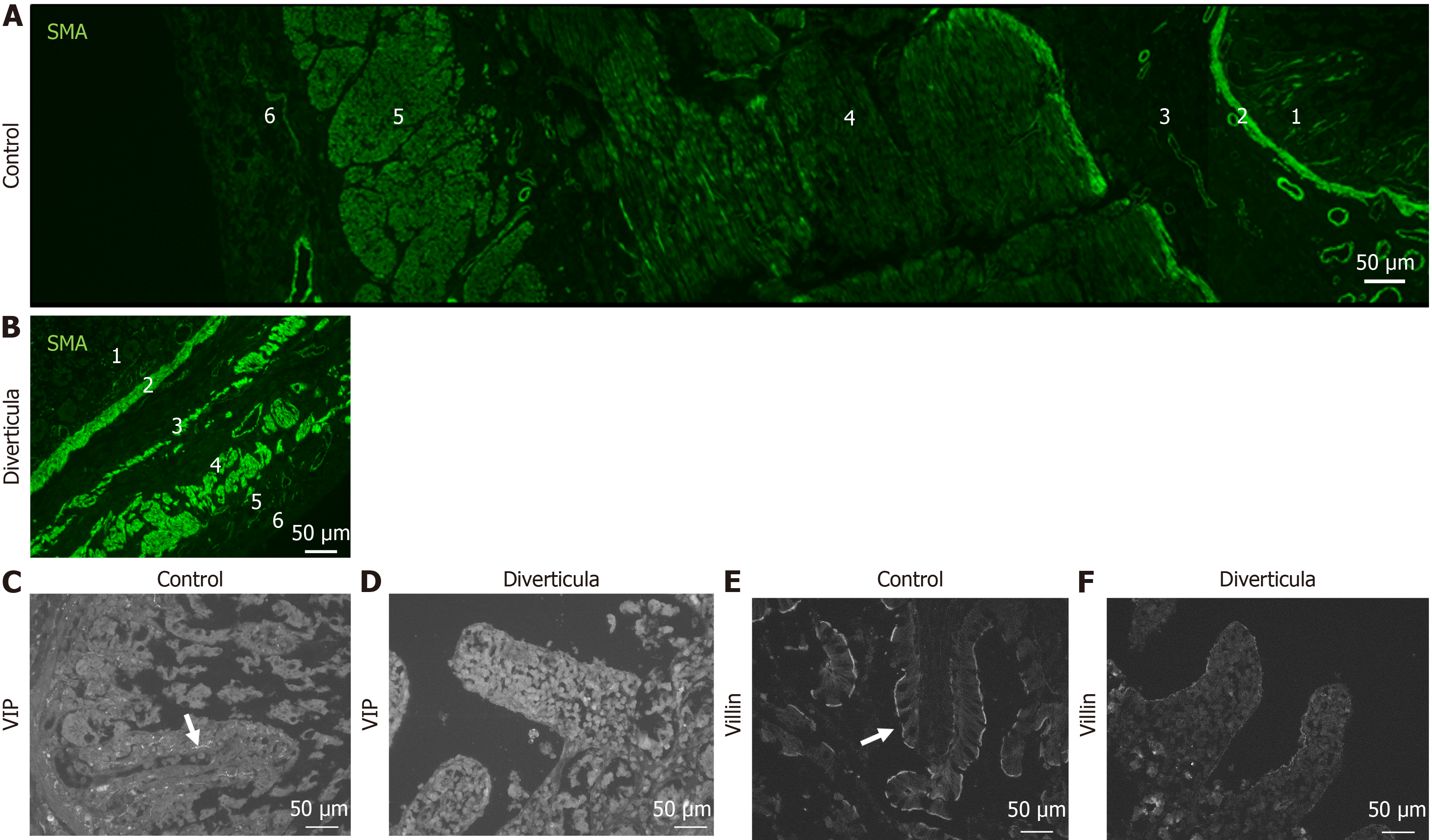
Figure 8 Immunohistochemical investigation of diverticula.
A and B: Immunohistochemical staining with smooth muscle actin antibody of control tissue (A) and diverticula (B); C-F: Investigation of intestinal villi in control tissue (C and E) and diverticula (D and F) utilizing vasoactive intestinal peptide (VIP; C and D) and villin (E and F) to mark VIP-positive fibers and brush border, respectively. 1: Intestinal villi; 2: Lamina muscularis mucosa; 3: Lamina submucosae; 4: Stratum circular; 5: Stratum longitudinale; 6: Adventitia/serosa.
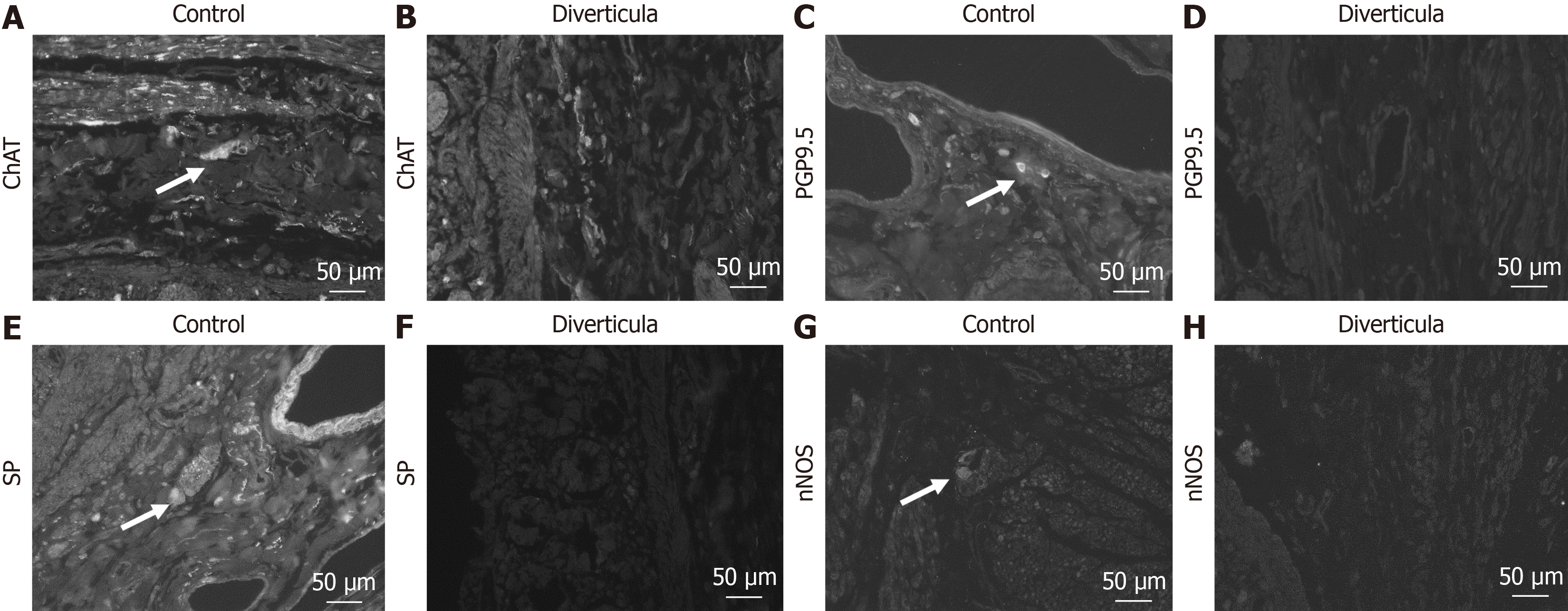
Figure 9 Immunohistochemical investigation of submucosal plexus in diverticula.
A-H: Detection of submucosal plexus in control tissue (A, C, E, and G) and diverticula (B, D, F, and H) utilizing choline acetyltransferase (ChAT) (A and B), protein gene product 9.5 (PGP9.5) (C and D), substance P (SP) (E and F) and neuronal nitric oxide synthase (nNOS) (G and H) antibody. Arrows mark positive cells in the plexus.
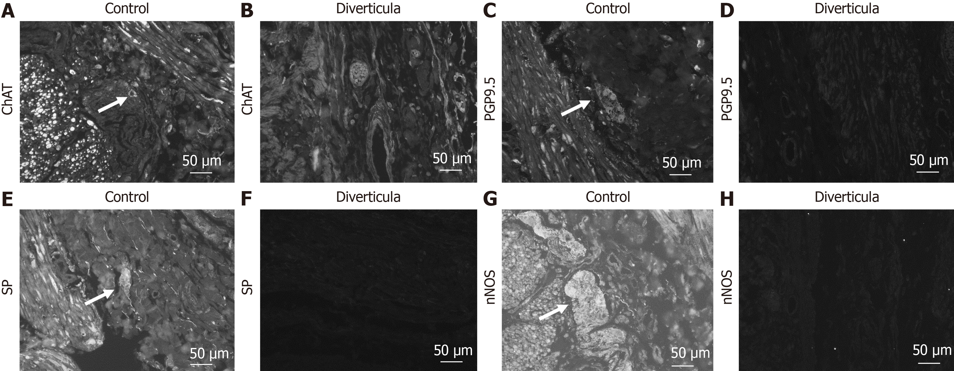
Figure 10 Immunohistochemical investigation of myenteric plexus in diverticula.
A-H: Detection of myenteric plexus in control tissue (A, C, E, and G) and diverticula (B, D, F, and H) utilizing choline acetyltransferase (ChAT) (A and B), protein gene product 9.5 (PGP9.5) (C and D), substance P (SP) (E and F) and neuronal nitric oxide synthase (nNOS) (G and H) antibody. Arrows mark positive cells in the plexus.
- Citation: Schmidt P, Perniss A, Nassenstein C, Keller H, Deckmann K. Multiple jejunal diverticulosis, from an anatomical and histological view: A case report. World J Gastrointest Surg 2025; 17(7): 104985
- URL: https://www.wjgnet.com/1948-9366/full/v17/i7/104985.htm
- DOI: https://dx.doi.org/10.4240/wjgs.v17.i7.104985


