Copyright
©The Author(s) 2025.
World J Gastrointest Surg. May 27, 2025; 17(5): 104893
Published online May 27, 2025. doi: 10.4240/wjgs.v17.i5.104893
Published online May 27, 2025. doi: 10.4240/wjgs.v17.i5.104893
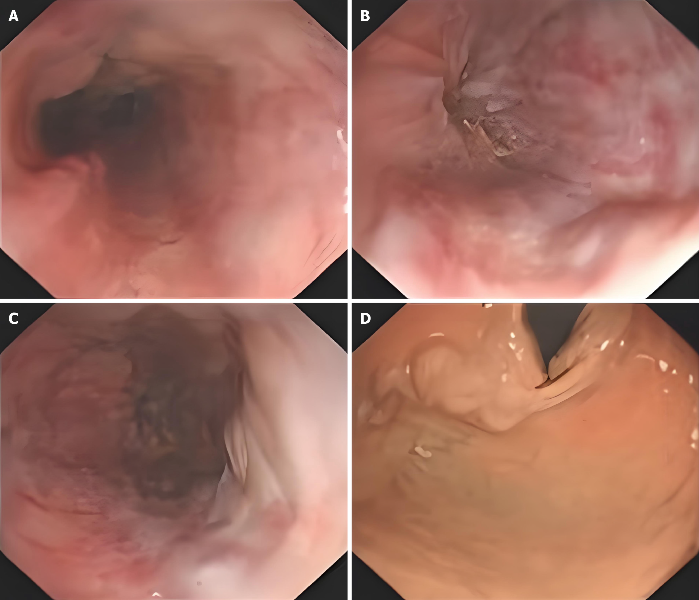
Figure 1 Gastroscopic findings.
Gastroscopy examination showed severe varices consistent with portal hypertensive gastropathy. A: Mid-esophageal varices; B and C: Lower esophageal varices; D: Gastric fundus varices.
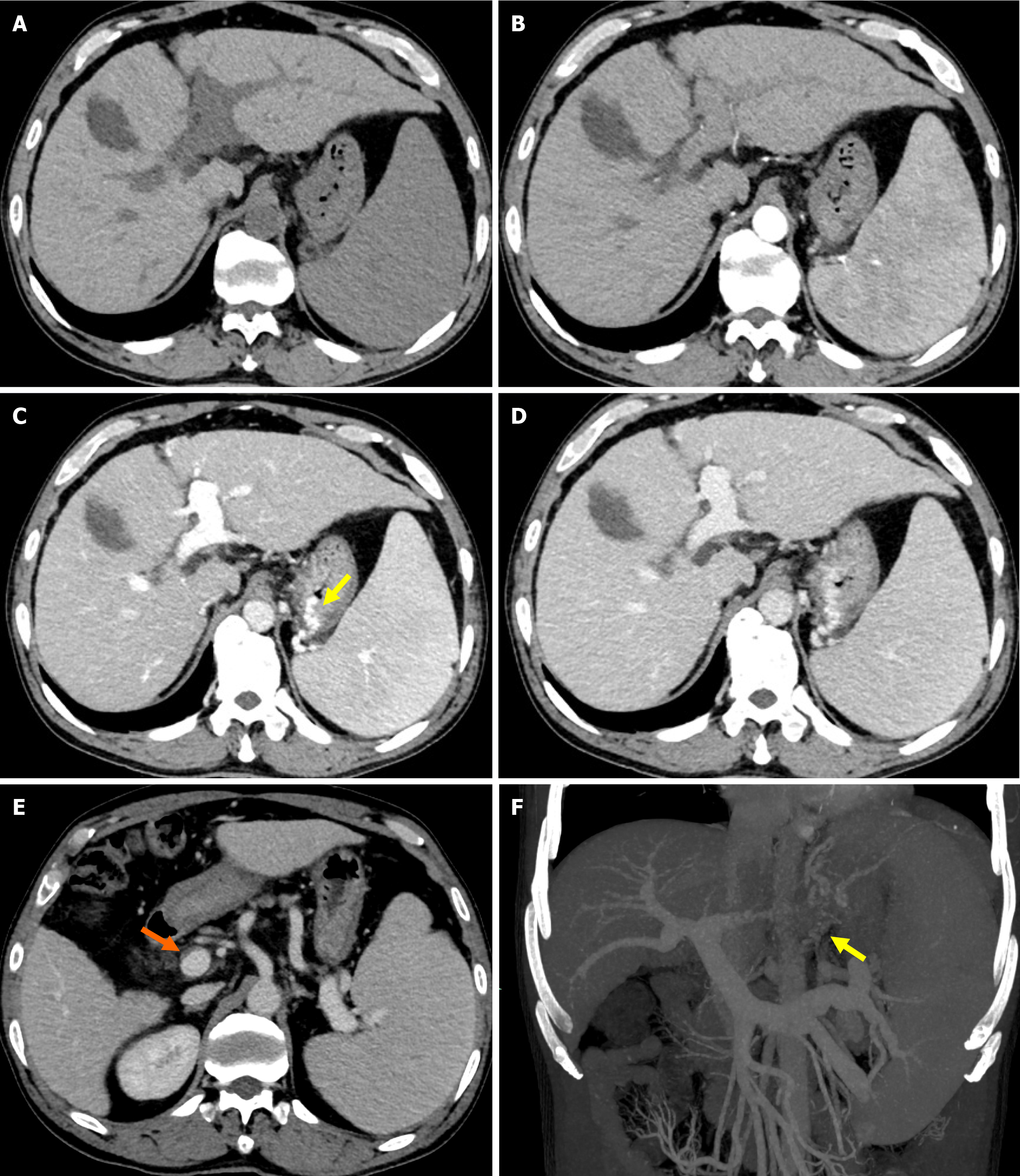
Figure 2 Abdominal computed tomography findings: Abdominal computed tomography revealed cirrhosis, splenomegaly, portal hypertension with collateral circulation formation (yellow arrow), and mural thrombi in the main portal vein (orange arrow), superior mesenteric vein, and splenic vein.
A: Unenhanced phase; B: Hepatic arterial phase; C: Portal venous phase; D: Delayed phase; E: Axial portal venous phase; F: Coronal reconstruction.
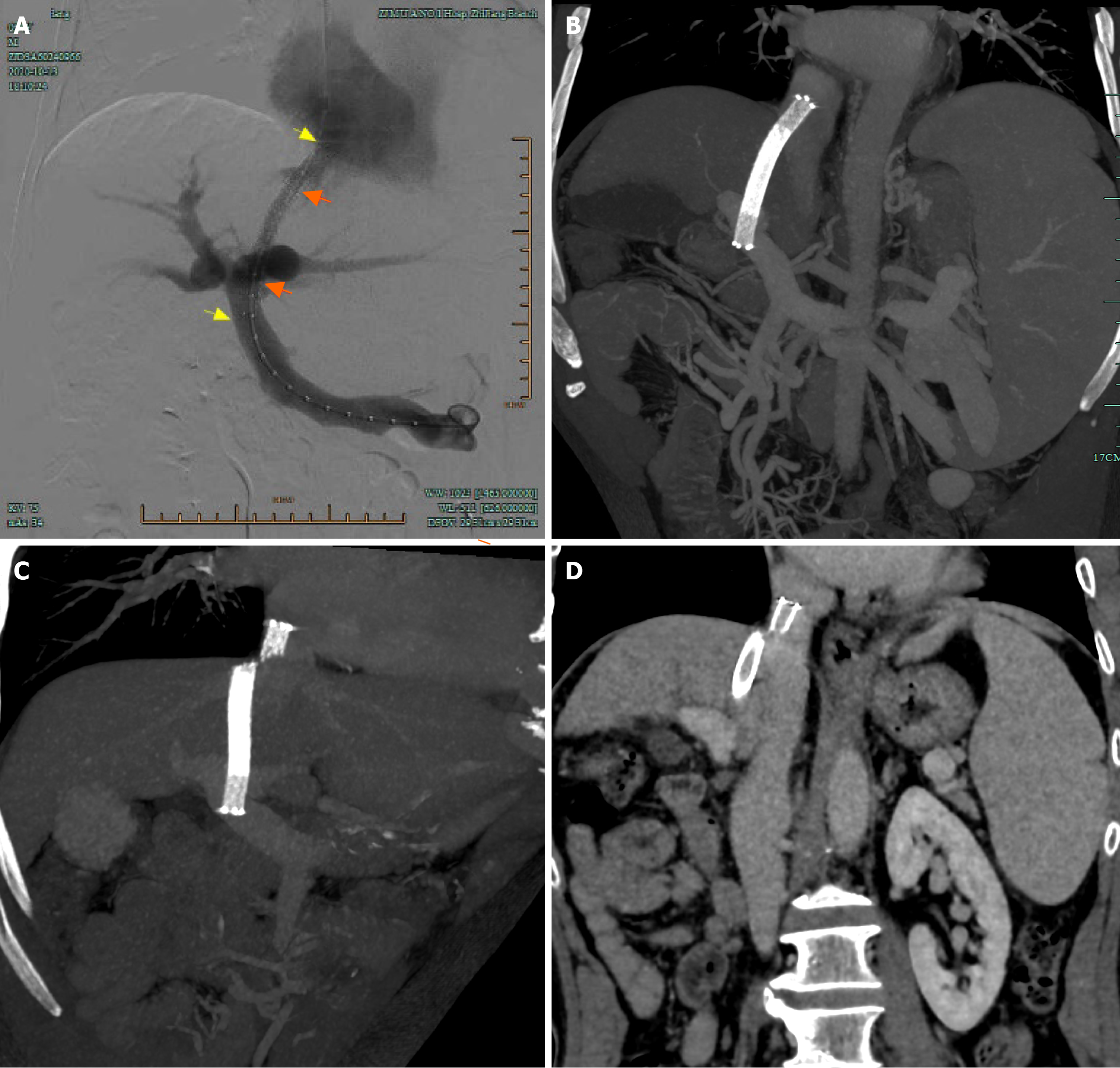
Figure 3 Imaging findings.
A: Portography immediately after TIPS creation shows a patent shunt. Orange arrows and yellow arrows indicate both ends of the stent-graft and the bare metal stent, respectively; B: Coronal contrast-enhanced CT at 1 month post-procedure confirms intact stent structural integrity; C and D: Fifteen-month follow-up coronal CT reveals fracture of the bare metal stent at the hepatocaval confluence (arrowheads), while the stent lumen remains patent.
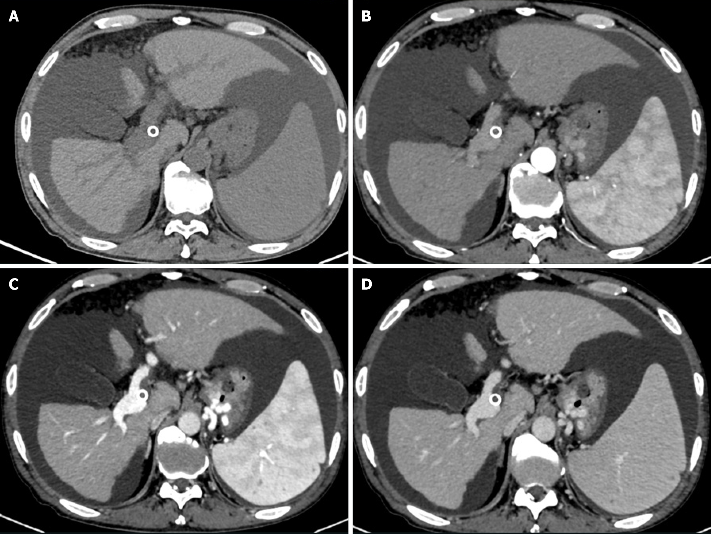
Figure 4 Thirty-month follow-up computed tomography after transjugular intrahepatic portosystemic shunt.
Computed tomography demonstrates shunt thrombosis accompanied by recurrent esophagogastric varices and massive ascites. A: Unenhanced phase; B: Hepatic arterial phase; C: Portal venous phase; D: Delayed phase.
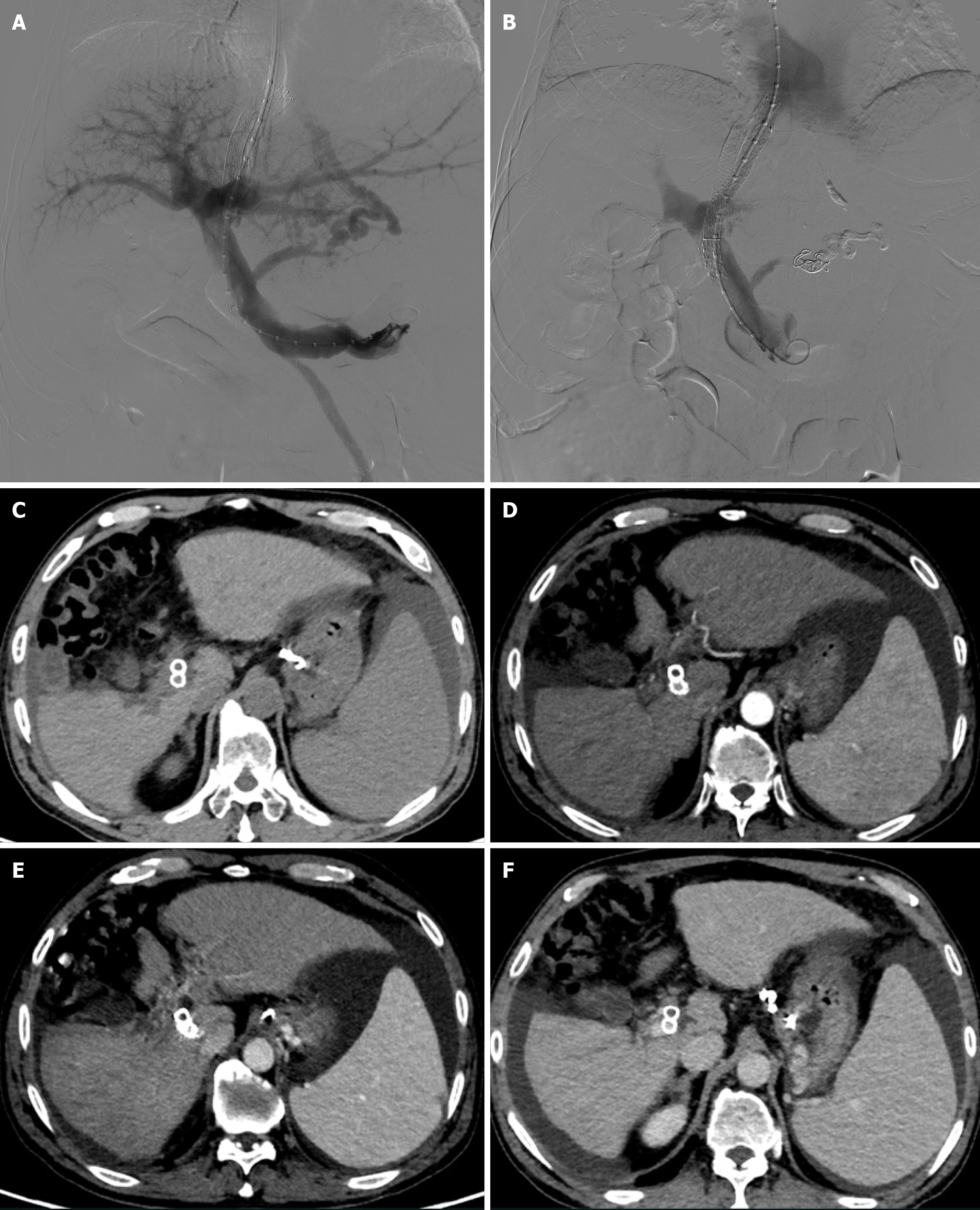
Figure 5 Treatment procedure.
A and B: A parallel transjugular intrahepatic portosystemic shunt (TIPS) was successfully created via the proximal end of the fractured stent. The esophagogastric varices were embolized with cyanoacrylate glue and microcoils due to persistent visibility of gastroesophageal varices post-shunt; C-F: One-month follow-up contrast-enhanced abdominal computed tomography after parallel TIPS demonstrated patent blood flow in the new shunt, complete variceal occlusion, and significant ascites reduction: Unenhanced phase (C); Hepatic arterial phase (D); Portal venous phase (E); Delayed phase (F).
- Citation: Zhou TY, Wang HL, Tao GF, Chen SQ. Stent fracture after transjugular intrahepatic portosystemic shunt: A case report. World J Gastrointest Surg 2025; 17(5): 104893
- URL: https://www.wjgnet.com/1948-9366/full/v17/i5/104893.htm
- DOI: https://dx.doi.org/10.4240/wjgs.v17.i5.104893


