Copyright
©The Author(s) 2025.
World J Gastrointest Surg. Apr 27, 2025; 17(4): 104102
Published online Apr 27, 2025. doi: 10.4240/wjgs.v17.i4.104102
Published online Apr 27, 2025. doi: 10.4240/wjgs.v17.i4.104102
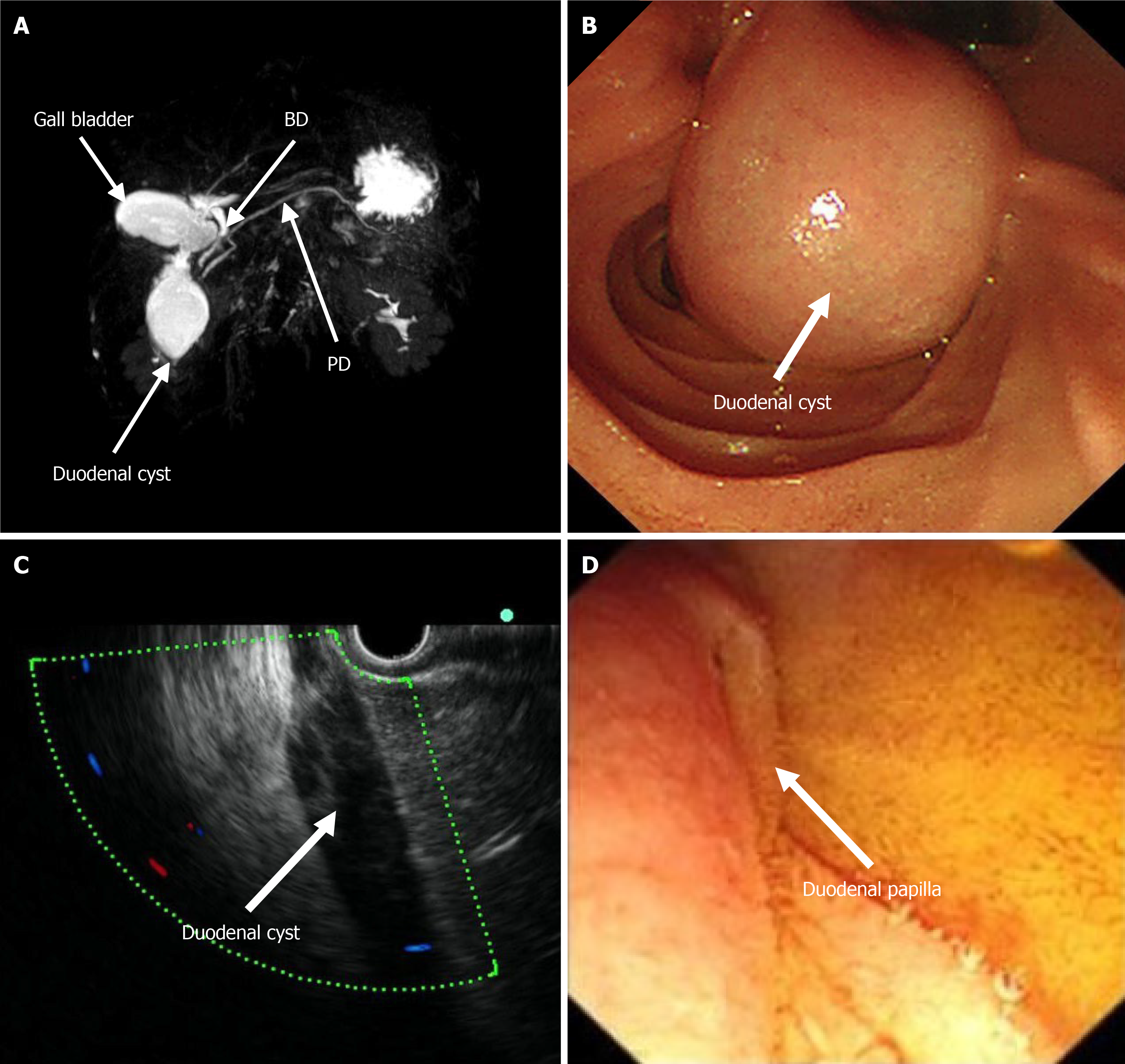
Figure 1 Imaging of the patient prior to treatment.
A: Magnetic resonance imaging revealed a duodenal cyst with normal bile and pancreatic ducts; B: Endoscopy revealed a cystic mass in the descending part of the duodenum; C: Endoscopic ultrasonography revealed a homogeneous hypoecho without blood flow; D: The duodenal papilla was pressed, and the bile flowed out under endoscopy. BD: Bile duct; PD: Pancreatic ducts.
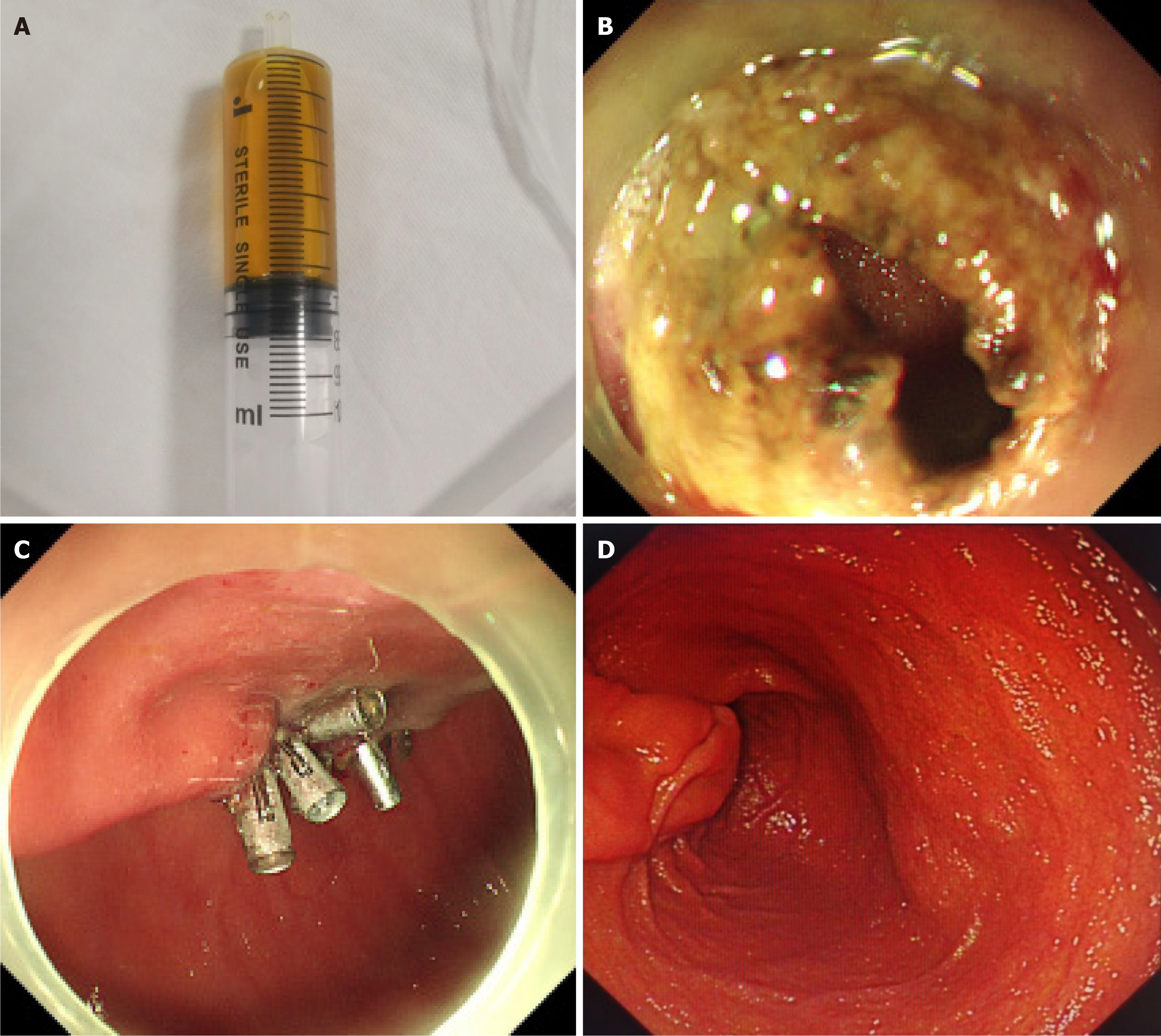
Figure 2 Images during patient treatment and follow-up.
A: Clear yellow liquid was aspirated from the cyst; B: Fenestration of the mucosa; C: Closure of the incision with tissue clips; D: Gastroduodenoscopy showed good recovery without recurrence three months after treatment.
- Citation: Wang ZM, Su S, Ling-Hu EQ, Chai NL. Type III choledochal cyst confirmed by aspiration and treated with endoscopic fenestration plus internal drainage: A case report. World J Gastrointest Surg 2025; 17(4): 104102
- URL: https://www.wjgnet.com/1948-9366/full/v17/i4/104102.htm
- DOI: https://dx.doi.org/10.4240/wjgs.v17.i4.104102


