Copyright
©The Author(s) 2025.
World J Gastrointest Surg. Mar 27, 2025; 17(3): 101940
Published online Mar 27, 2025. doi: 10.4240/wjgs.v17.i3.101940
Published online Mar 27, 2025. doi: 10.4240/wjgs.v17.i3.101940
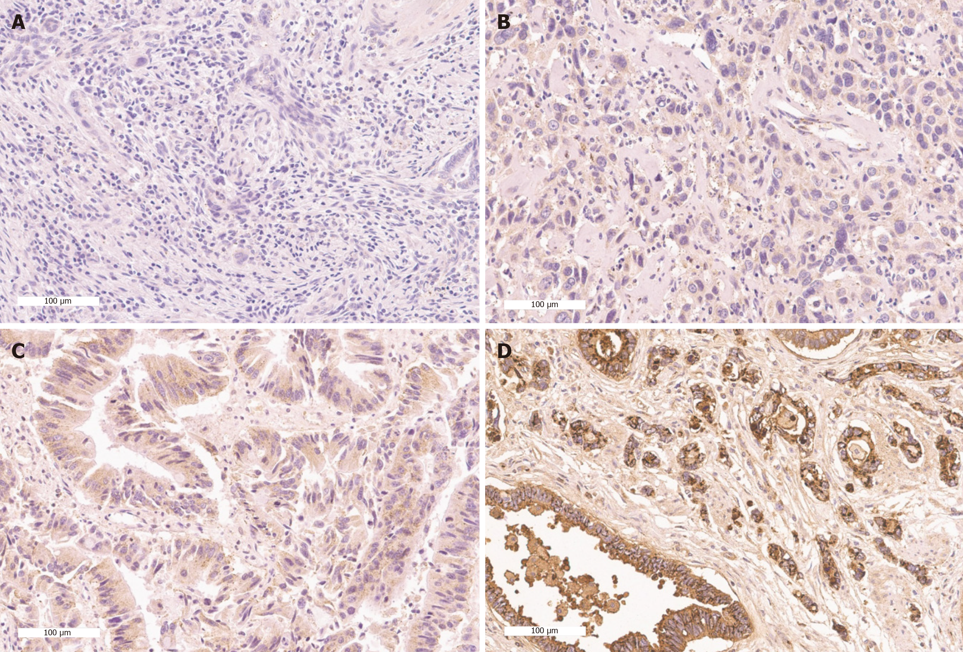
Figure 1 Representative figures of prion protein expression by immunohistochemical staining.
A: No staining (0); B: Weak staining (1+); C: Moderate staining (2+); D: Strong staining (3+).
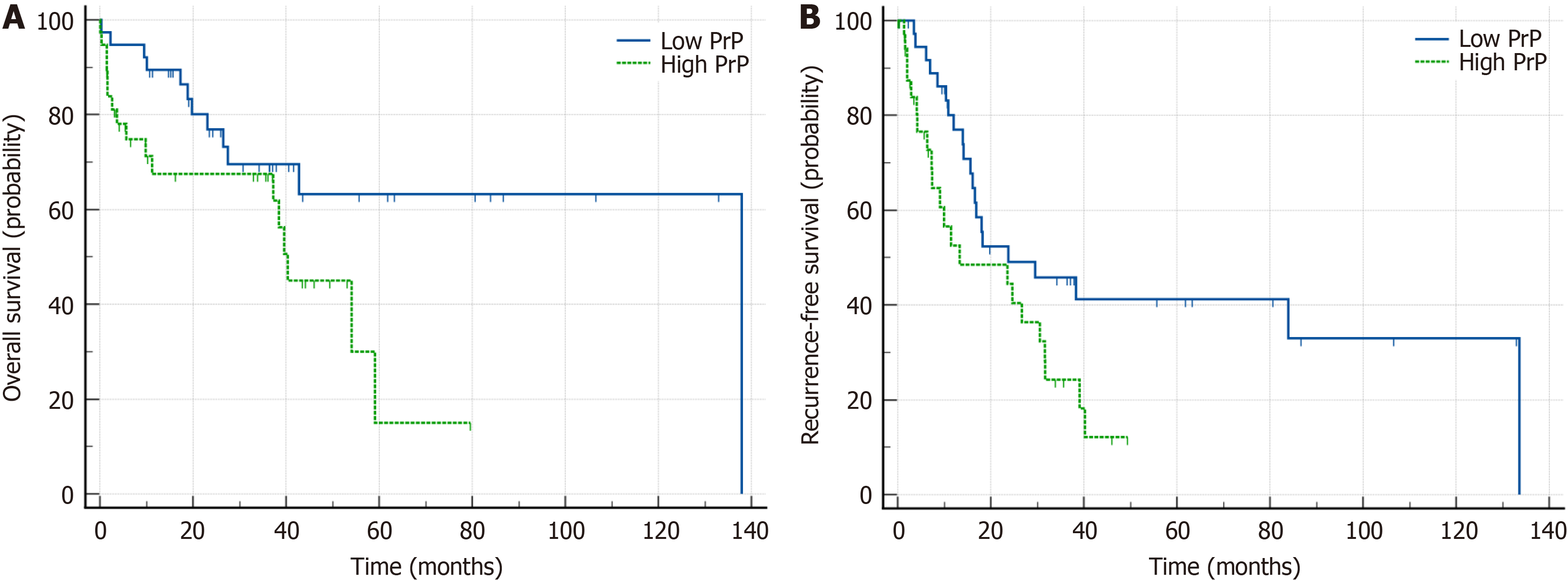
Figure 2 Kaplan Meier survival curves according to prion protein expression.
A: Overall survival; B: Recurrence free survival.
- Citation: Shin DW, Cho YA, Moon SH, Kim TH, Park JW, Lee JW, Choe JY, Kim MJ, Kim SE. High cellular prion protein expression in cholangiocarcinoma: A marker for early postoperative recurrence and unfavorable prognosis. World J Gastrointest Surg 2025; 17(3): 101940
- URL: https://www.wjgnet.com/1948-9366/full/v17/i3/101940.htm
- DOI: https://dx.doi.org/10.4240/wjgs.v17.i3.101940


