©The Author(s) 2025.
World J Gastrointest Surg. Feb 27, 2025; 17(2): 101775
Published online Feb 27, 2025. doi: 10.4240/wjgs.v17.i2.101775
Published online Feb 27, 2025. doi: 10.4240/wjgs.v17.i2.101775
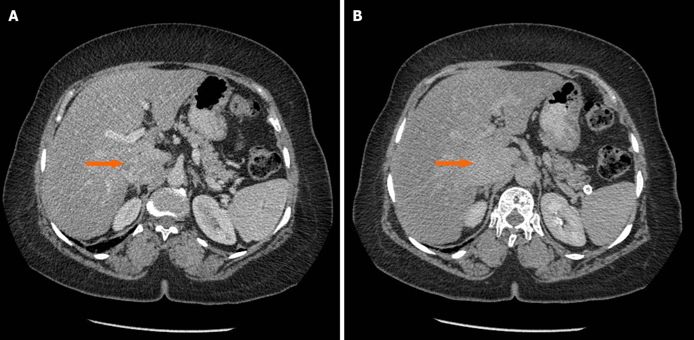
Figure 1 Computed tomography abdomen and pelvis showing heterogeneously enhancing lobulated soft tissue lesion enhancing.
A: The portal phase; B: Delayed phase.
Figure 2 Histopathology images of the final specimen.
A: Tumor cells seen within the inferior vena cava. Astrex denoting wall muscle wall; B: Bizarre shaped cells with multiple mitotic figures; C: Tumor cells invading the liver with a background of hepatic steatosis.
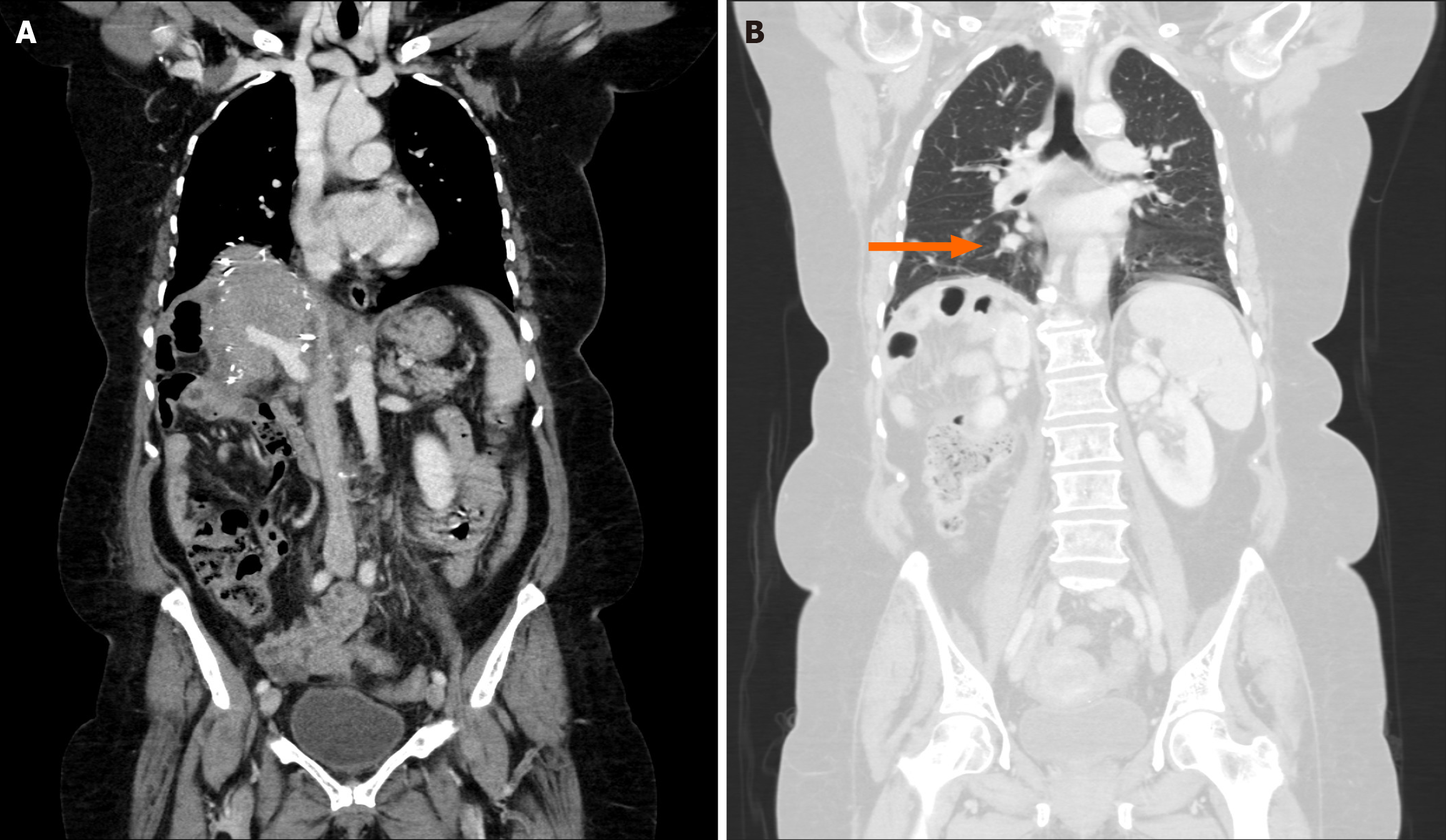
Figure 3 Follow up computed tomography scan, obtained 1 month post op.
A: No evidence of oral recurrence and patent inferior vena cava graft; B: Newly found lung nodule.
- Citation: AlOmran HA, AlMatar B, AlMonsained M, Bojal S, Momani H, AlQahtani MS. Novel surgical approach - cadaveric inferior vena cava graft reconstruction following leiomyosarcoma resection: A case report. World J Gastrointest Surg 2025; 17(2): 101775
- URL: https://www.wjgnet.com/1948-9366/full/v17/i2/101775.htm
- DOI: https://dx.doi.org/10.4240/wjgs.v17.i2.101775


