©The Author(s) 2025.
World J Gastrointest Surg. Feb 27, 2025; 17(2): 100244
Published online Feb 27, 2025. doi: 10.4240/wjgs.v17.i2.100244
Published online Feb 27, 2025. doi: 10.4240/wjgs.v17.i2.100244
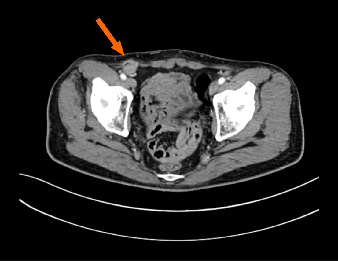
Figure 1
A contrast-enhanced computed tomography image of the abdomen showed a soft tissue shadow in the right inguinal area.
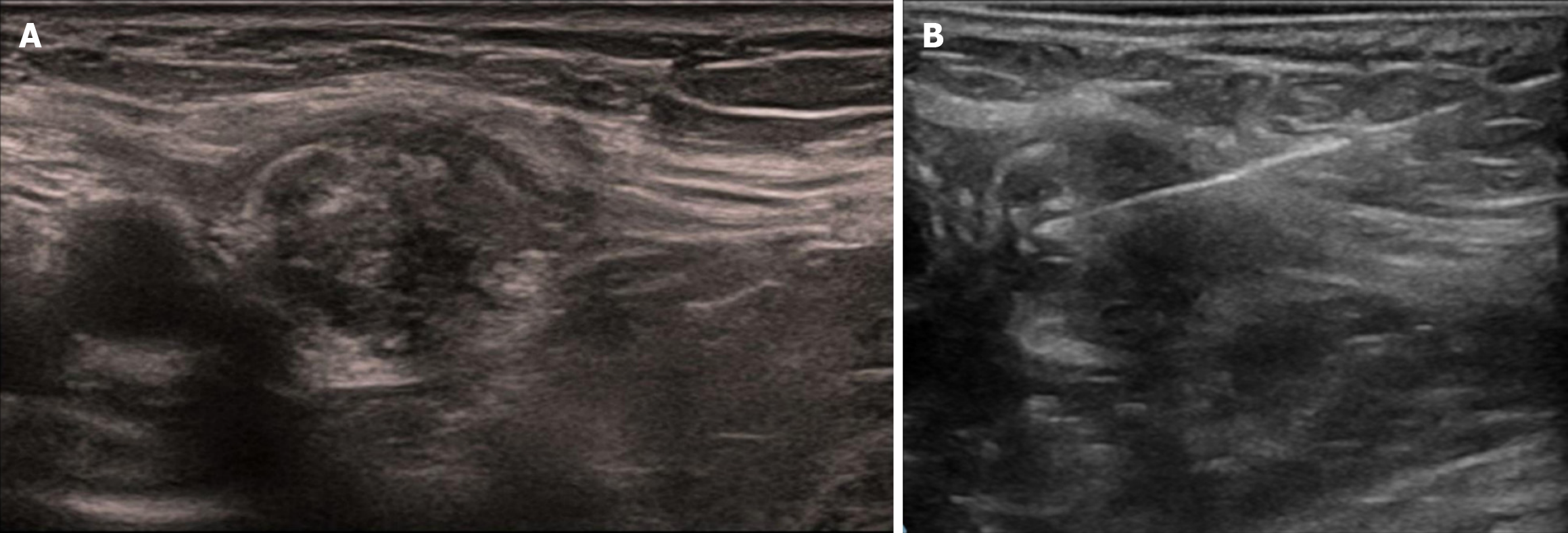
Figure 2 Ultrasound imaging and ultrasound-guided biopsy of the right inguinal mass.
A: Color doppler ultrasound of the right inguinal mass showed a heterogeneous echo pattern; B: Fine needle aspiration of the right inguinal mass under the guidance of Color doppler ultrasound.
Figure 3 Pathological examination of fine needle aspiration biopsy showed adenocarcinoma.
A: Hematoxylin and eosin-stained section; B: Villin positive.
- Citation: Hao JQ, Hu SY, Zhuang ZX, Zhang YJ, Zhang JW, He FJ, Zhuang W, Wang MJ. Distant metastasis in the right inguinal area from gastric cancer: A case report. World J Gastrointest Surg 2025; 17(2): 100244
- URL: https://www.wjgnet.com/1948-9366/full/v17/i2/100244.htm
- DOI: https://dx.doi.org/10.4240/wjgs.v17.i2.100244


