©The Author(s) 2025.
World J Gastrointest Surg. Nov 27, 2025; 17(11): 111285
Published online Nov 27, 2025. doi: 10.4240/wjgs.v17.i11.111285
Published online Nov 27, 2025. doi: 10.4240/wjgs.v17.i11.111285
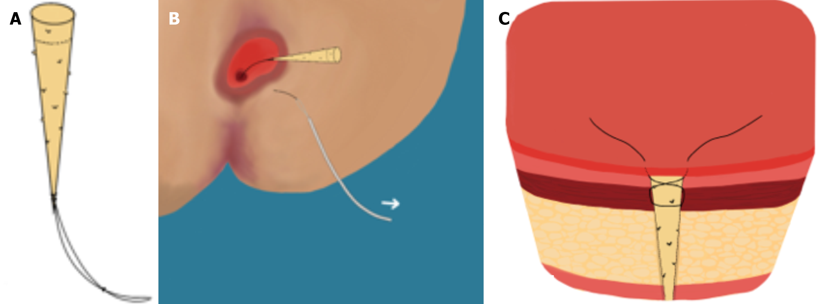
Figure 1 Schematic application of the over the scope clip.
A: Circumferential excision (approximately 2 cm diameter) of tissue surrounding internal opening and insertion of U-shaped sutures, positioned to allow for subsequent tensioning and guidance; B: Correct positioning of over the scope clip (OTSC) proctology applicator and subsequent advancement toward internal opening with sutures as guidance. Applicator cap should be accurately aligned to internal opening and overall orientation of applicator is parallel to axis of the anal canal; C: OTSC proctology clip application in situ.
Figure 2 Schematic deployment of anal fistula plug.
A: Suture attached to the tail of the fistula plug; B: Plug is pulled into the tract via the suture, tail first, until internal opening is fully occluded and plug is tightly fit within tract; C: Secure suturing of plug inner tail with a “figure of 8” stitch which should seal the internal opening.
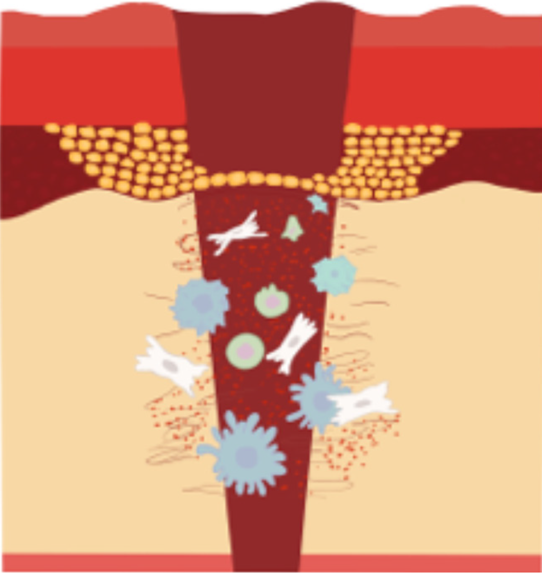
Figure 3 Mechanism of action of stem cells in anal fistulae treatment.
(1) Regeneration and proliferation; and (2) Anti-inflammatory, immunomodulatory effects.
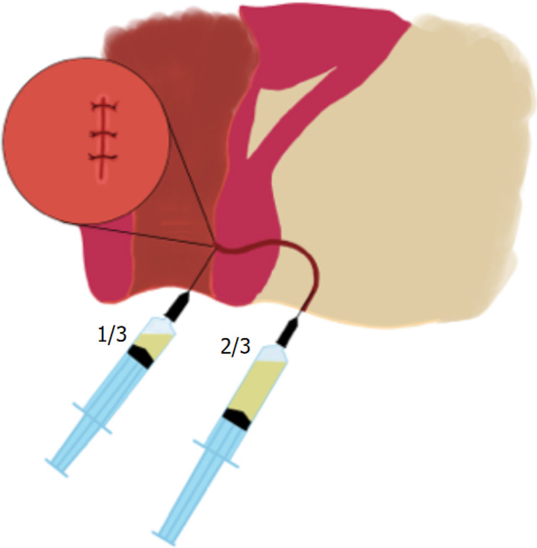
Figure 4
Mesenchymal stem cell suspension injection process (1/3 into submucosa surrounding internal opening, 2/3 through external opening intramurally along course of fistula).
- Citation: Chua KHL, Lee DJK. Evidence outside the box: Minimally invasive treatment for anal fistula. World J Gastrointest Surg 2025; 17(11): 111285
- URL: https://www.wjgnet.com/1948-9366/full/v17/i11/111285.htm
- DOI: https://dx.doi.org/10.4240/wjgs.v17.i11.111285


