Copyright
©The Author(s) 2025.
World J Gastrointest Surg. Oct 27, 2025; 17(10): 110017
Published online Oct 27, 2025. doi: 10.4240/wjgs.v17.i10.110017
Published online Oct 27, 2025. doi: 10.4240/wjgs.v17.i10.110017
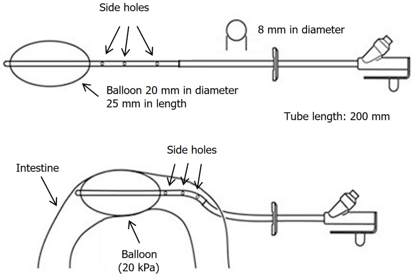
Figure 1
Schematic diagram of a modified ileostomy tube with a balloon.
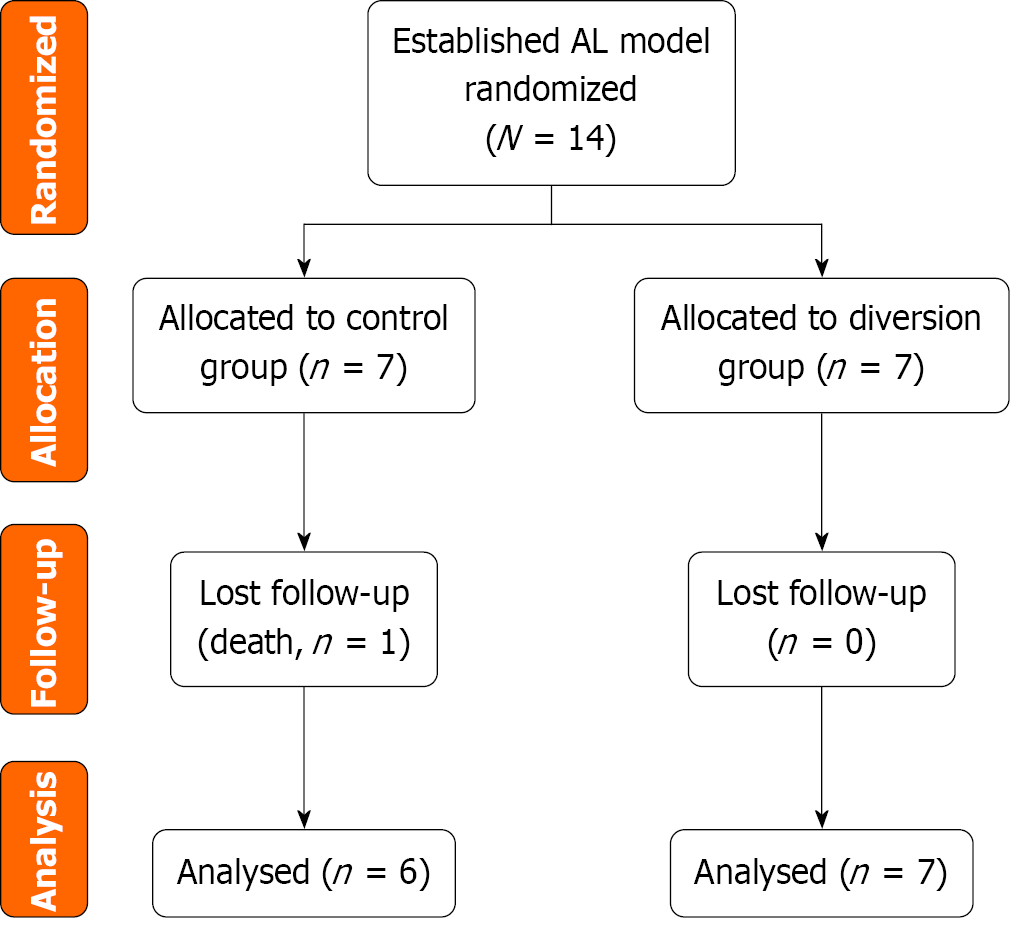
Figure 2 Flow diagram of the experimental pig model allocation and follow-up in the anastomotic leakage study.
AL: Anastomotic leakage.
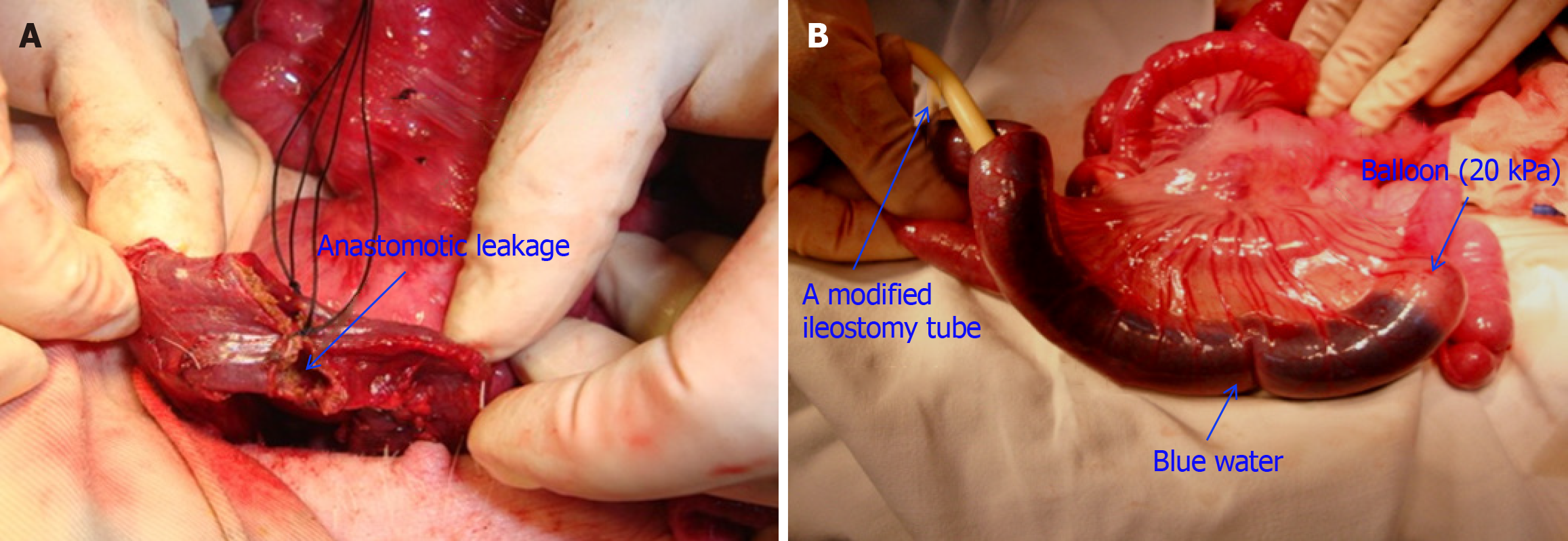
Figure 3 Intraoperative operation diagram.
A: Macroscopic aspect of a descending colostomy with ligation of supplying vessels at 1 cm above the anastomotic segment. Local ischaemia at the anastomotic site was created by ligating the supplying mesocolic vessels 1 cm above the anastomotic segment; B: Leakage test: After placing the tube, the balloon was infused with water to a pressure of 20 kPa. The water mixed with methylene blue did not leak into the proximal intestine.
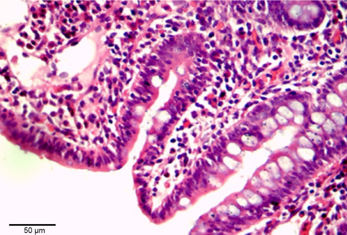
Figure 4 Haematoxylin and eosin staining.
Histological findings in the balloon-pressed intestinal tissue on postoperative day 7, showing mild mucosal injury features, such as subepithelial space extension (magnification × 400).
- Citation: Hu T, Wang J, Yu NH. Complete prevention of anastomotic leakage using total enteric flow diversion. World J Gastrointest Surg 2025; 17(10): 110017
- URL: https://www.wjgnet.com/1948-9366/full/v17/i10/110017.htm
- DOI: https://dx.doi.org/10.4240/wjgs.v17.i10.110017


