Copyright
©The Author(s) 2025.
World J Gastrointest Surg. Oct 27, 2025; 17(10): 109062
Published online Oct 27, 2025. doi: 10.4240/wjgs.v17.i10.109062
Published online Oct 27, 2025. doi: 10.4240/wjgs.v17.i10.109062
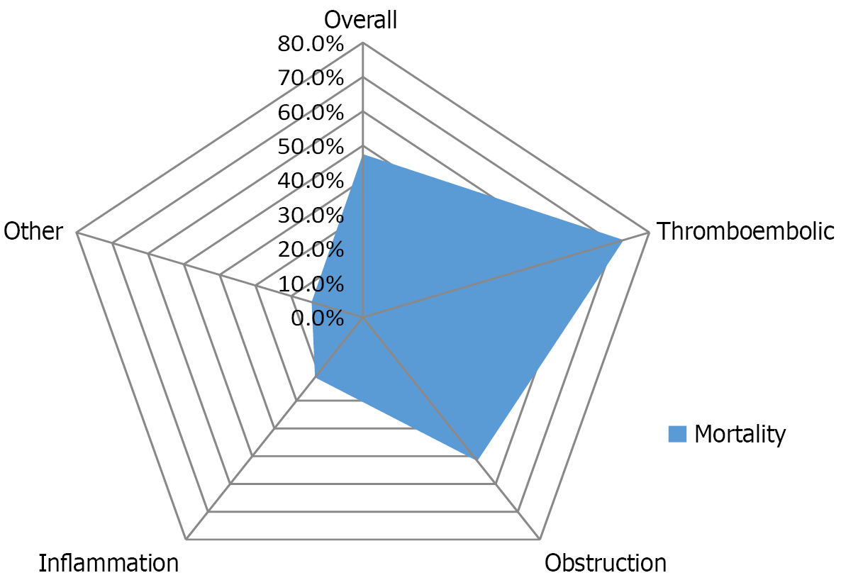
Figure 1
The comparison of mortality rates across etiologies.
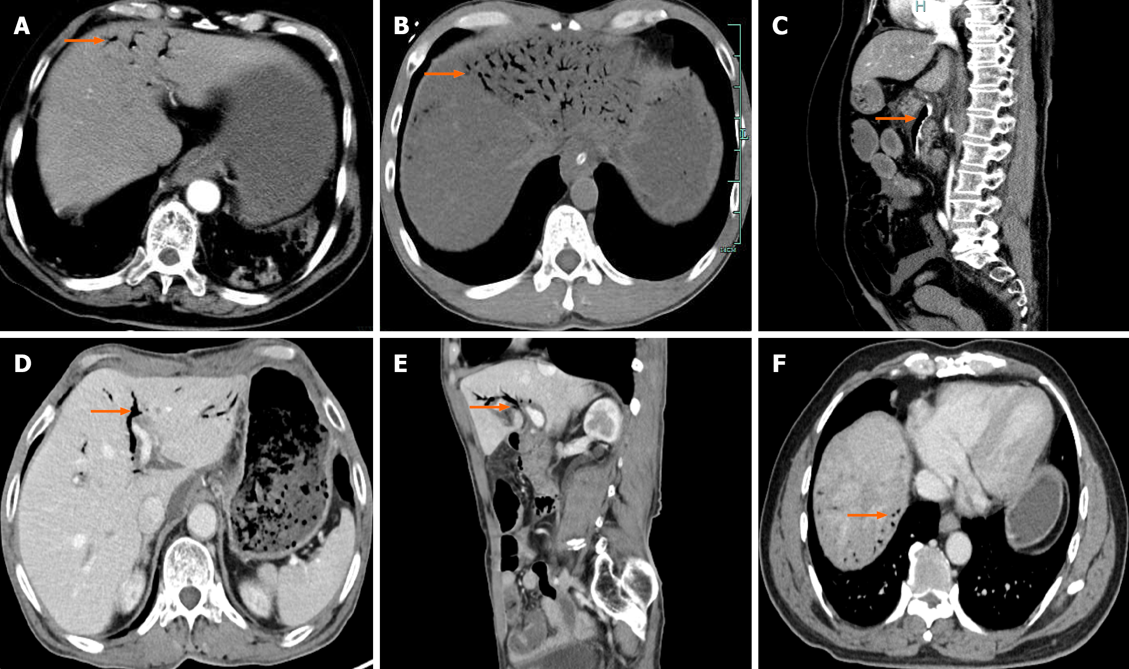
Figure 2 Contrast-enhanced abdominal computed tomography.
A: Hepatic portal venous gas (HPVG) is localized in the left lobe of the liver, and closer to the liver capsule (the arrow shows the gas); B: HPVG exhibits the classic “withered branches” expansion (the arrow shows the gas); C: HPVG involves the superior mesenteric vein and portal vein (the arrow shows the gas); D and E: Pneumobilia is typically centrally located within the liver (the arrow shows the gas); F: The rare presence of gas shadows closing to the liver capsule in the Pneumobilia (the arrow shows the gas).
- Citation: Zhou QY, Zheng ZH, Lv XL, Guo JQ, Zhang K, Xu HT. Advancement in the diagnosis and treatment of hepatic portal venous gas. World J Gastrointest Surg 2025; 17(10): 109062
- URL: https://www.wjgnet.com/1948-9366/full/v17/i10/109062.htm
- DOI: https://dx.doi.org/10.4240/wjgs.v17.i10.109062


