©The Author(s) 2024.
World J Gastrointest Surg. Jul 27, 2024; 16(7): 2343-2350
Published online Jul 27, 2024. doi: 10.4240/wjgs.v16.i7.2343
Published online Jul 27, 2024. doi: 10.4240/wjgs.v16.i7.2343
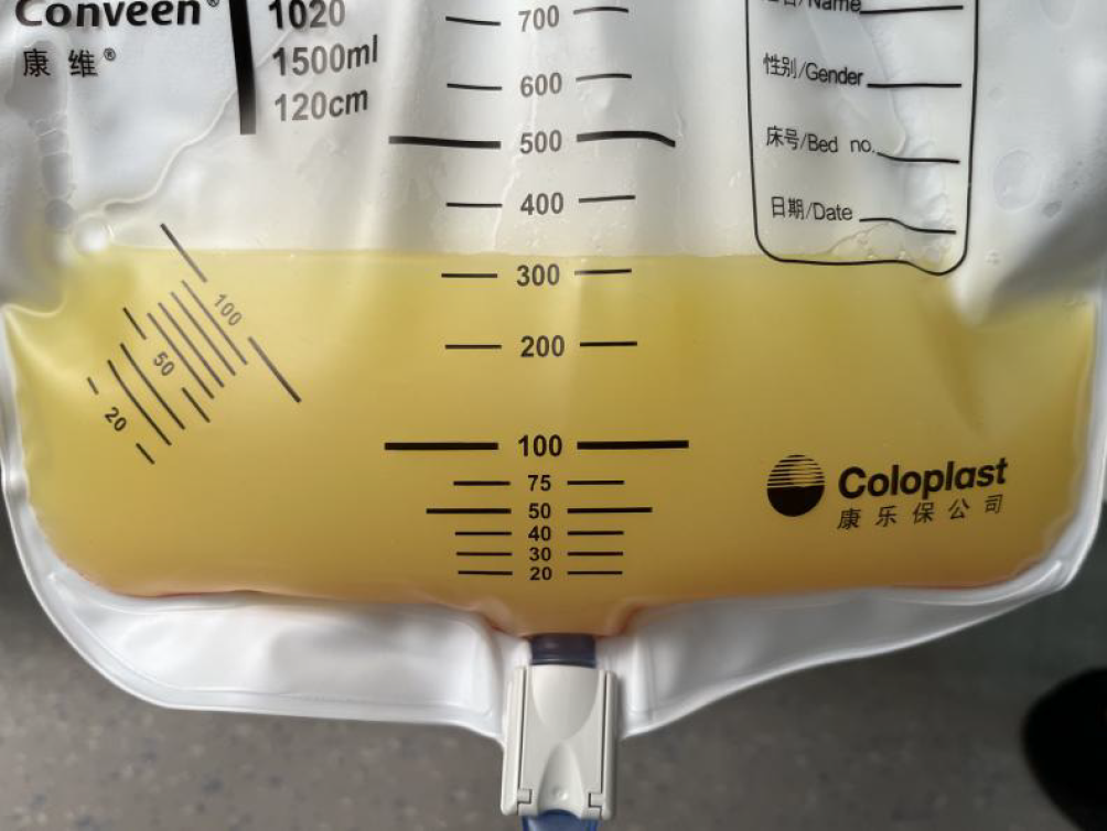
Figure 1 The abdominal puncture fluid was composed of chylous ascites.
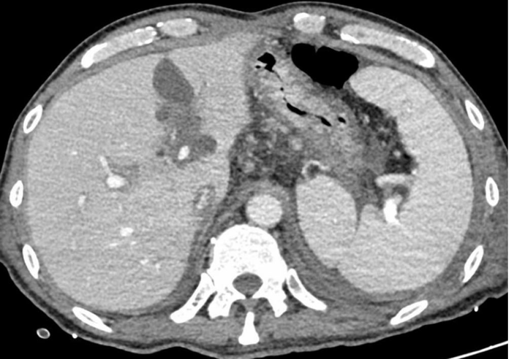
Figure 2 Abdominal contrast-enhanced computed tomography venous phase image showing dilated liver lymphatic vessels.
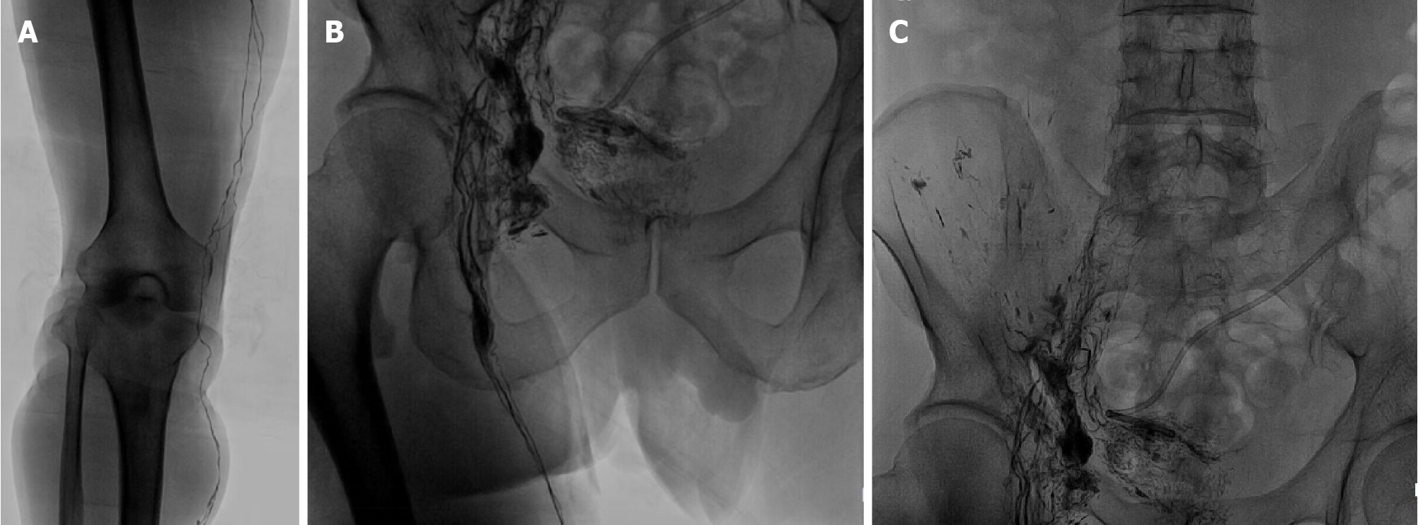
Figure 3 Lymphoscintigraphy showing diffuse and uneven increases in radioactivity distribution.
A: Lower extremities; B: Abdomen; C: Pelvic region.
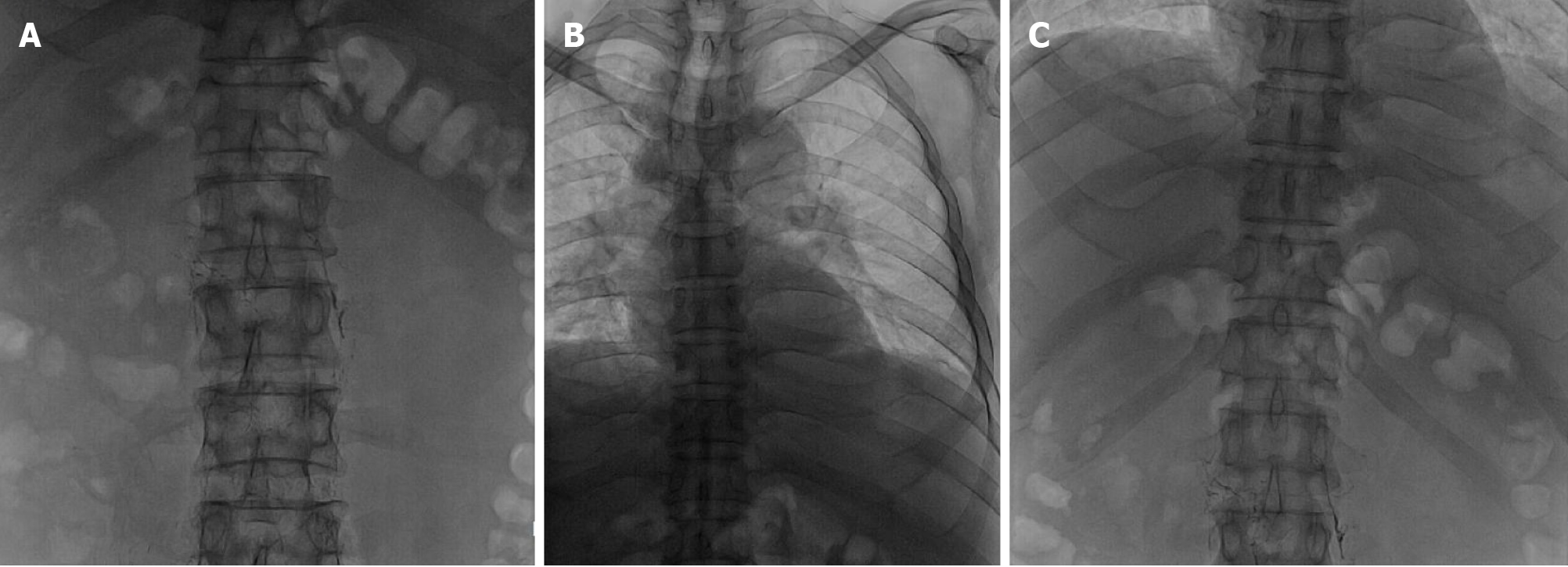
Figure 4 Lymphangiography revealing lymphatic drainage obstruction above the L3 level.
A: L1-L4 level; B: T1-T9 level; C: T10-T12 level.
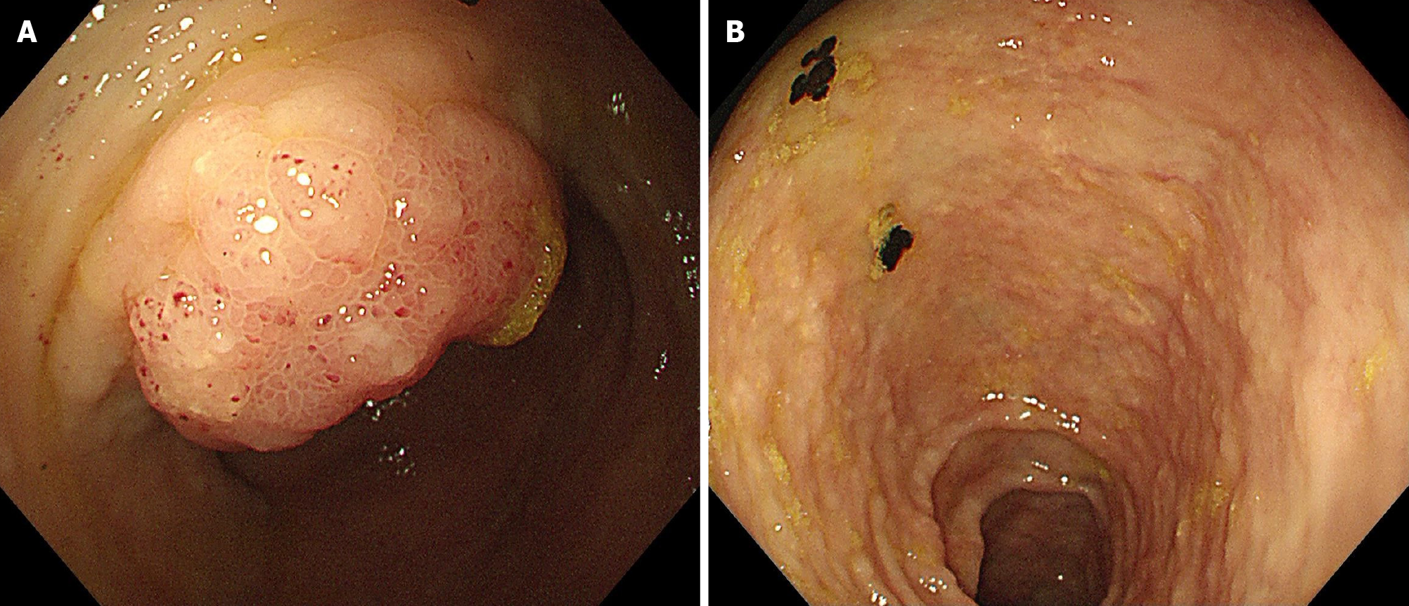
Figure 5 Enteroscopy.
A: Edema of the mucosa in the ileocecal region; B: Granular changes in the colon.
Figure 6 A biopsy of the ileocecal region suggesting atypical signet ring cells in the ileocecal valve (hematoxylin and eosin staining, 200 ×).
A: Region 1 in the ileocecal valve area; B: Region 2 in the ileocecal valve area.
- Citation: Li Y, Tai Y, Wu H. Colon signet-ring cell carcinoma with chylous ascites caused by immunosuppressants following liver transplantation: A case report. World J Gastrointest Surg 2024; 16(7): 2343-2350
- URL: https://www.wjgnet.com/1948-9366/full/v16/i7/2343.htm
- DOI: https://dx.doi.org/10.4240/wjgs.v16.i7.2343


