Copyright
©The Author(s) 2024.
World J Gastrointest Surg. Nov 27, 2024; 16(11): 3578-3583
Published online Nov 27, 2024. doi: 10.4240/wjgs.v16.i11.3578
Published online Nov 27, 2024. doi: 10.4240/wjgs.v16.i11.3578
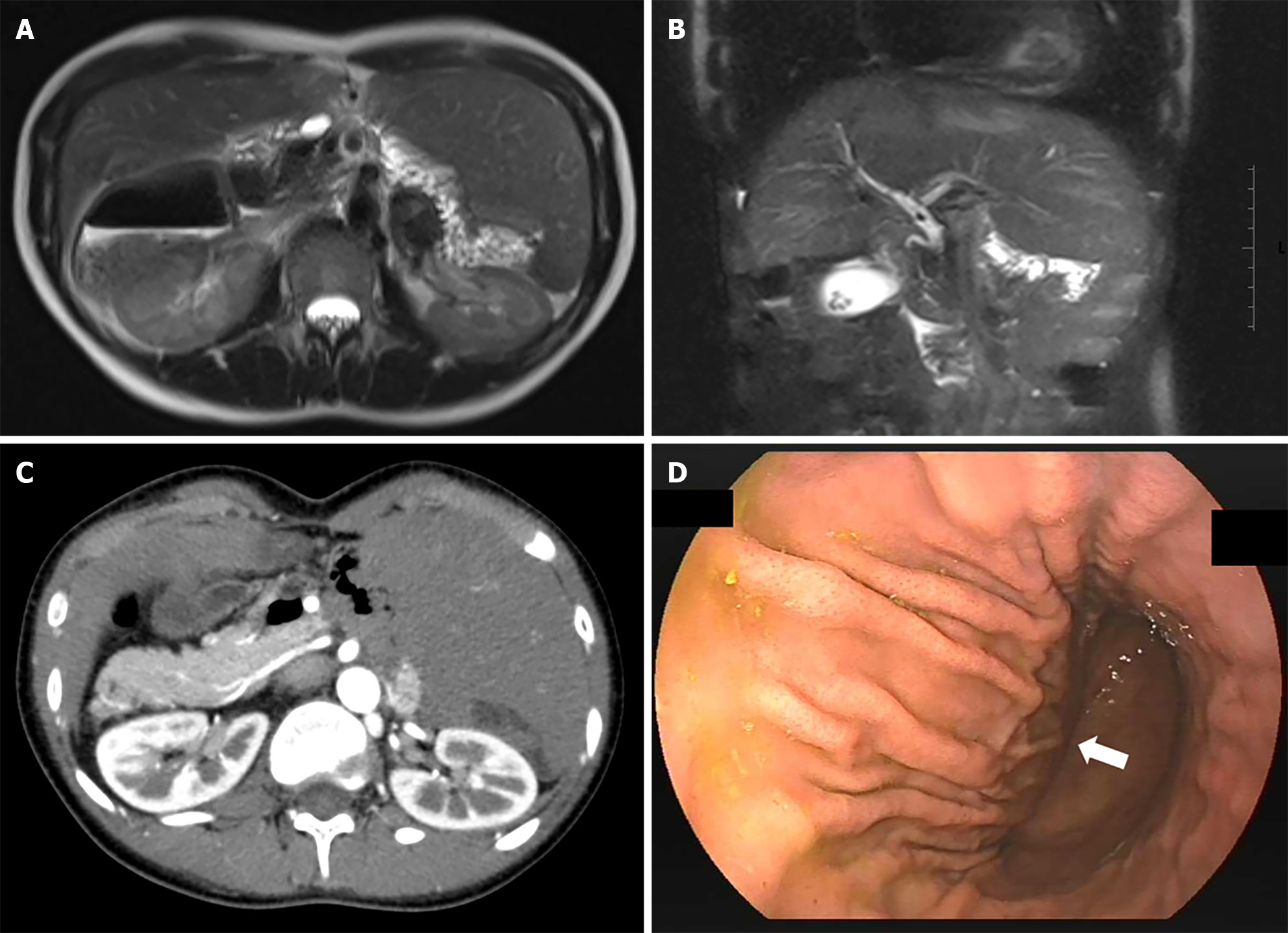
Figure 1 Evaluation before endoscopic retrograde cholangiopancreatography.
A: Magnetic resonance imaging revealed a right-sided stomach and midline liver placement; B: Computed tomography revealed a right-sided pancreas and left-sided inferior vena cava; C: Magnetic resonance cholangiopancreatography, indicated a midline liver placement and a stone measuring 0.6 cm in the common bile duct; D: Gastroscopy revealed inverted greater and lesser curvatures (view at the cardia, white arrow: Direction of the antrum).
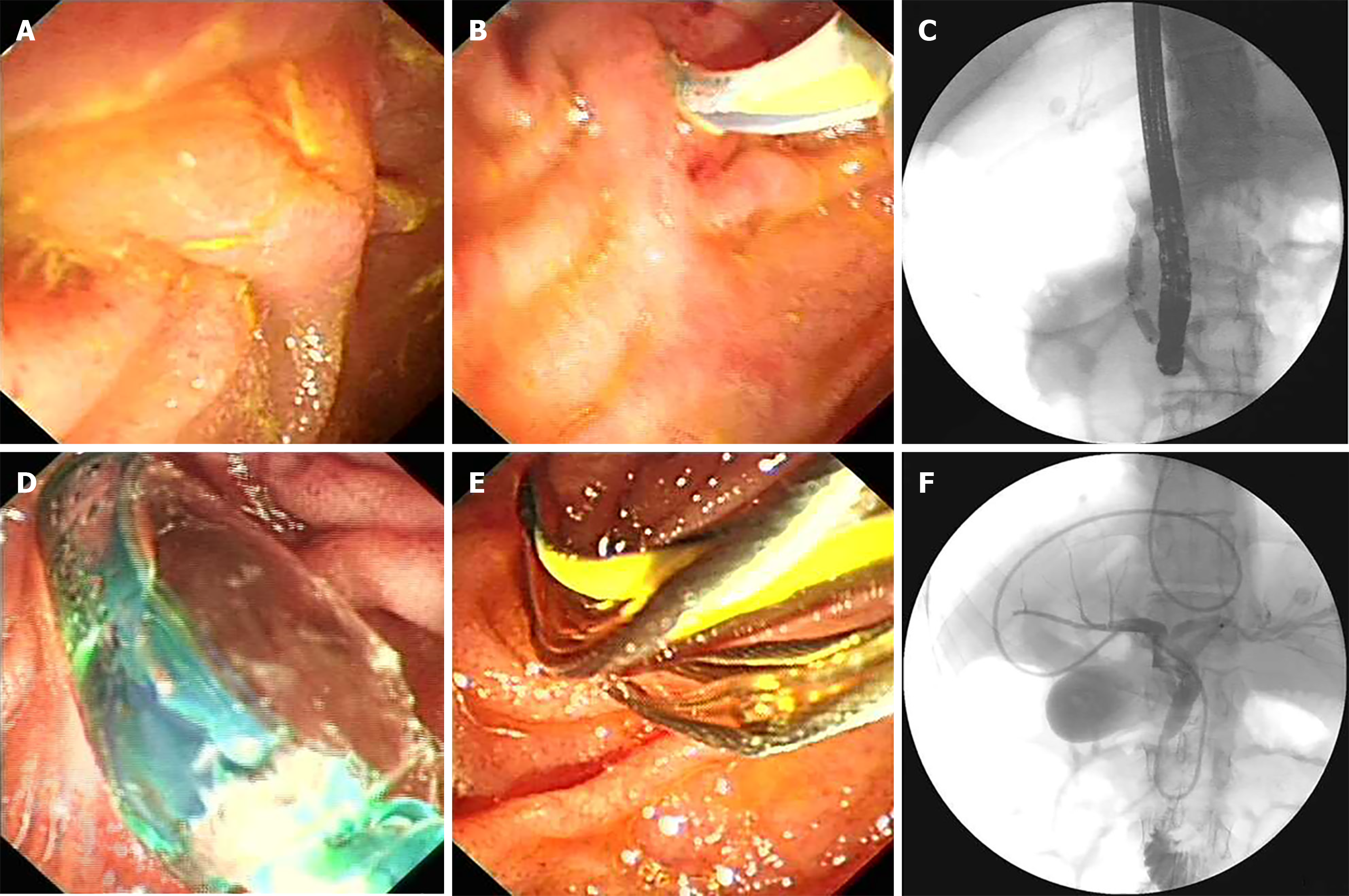
Figure 2 Endoscopic retrograde cholangiopancreatography procedure.
A: Papillary oriented at 11 o’clock; B: Accessing the common bile duct (CBD) with an endoscopic retrograde cholangiopancreatography cannula; C: Cholangiography revealed mild CBD dilation with the impacted stone measuring 0.6 cm; D: Endoscopic papillary balloon dilation (6 mm, maximum pressure 35 standard atmospheric pressure); E: Basket used to remove the stone; F: Postoperative cholangiography showed no residual stones and an inverted stomach shape.
- Citation: Zhang YY, Ruan J, Fu Y. Therapeutic endoscopic retrograde cholangiopancreatography in a patient with asplenia-type heterotaxy syndrome: A case report. World J Gastrointest Surg 2024; 16(11): 3578-3583
- URL: https://www.wjgnet.com/1948-9366/full/v16/i11/3578.htm
- DOI: https://dx.doi.org/10.4240/wjgs.v16.i11.3578


