Copyright
©The Author(s) 2023.
World J Gastrointest Surg. Feb 27, 2023; 15(2): 287-293
Published online Feb 27, 2023. doi: 10.4240/wjgs.v15.i2.287
Published online Feb 27, 2023. doi: 10.4240/wjgs.v15.i2.287
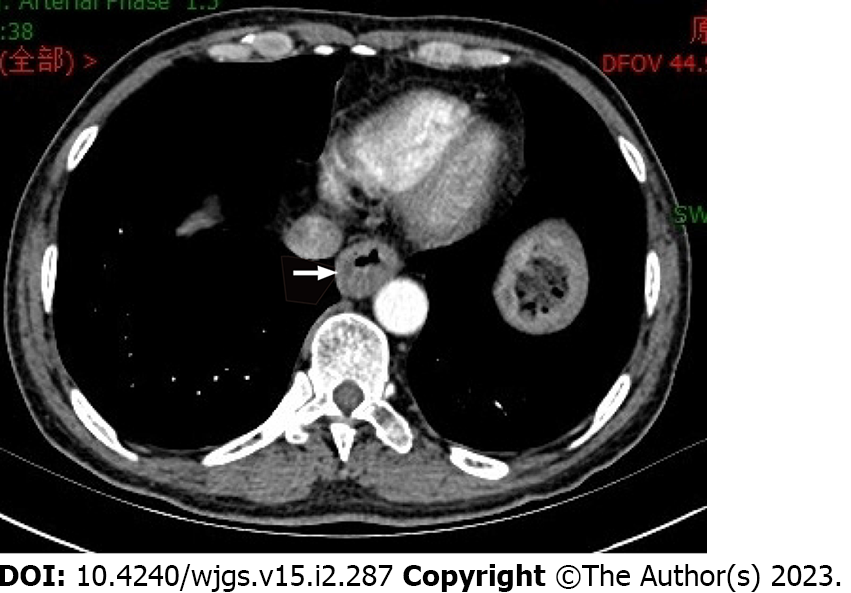
Figure 1
Computed tomography indicates thickening of the esophageal wall.
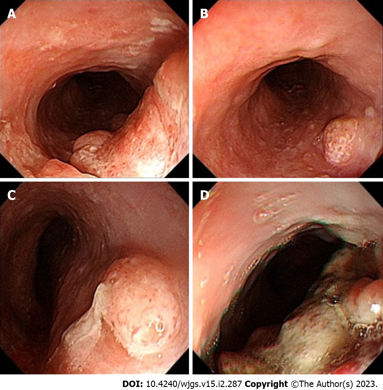
Figure 2 Gastroscopy suggested multiple lesions of the esophagus.
A: 23–27 cm from the incisor; B: 29 cm from the incisor; C: 32 cm from the incisor; D: 39–43 cm from the incisor.
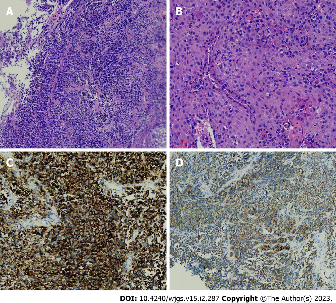
Figure 3 Endoscopic biopsy.
A: HE staining of malignant melanoma; B: HE-stained squamous cell carcinoma; C: Immunohistochemical HMB45 positivity; D: Immunohistochemical Melan-A positivity.
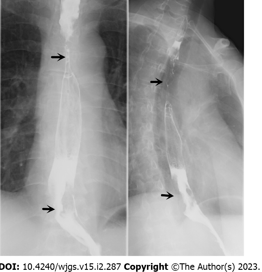
Figure 4
Esophageal barium swallow (the arrow indicates the location of the lesion).
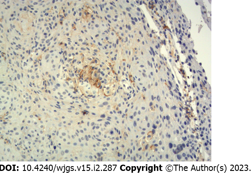
Figure 5
Programmed death ligand-1 immunohistochemistry 22C3: The tumour proportion score results were 1%-2%.
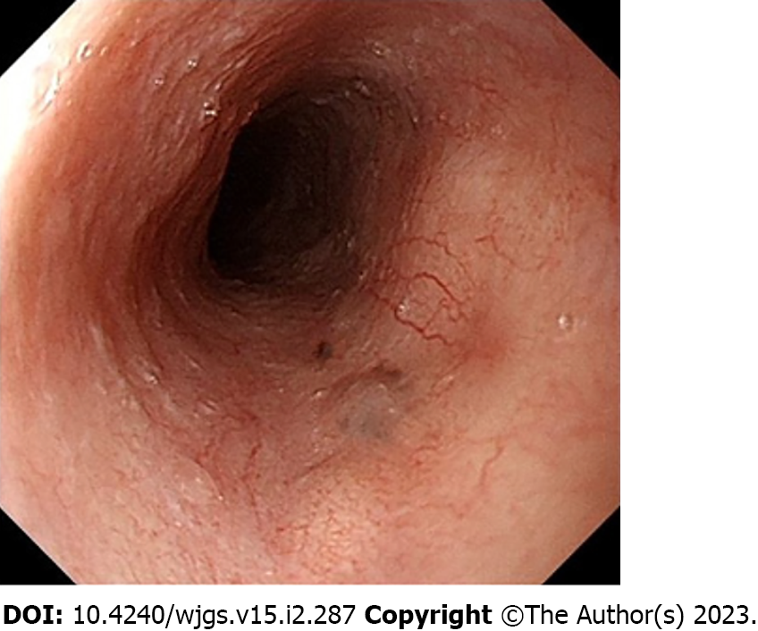
Figure 6
Gastroscopy review.
- Citation: Zhu ML, Wang LY, Bai XQ, Wu C, Liu XY. Primary malignant melanoma of the esophagus combined with squamous cell carcinoma: A case report. World J Gastrointest Surg 2023; 15(2): 287-293
- URL: https://www.wjgnet.com/1948-9366/full/v15/i2/287.htm
- DOI: https://dx.doi.org/10.4240/wjgs.v15.i2.287


