Copyright
©The Author(s) 2022.
World J Gastrointest Surg. Dec 27, 2022; 14(12): 1329-1339
Published online Dec 27, 2022. doi: 10.4240/wjgs.v14.i12.1329
Published online Dec 27, 2022. doi: 10.4240/wjgs.v14.i12.1329
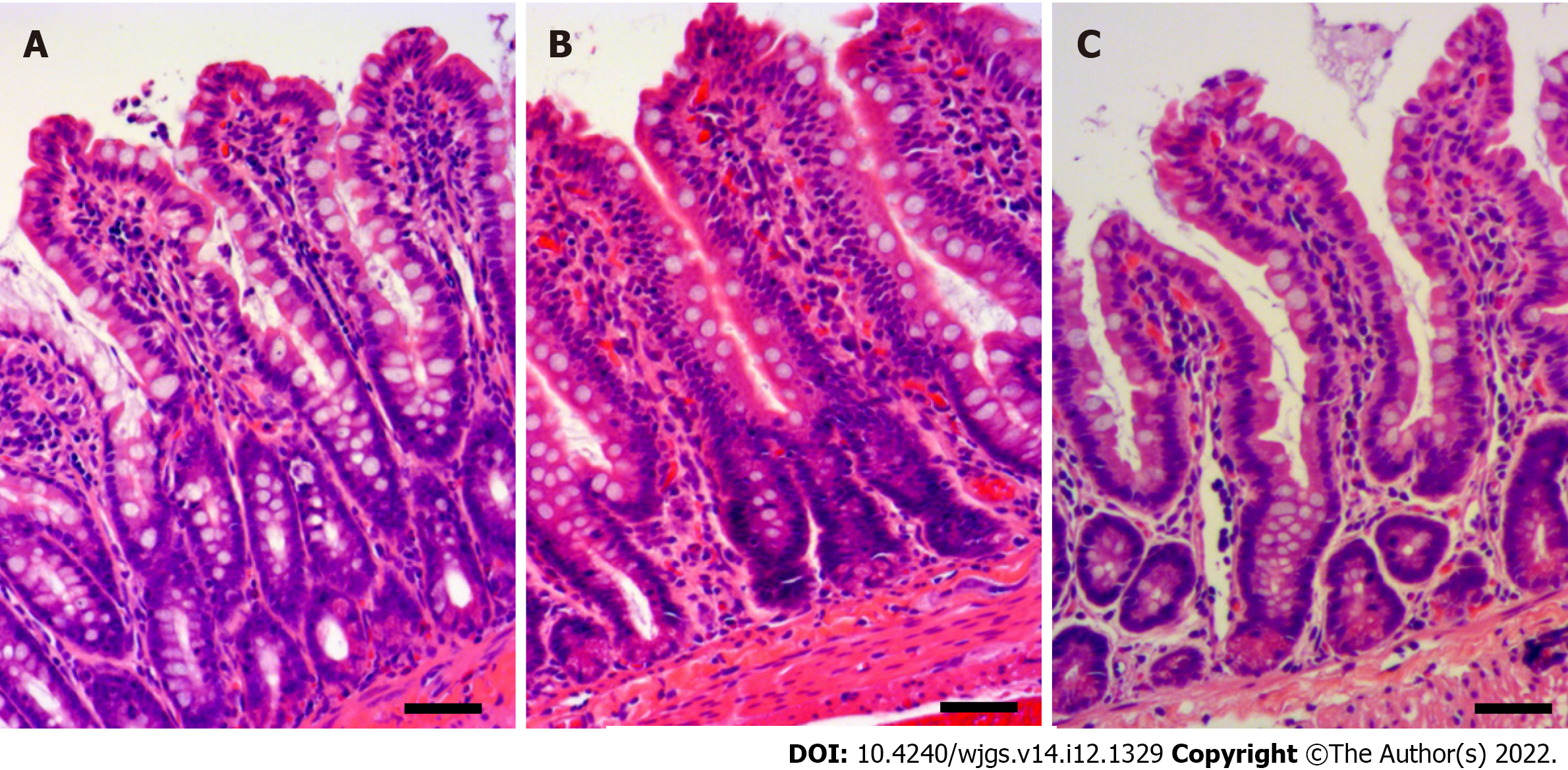
Figure 1 Intestinal mucosal injury with ischemia-reperfusion injury.
A: Histological sections with H&E stain showed pyknosis in the epithelial layer in the ischemia group; B: Extensive pyknosis in the epithelial layer, denudation of the tip of the villi, and capillary congestion in the reperfusion group; C: Mild epithelial pyknosis and denudation of the villi in the hydrogen group. Scale bar: 50 μm.
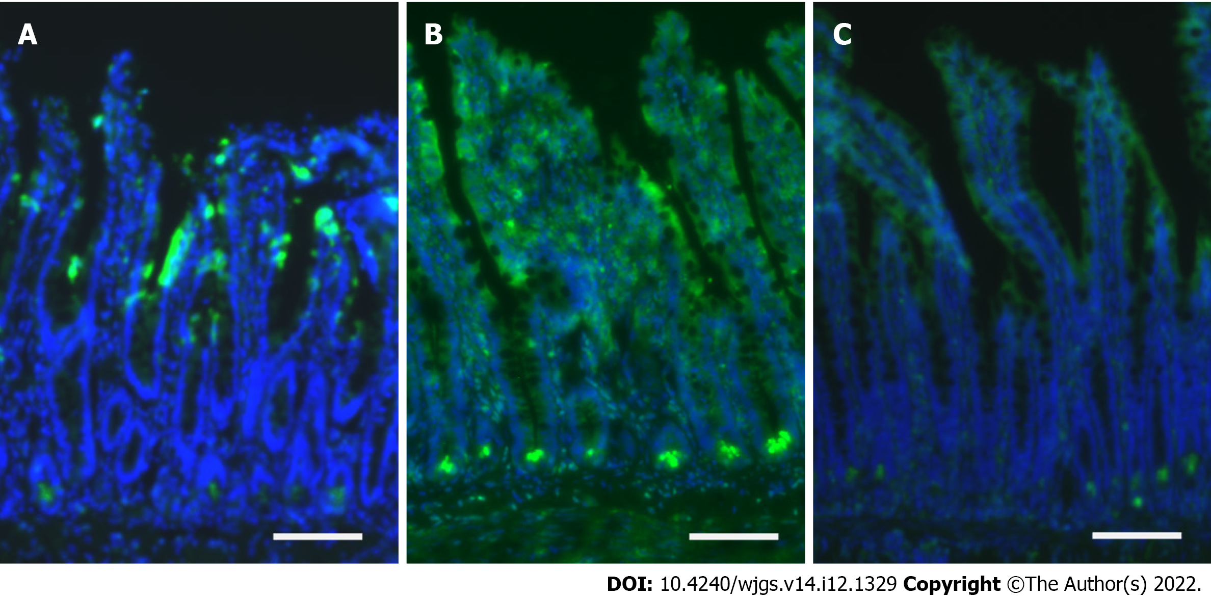
Figure 2 Intestinal stem cell injury by ischemia-reperfusion injury.
A: Immunofluorescence staining with caspase-3 (green) revealed mucosal injury at the epithelial layer closed to the tip of the villi in the ischemia group; B: Extensive apoptosis was identified at the crypt base and the whole epithelial layer in the reperfusion group; C: Apoptosis at the crypt base was limited in the hydrogen group. Scale bar: 100 μm.
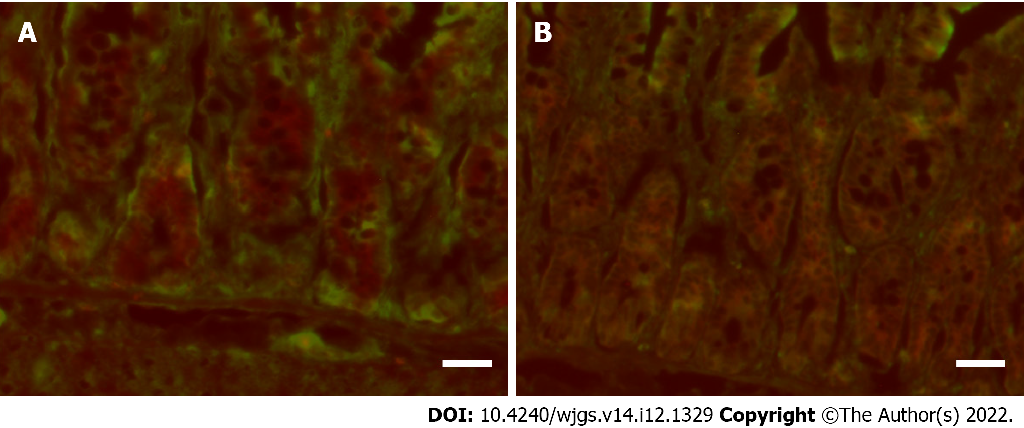
Figure 3 Intestinal stem cell with double immunofluorescence staining.
A: Double immunofluorescence staining with caspase-3 (red) and leucine-rich repeat-containing G-protein-coupled 5 (LGR5) (green) showed that LGR5-positive cells at the crypt base did not undergo apoptosis in the ischemia group (caspase-3 and LGR5 were separately stained); B: Multiple LGR5-positive cells underwent apoptosis after reperfusion (there were double-stained cells with caspase-3 and LGR5). Scale bar: 50 μm.
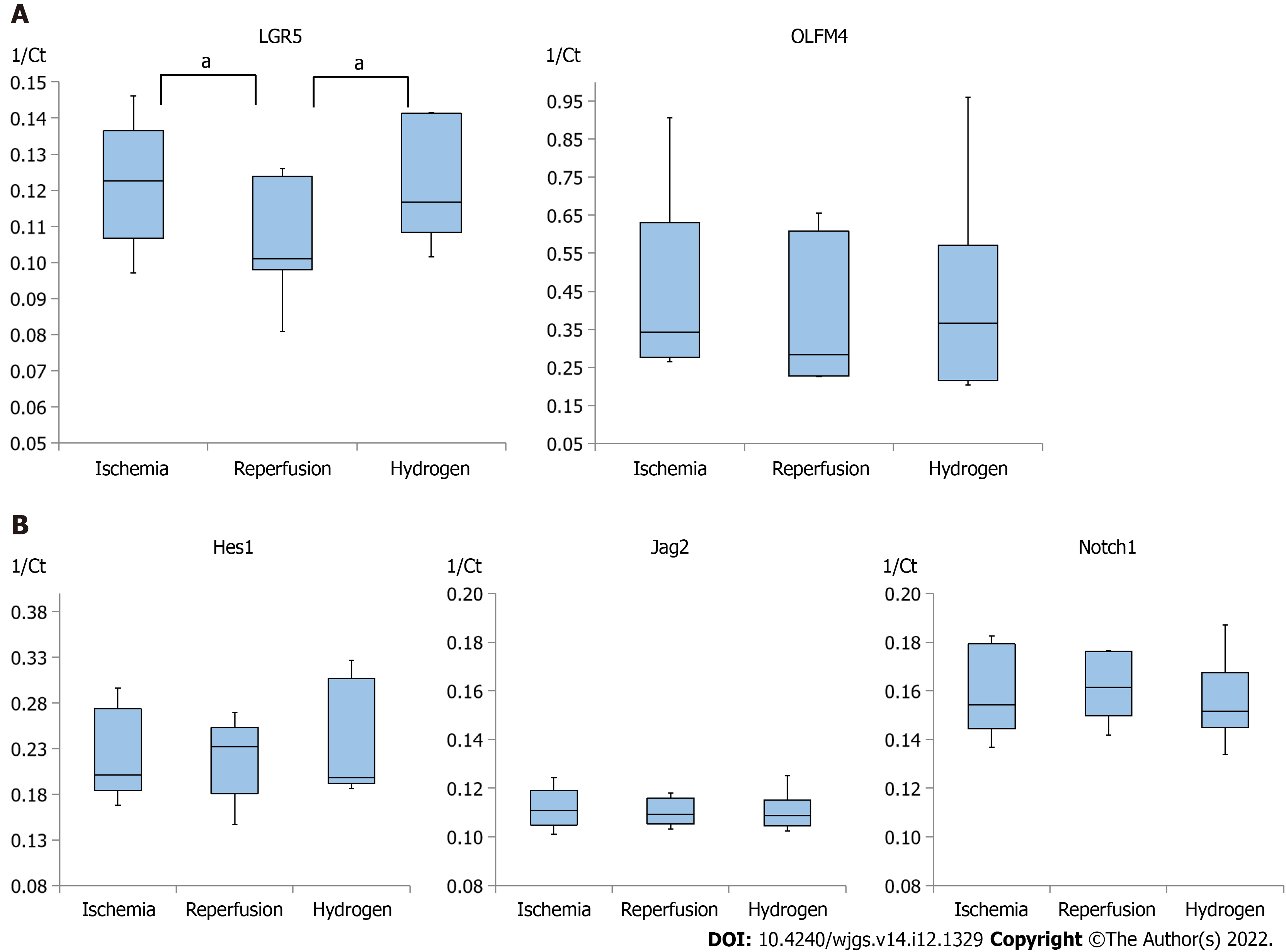
Figure 4 Quantitative analysis of RNA.
A: Leucine-rich repeat-containing G-protein-coupled 5 expression was significantly lower in the reperfusion group than in the ischemia group, but it was significantly higher in the hydrogen group than in the reperfusion group. Olfactomedin 4 expression was also relatively higher in the hydrogen group than in the reperfusion group, although the differences were not significant; B: Hes1 expression was relatively high in the reperfusion group than in the ischemia and hydrogen groups, whereas Jag2 and Notch1 expressions were comparable between the ischemia, reperfusion, and hydrogen groups. aP < 0.05. LGR5: Leucine-rich repeat-containing G-protein-coupled 5; OLFM4: Olfactomedin 4; Hes1: Hairy and enhancer of split 1; Jag2: Jagged 2; Notch1: Neurogenic locus notch homolog protein 1.
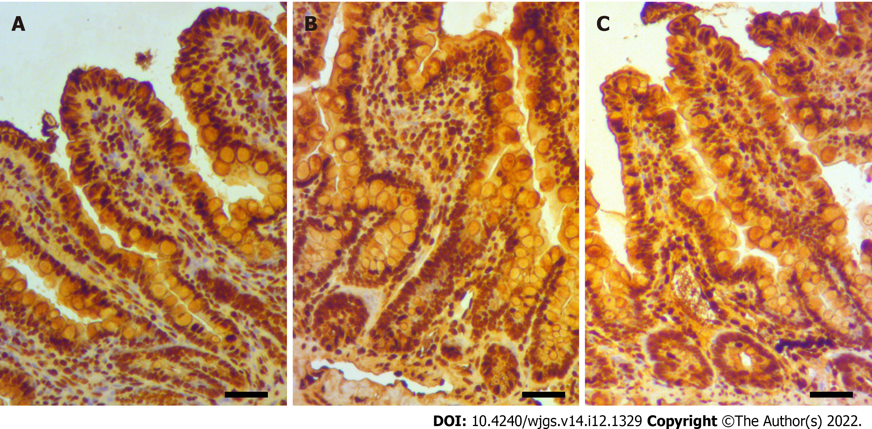
Figure 5 Mucosal oxidative injury in the ischemia-reperfusion model.
A: Immunostaining for 8-hydroxy-2'-deoxyguanosine showed limited oxidative stress at the mucosa in the ischemia group, although there was considerable pyknosis at the tip of the villi; B: Extensive oxidative injury throughout the epithelium, including the crypt base, in the reperfusion group; C: Mild-to-moderate oxidative injury at the mucosa of the crypt base, along with pyknosis with the denudation at the tip of the villi, in the hydrogen group. Scale bar: 50 μm.
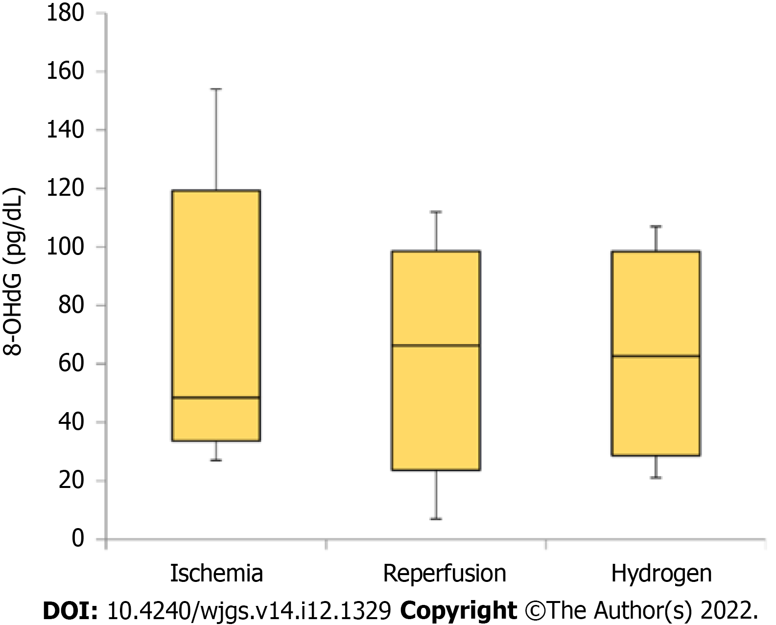
Figure 6 Systematic oxidative injury in the ischemia-reperfusion model.
Serum 8-hydroxy-2'-deoxyguanosine (8-OHdG) concentration immediately after each intervention (ischemia, ischemia-reperfusion, and ischemia-reperfusion under hydrogen gas inhalation) was measured. Although not significant, the 8-OHdG concentration was higher in the reperfusion and hydrogen groups than in the ischemia group. 8-OHdG: 8-hydroxy-2'-deoxyguanosine.
- Citation: Yamamoto R, Suzuki S, Homma K, Yamaguchi S, Sujino T, Sasaki J. Hydrogen gas and preservation of intestinal stem cells in mesenteric ischemia and reperfusion. World J Gastrointest Surg 2022; 14(12): 1329-1339
- URL: https://www.wjgnet.com/1948-9366/full/v14/i12/1329.htm
- DOI: https://dx.doi.org/10.4240/wjgs.v14.i12.1329


