Copyright
©The Author(s) 2022.
World J Gastrointest Surg. Jan 27, 2022; 14(1): 36-45
Published online Jan 27, 2022. doi: 10.4240/wjgs.v14.i1.36
Published online Jan 27, 2022. doi: 10.4240/wjgs.v14.i1.36
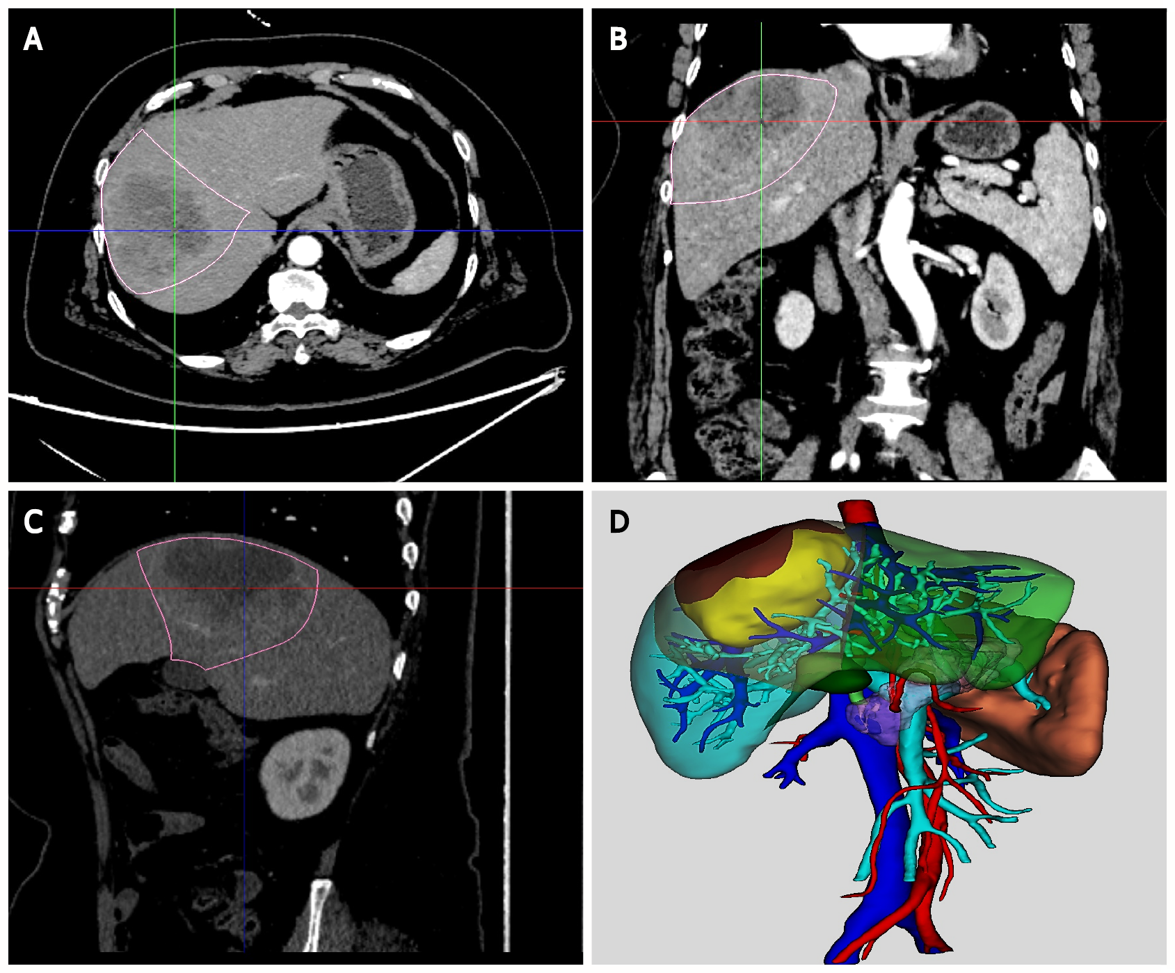
Figure 1 Two-dimensional imaging and three-dimensional reconstruction.
A-C: Two-dimensional imaging (2D) abdominal enhanced computed tomography images of a patient with hepatocellular carcinoma; D: Three-dimensional (3D) hologram reconstructed by mixed reality software.
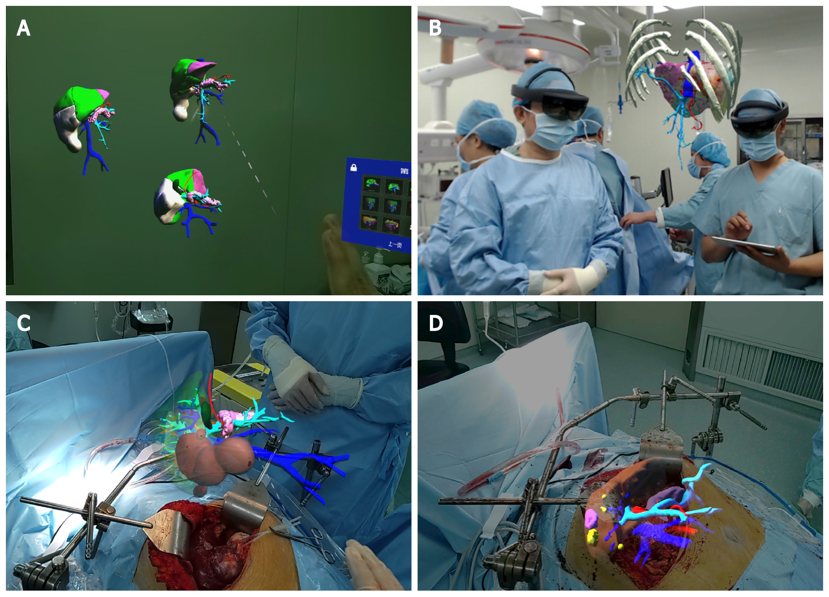
Figure 2 Mixed reality-assisted hepatectomy guided by three-dimensional holograms.
A: Three-dimensional (3D) holograms were observed with the mixed reality head-mounted display in the operating room; B: The surgeon observed the tumor location and vascular anatomy with a 3D hologram and determined the surgical planning again; C: 3D hologram was placed above the surgical field; D: 3D holograms were fused with the patient's liver.
- Citation: Zhu LY, Hou JC, Yang L, Liu ZR, Tong W, Bai Y, Zhang YM. Application value of mixed reality in hepatectomy for hepatocellular carcinoma. World J Gastrointest Surg 2022; 14(1): 36-45
- URL: https://www.wjgnet.com/1948-9366/full/v14/i1/36.htm
- DOI: https://dx.doi.org/10.4240/wjgs.v14.i1.36


