©The Author(s) 2020.
World J Gastrointest Surg. Dec 27, 2020; 12(12): 534-548
Published online Dec 27, 2020. doi: 10.4240/wjgs.v12.i12.534
Published online Dec 27, 2020. doi: 10.4240/wjgs.v12.i12.534
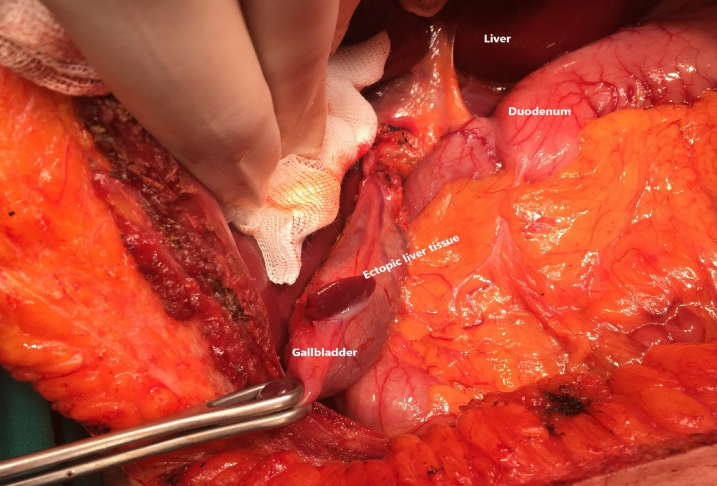
Figure 1 Intraoperative view of the ectopic liver tissue located in the gallbladder.
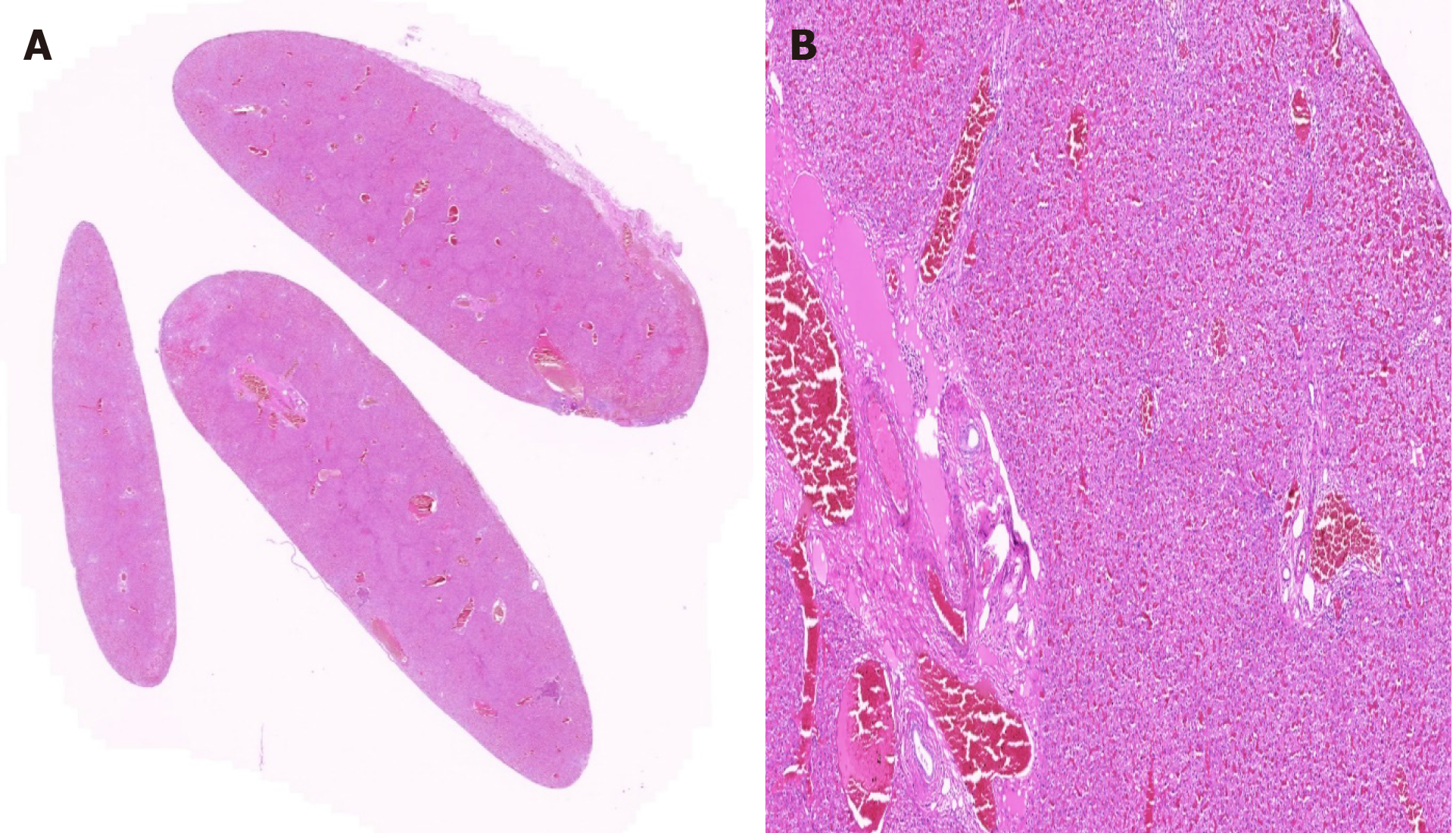
Figure 2 Encapsulated liver tissue with normal histological features.
A: HE × 1; B: HE × 2.5.
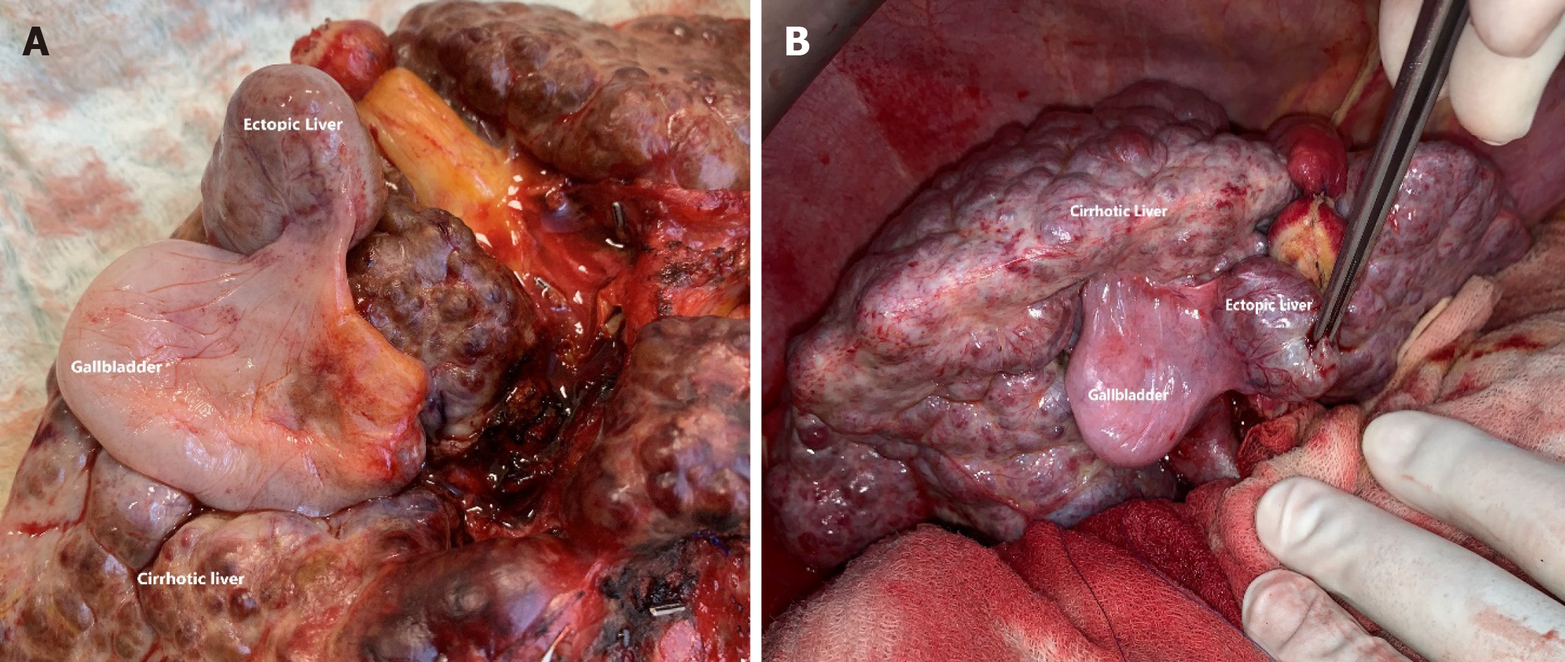
Figure 3 Intraoperative view of the ectopic liver tissue located in the gallbladder mesentery along with the main cirrhotic liver.
A and B: The ectopic liver tissue was showed a cirrhotic appearance similar to the main liver tissue.
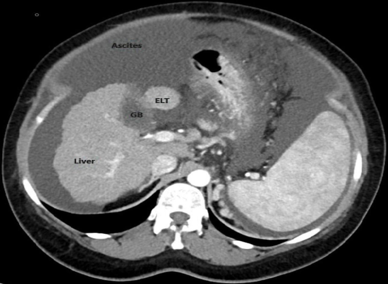
Figure 4 Axial contrast-enhanced multidetector computed tomography section shows an ectopic liver tissue-like nodular lesion associated with the gallbladder.
- Citation: Akbulut S, Demyati K, Ciftci F, Koc C, Tuncer A, Sahin E, Karadag N, Yilmaz S. Ectopic liver tissue (choristoma) on the gallbladder: A comprehensive literature review. World J Gastrointest Surg 2020; 12(12): 534-548
- URL: https://www.wjgnet.com/1948-9366/full/v12/i12/534.htm
- DOI: https://dx.doi.org/10.4240/wjgs.v12.i12.534


