©The Author(s) 2019.
World J Gastrointest Surg. Dec 27, 2019; 11(12): 433-442
Published online Dec 27, 2019. doi: 10.4240/wjgs.v11.i12.433
Published online Dec 27, 2019. doi: 10.4240/wjgs.v11.i12.433
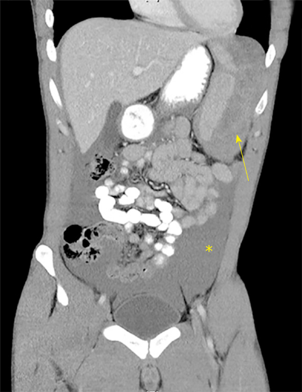
Figure 1 Coronal slice of abdominal computed tomography with intravenous contrast in the portal venous phase demonstrating haemoperitoneum (asterisk) and splenic subcapsular haematoma (arrow).
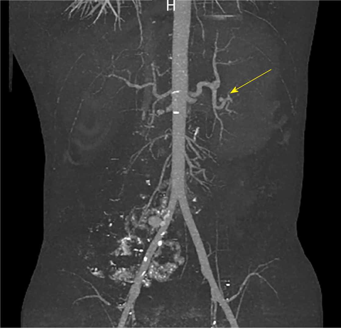
Figure 2 Coronal slice of computed tomography angiogram demonstrating splenic artery pseudoaneurysm (arrow) with no evidence of active bleeding (measurements 9.
1 × 8.3 × 8.1 mm).
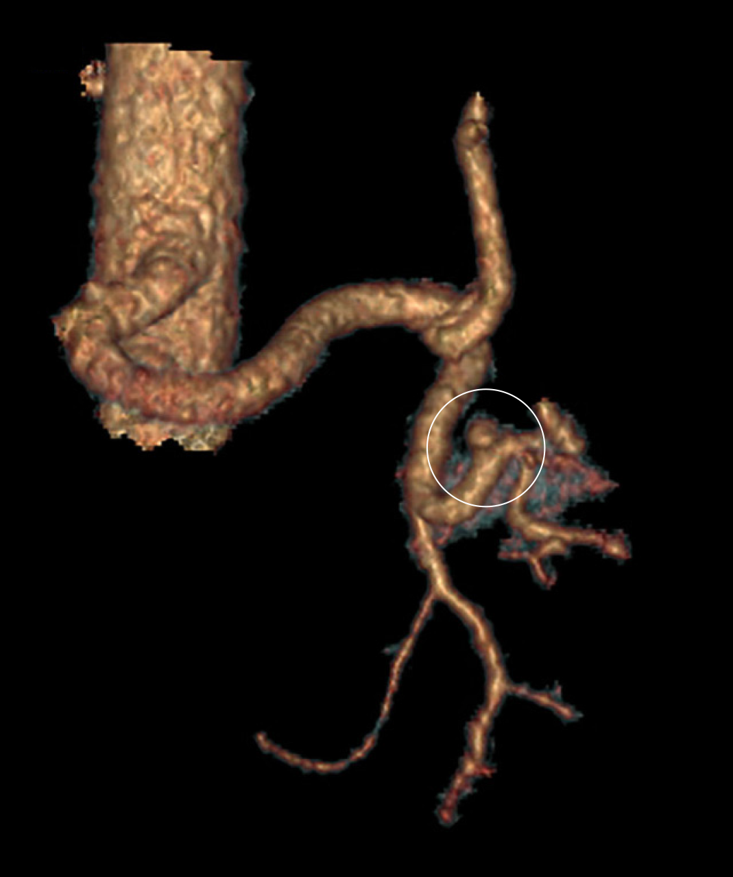
Figure 3 3D reconstruction of computed tomography angiogram demonstrating splenic artery pseudoaneurysm (circled).
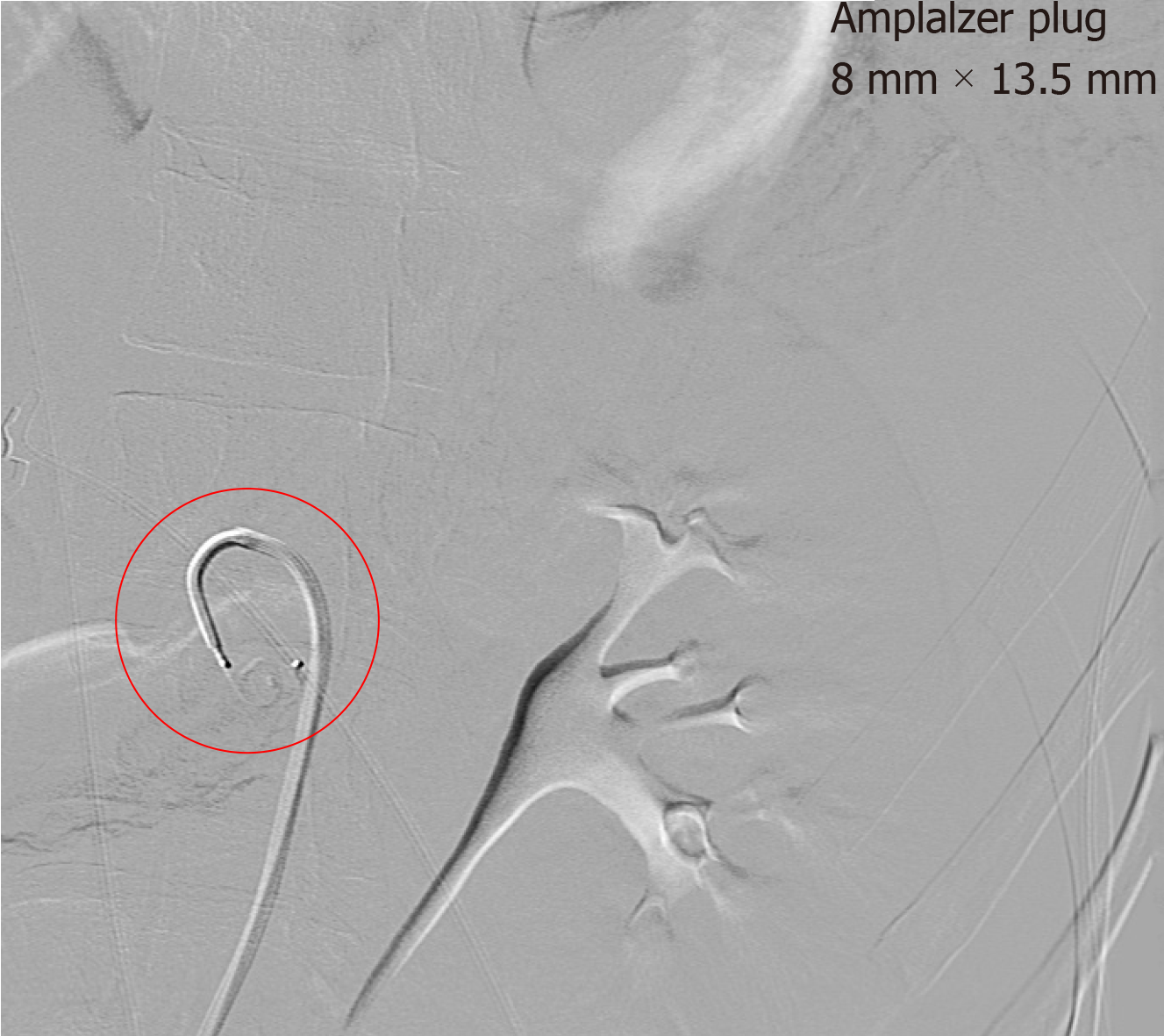
Figure 4 Still image from formal angiogram demonstrating intra-arterial catheter and deployment of endovascular plug (circled, visible beyond the tip of the catheter).
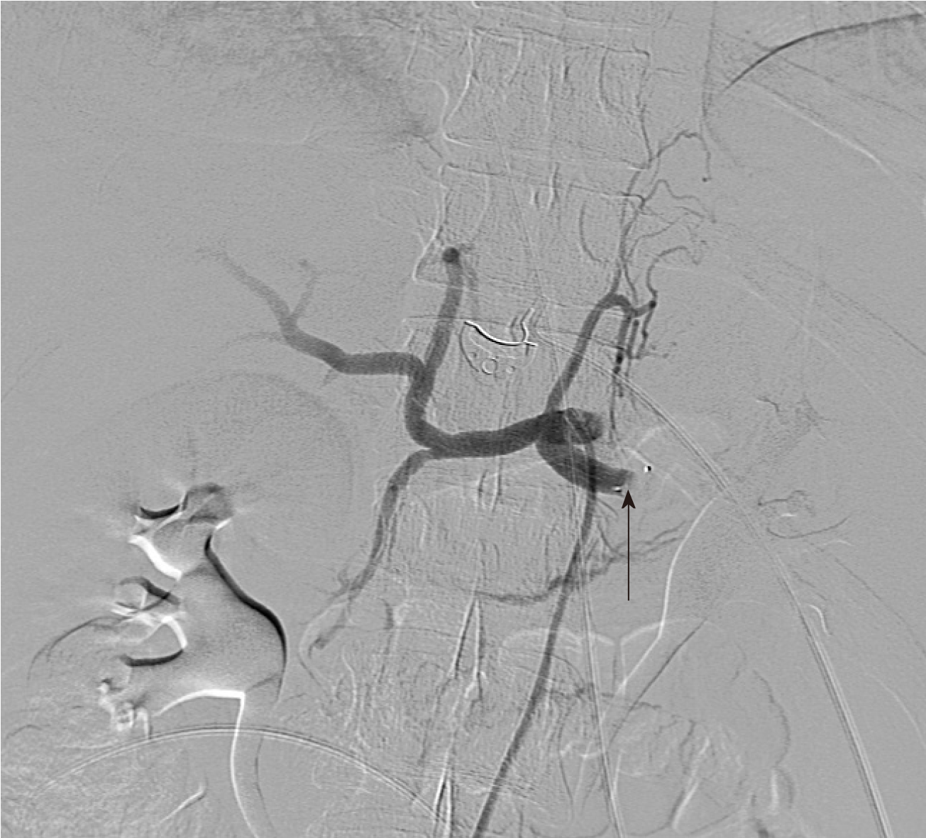
Figure 5 Coeliac angiography after deployment of endovascular plug.
Note the abrupt cessation of blood flow in the proximal splenic artery (arrow).
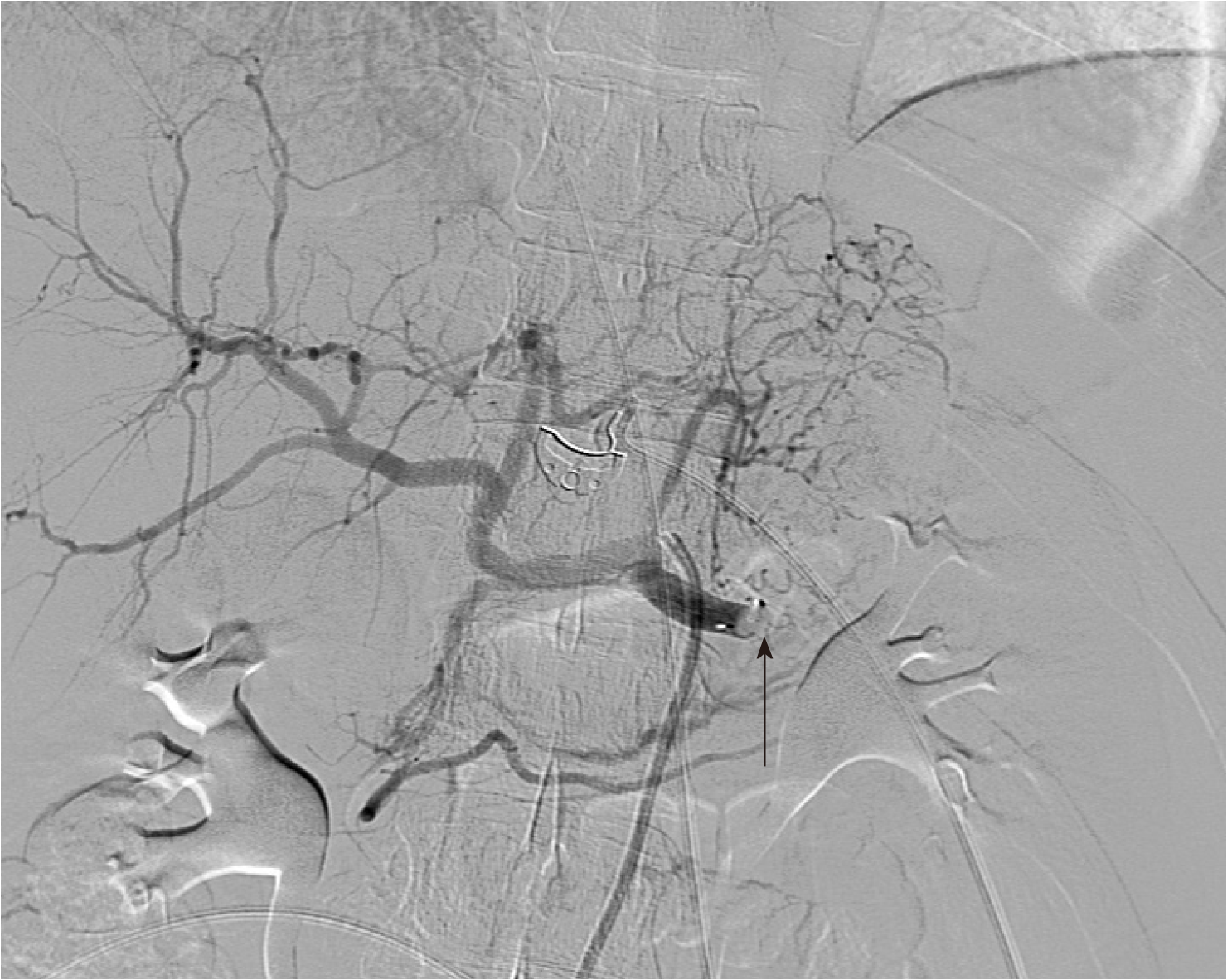
Figure 6 Delayed still image from coeliac angiogram confirming complete occlusion of the proximal splenic artery by endovascular plug (arrow).
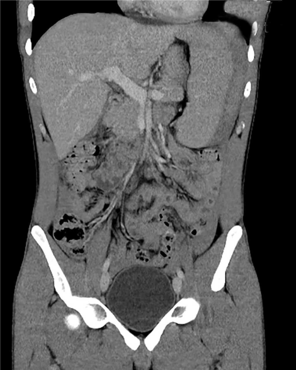
Figure 7 Coronal slice of abdominal computed tomography on day 5 of admission, demonstrating reduction in size of both haemoperitoneum and splenic subcapsular haematoma.
- Citation: Kwok AMF. Atraumatic splenic rupture after cocaine use and acute Epstein-Barr virus infection: A case report and review of literature. World J Gastrointest Surg 2019; 11(12): 433-442
- URL: https://www.wjgnet.com/1948-9366/full/v11/i12/433.htm
- DOI: https://dx.doi.org/10.4240/wjgs.v11.i12.433


