Copyright
©The Author(s) 2015.
World J Diabetes. Sep 25, 2015; 6(12): 1223-1242
Published online Sep 25, 2015. doi: 10.4239/wjd.v6.i12.1223
Published online Sep 25, 2015. doi: 10.4239/wjd.v6.i12.1223
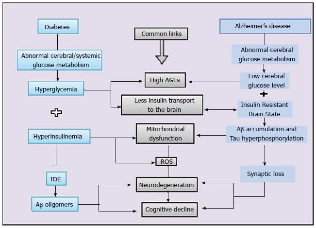
Figure 1 Schematic representation of commonalities between diabetes and Alzheimer’s disease.
Hyperglycemia and hyperinsulinemia are hallmark features of diabetes which leads to advanced glycation end product, reduced insulin supply to brain as well as mitochondrial dysfunction, which further leads to vicious cycle of oxidative stress. On the other side, any defect in glucose metabolism and insulin signaling in brain is one metabolic status of Alzheimer’s disease brain which translates into insulin resistant brain status and converges to all common interfaces of mitochondrial dysfunction, oxidative stress and neurodegenrtaion. IDE: Insulin degrading enzyme; Aβ: Amyloid beta; AGEs: Advanced glycation end products; ROS: Reactive oxygen species.
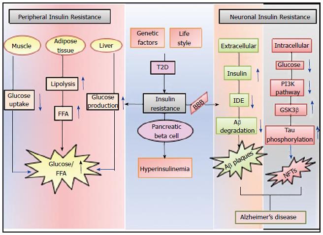
Figure 2 Diagrammatic representation of peripheral and neuronal complications of insulin resistance in case of type 2 diabetes.
Insulin signaling dysfunction in peripheral system affect muscle, adipose tissue and liver (by decreasing glucose uptake, increasing free fatty acids) and by increasing glucose production respectively. When this dysfunction appears in CNS as a diabetes complication (by limited insulin supply to brain), it leads to deposition of Aβ plaques and NFTs in extracellular and intracellular milieu of neurons respectively and represents AD type brain status. AD: Alzheimer’s disease; T2D: Type 2 diabetes; IDE: Insulin degrading enzyme; PI3K: Phosphoinositide 3-kinase; Aβ: Amyloid beta; NFTs: Neurofibrillary tangles; GSK3β: Glycogen synthase kinase 3 beta; FFA: Free fatty acids; BBB: Blood brain barrier.
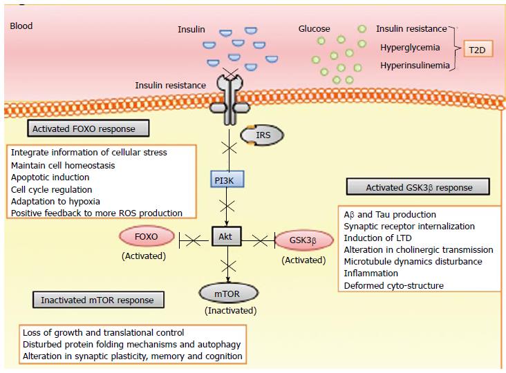
Figure 3 Diagrammatic representation of molecular orchestra downstream of insulin receptor.
IRs activation leads to downstream PI3K signaling pathway with Akt as a central hub which diverges into three main branches including FOXO, GSK3β and mTOR. Akt inhibition leads to activated FOXO and GSK3β response while inactivated mTOR response. FOXO activation leads to cellular stress response, mTOR dysfunction leads to loss of translational control and altered cognition while GSK3β activation leads to Aβ and tau production. T2D: Type 2 diabetes; PI3K: Phosphatidylinositide 3-kinases; Aβ: Amyloid beta; GSK3β: Glycogen synthase kinase 3 beta; FOXO: Forkhead box O; mTOR: Mammalian target of rapamycin; LTD: Long term depression.
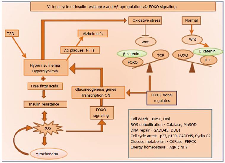
Figure 4 Diagrammatic representation of vicious cycle of insulin resistance and amyloid beta up regulation via forkhead box O signaling.
FOXO signaling is a protective mechanism adopted by body to cope up with any cellular stress by ROS detoxification. In case of DM whenever there is any metabolic disturbance like hyperinsulinemia and hyperglycemia, it leads to further oxidative stress and ROS generation which promotes further trigger for FOXO signaling and promotes a vicious cycle of ROS generation leading the cell towards apoptosis. T2D: Type 2 diabetes; Aβ: Amyloid beta; FOXO: Forkhead box O; ROS: Reactive oxygen species; TCF: T cell factor; DM: Diabetes mellitus.
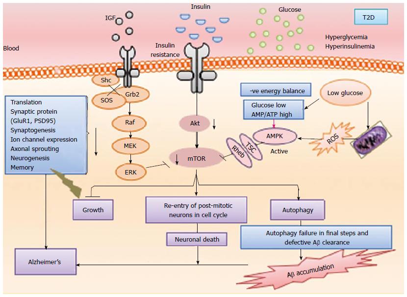
Figure 5 Diagrammatic representation of mammalian target of rapamycin pathway: A crucial intersection of type 2 diabetes and Alzheimer’s disease.
Diabetes results in dysfunctional insulin signaling which brings mTOR pathway down and ultimately results in autophagy failure to accumulate Aβ, inhibit re-entry of post mitotic neurons in cell cycle, stimulate aberrant growth pathways, lose of translational control and impaired neurogenesis, etc. T2D: Type 2 diabetes; IGF: Insulin like growth factor; mTOR: Mammalian target of rapamycin; Aβ: Amyloid beta; ROS: Reactive oxygen species.
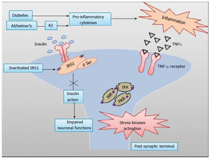
Figure 6 Diagrammatic representation of insulin signal dysfunction in Alzheimer’s disease and diabetes mellitus via common inflammatory cascade.
Diabetes and AD lead to production of pro-inflammatory cytokines in inflammatory response. These stress kinases inhibit IRs1, an adaptor protein for insulin receptor signaling and result into defective insulin signaling in brain. Aβ: Amyloid beta; IRs: Insulin receptors; JNK: c-Jun N-terminal kinases.
- Citation: Sandhir R, Gupta S. Molecular and biochemical trajectories from diabetes to Alzheimer’s disease: A critical appraisal. World J Diabetes 2015; 6(12): 1223-1242
- URL: https://www.wjgnet.com/1948-9358/full/v6/i12/1223.htm
- DOI: https://dx.doi.org/10.4239/wjd.v6.i12.1223


