Copyright
©2014 Baishideng Publishing Group Co.
World J Diabetes. Feb 15, 2014; 5(1): 59-68
Published online Feb 15, 2014. doi: 10.4239/wjd.v5.i1.59
Published online Feb 15, 2014. doi: 10.4239/wjd.v5.i1.59
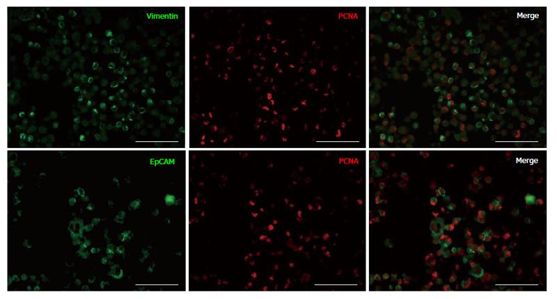
Figure 1 Double immunofluorescence staining of islet depleted pancreatic tissue expanded cells after 14 d in culture.
Cells are positive for vimentin (green), EpCAM (green) and PCNA (red). Both vimentin and EpCAM positive cells are positive for PCNA staining (merged) and proliferating. Scale bars are 50 μm. EpCAM: Epithelial cell adhesion molecule; PCNA: Proliferating cell nuclear antigen.
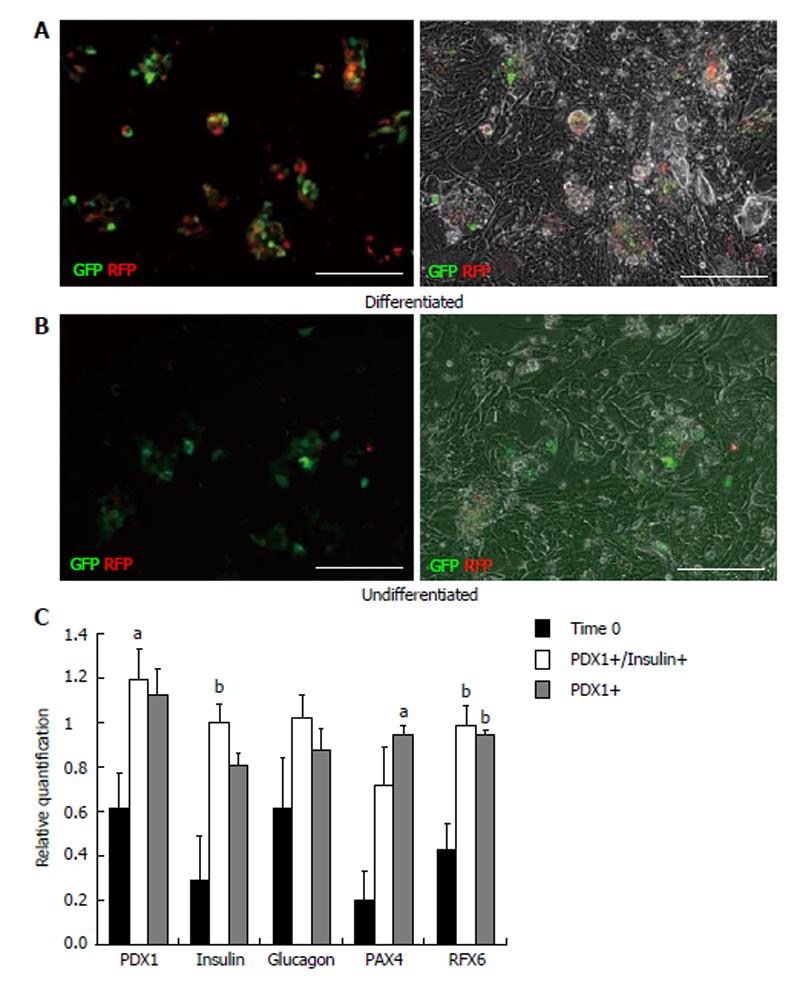
Figure 2 Comparison of differentiated and undifferentiated islet cells infected with PDX1-monomeric red fluorescent protein/insulin 1-enhanced green fluorescent protein.
Islet cells cultured in differentiation medium (A) form adherent cell aggregates within the cell monolayer and insulin+ (GFP) and PDX1+ (RFP) expressing cells are localized within these cell aggregates. Islet cells cultured in control medium (B, undifferentiated) have fewer cell aggregates and insulin (GFP) and PDX1+ (RFP) cells. Scale bars are 100 μm. Gene expression (C) of PDX1-monomeric red fluorescent protein (mRFP)/insulin-enhanced green fluorescent protein islet cells (PDX1+ insulin+, white bars; n = 7) and PDX1-mRFP infected islet cells (PDX1+, grey bars; n = 4) post-differentiation compared to starting islet tissue (Time 0, black bars; n = 8) measured by real-time PCR. aP < 0.05 and bP < 0.01 compared to Time 0. RFP: Red fluorescent protein.
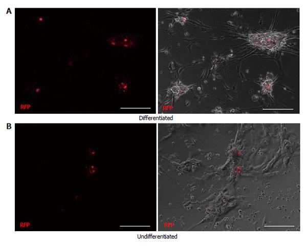
Figure 3 Comparison of differentiated (A) and undifferentiated (B) cells from the islet depleted pancreatic tissue infected with PDX1-monomeric red fluorescent protein.
A few PDX1+ cells (RFP) are visible within the adhered aggregates in the differentiated condition (A). Cell aggregates are absent in the undifferentiated cell conditions (B) and PDX1+ cells are within the monolayer. Scale bars are 100 μm. RFP: Red fluorescent protein.
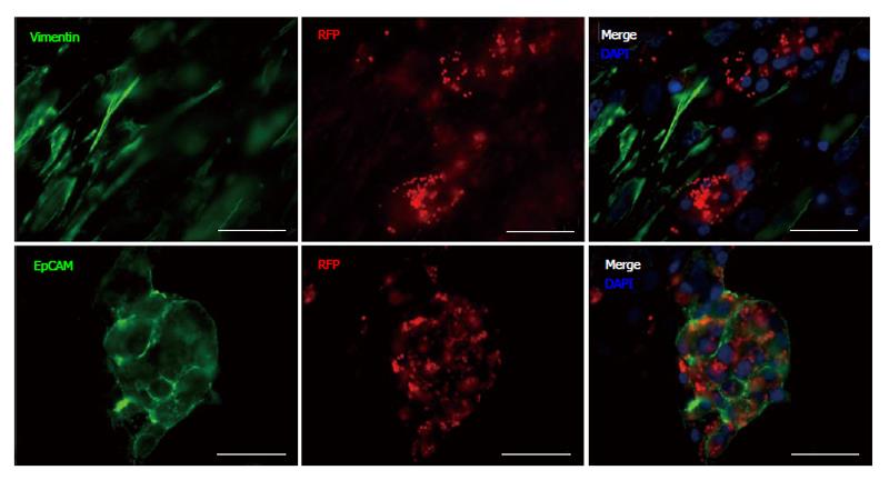
Figure 4 Immunofluorescence staining of differentiated PDX1- monomeric red fluorescent protein infected islet cells with primary antibodies to vimentin and epithelial cell adhesion molecule, with secondary antibodies conjugated to Alexa-488 (green).
PDX1+ positive cells (RFP) are not co-localized with vimentin (merged), but are co-localized with epithelial cell adhesion molecule. Nuclei are stained blue with DAPI. Scale bars are 20 μm. RFP: Red fluorescent protein; DAPI: 4',6-diamidino-2-phenylindole.
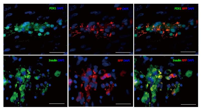
Figure 5 Immunofluorescence staining of differentiated PDX1-monomeric red fluorescent protein infected islets with primary PDX1 and insulin antibodies with secondary antibodies conjugated to Alexa-Fluor488 (green).
PDX1+ cells (RFP) nuclei stain positive with PDX1/Alexa-488 antibody confirming lentiviral expression. Insulin/Alexa-Fluor488 (green) stains insulin within PDX1+ infected cells (yellow). Nuclei are stained blue with DAPI. Scale bars are 20 μm. RFP: Red fluorescent protein.
- Citation: Seeberger KL, Anderson SJ, Ellis CE, Yeung TY, Korbutt GS. Identification and differentiation of PDX1 β-cell progenitors within the human pancreatic epithelium. World J Diabetes 2014; 5(1): 59-68
- URL: https://www.wjgnet.com/1948-9358/full/v5/i1/59.htm
- DOI: https://dx.doi.org/10.4239/wjd.v5.i1.59


