Copyright
©The Author(s) 2025.
World J Diabetes. Mar 15, 2025; 16(3): 97336
Published online Mar 15, 2025. doi: 10.4239/wjd.v16.i3.97336
Published online Mar 15, 2025. doi: 10.4239/wjd.v16.i3.97336
Figure 1 Glabridin and gymnemic acid have ability to reverse histopathological changes in the choroid thickness of diabetic rats.
Light micrographs of the choroidal and scleral layers and the extra ocular muscle of the eye. The choroid and sclera can be separated by choroidoscleral junctions (black arrows). A-E: Hematoxylin eosin stained; F-J: Masson’s trichrome in all five groups of rats. The thickness of the choroidal layer in each group is presented with the white line. The choroidal layer is composed of choroid arteries (blue arrow heads). Changes in the histological characteristics of the choroidal layer were observed in the diabetic group, involving the thickness of the choroidal layer and the choroidal arterial wall, particularly in the region of the tunica adventitia (white roundness). Scale bar = 20 μm. SC: Scleral; DM: Diabetic; DM + GB: Diabetic rats treated with glabridin 40 mg/kg body weight; DM + GM: Diabetic rats treated with gymnemic acid 400 mg/kg body weight; DM + GR: Diabetic rats treated with glyburide 4 mg/kg body weight.
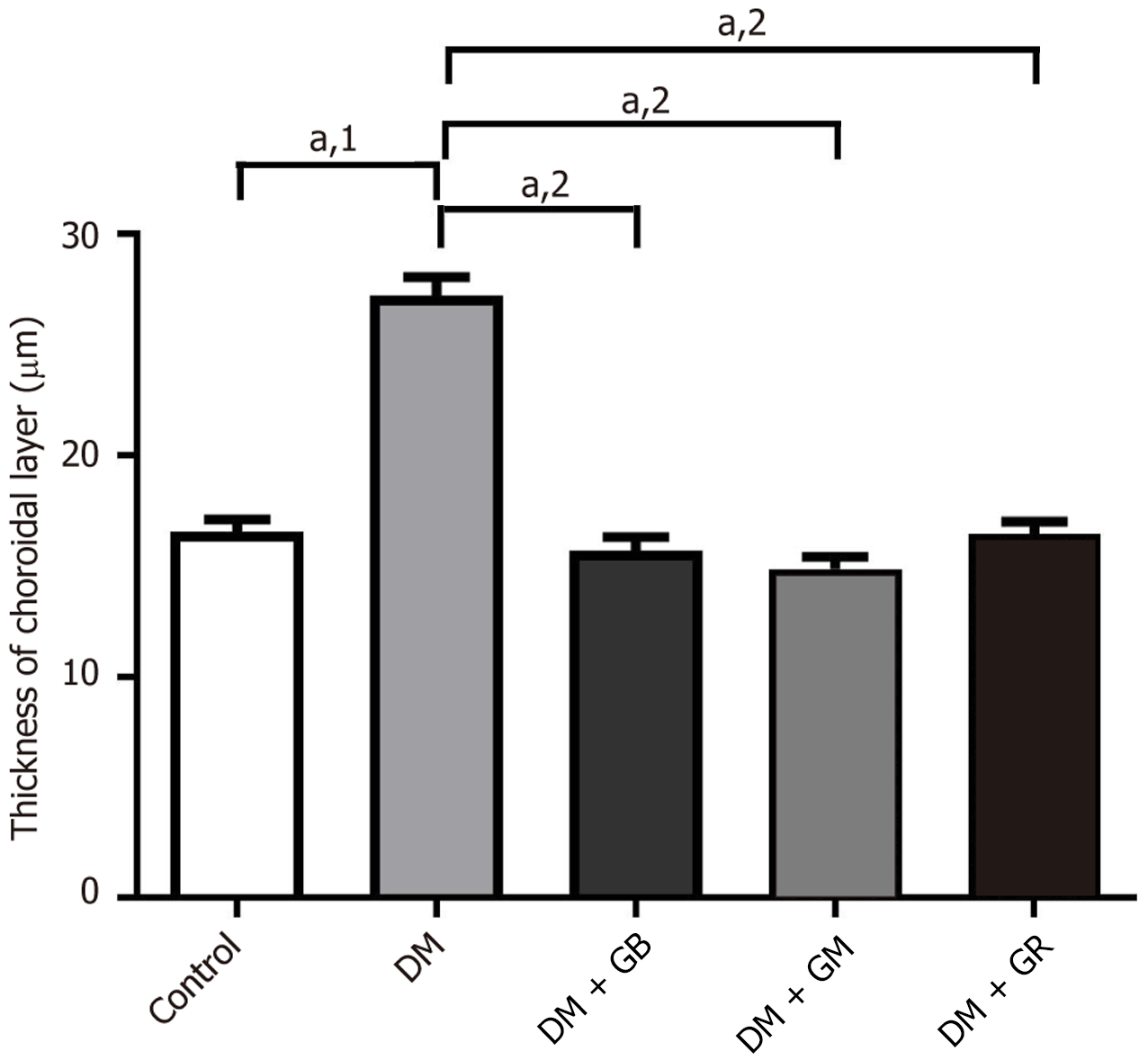
Figure 2 The effect of glabridin and gymnemic acid on the thickness of the choroidal layer in five groups of rats at 8 weeks.
aP < 0.001. 1P calculated as representative graph presents the significant increases in the thickness of the choroidal layer in the diabetic group compared with the control rats. 2P calculated after supplementation with glabridin, gymnemic acid, and glyburide, the thickness of choroidal layer decreased in the diabetic rats treated with glabridin 40 mg/kg body weight, diabetic rats treated with gymnemic acid 400 mg/kg body weight, and diabetic rats treated with glyburide 4 mg/kg body weight groups compared with the diabetic rats. DM: Diabetic; DM + GB: Diabetic rats treated with glabridin 40 mg/kg body weight; DM + GM: Diabetic rats treated with gymnemic acid 400 mg/kg body weight; DM + GR: Diabetic rats treated with glyburide 4 mg/kg body weight.
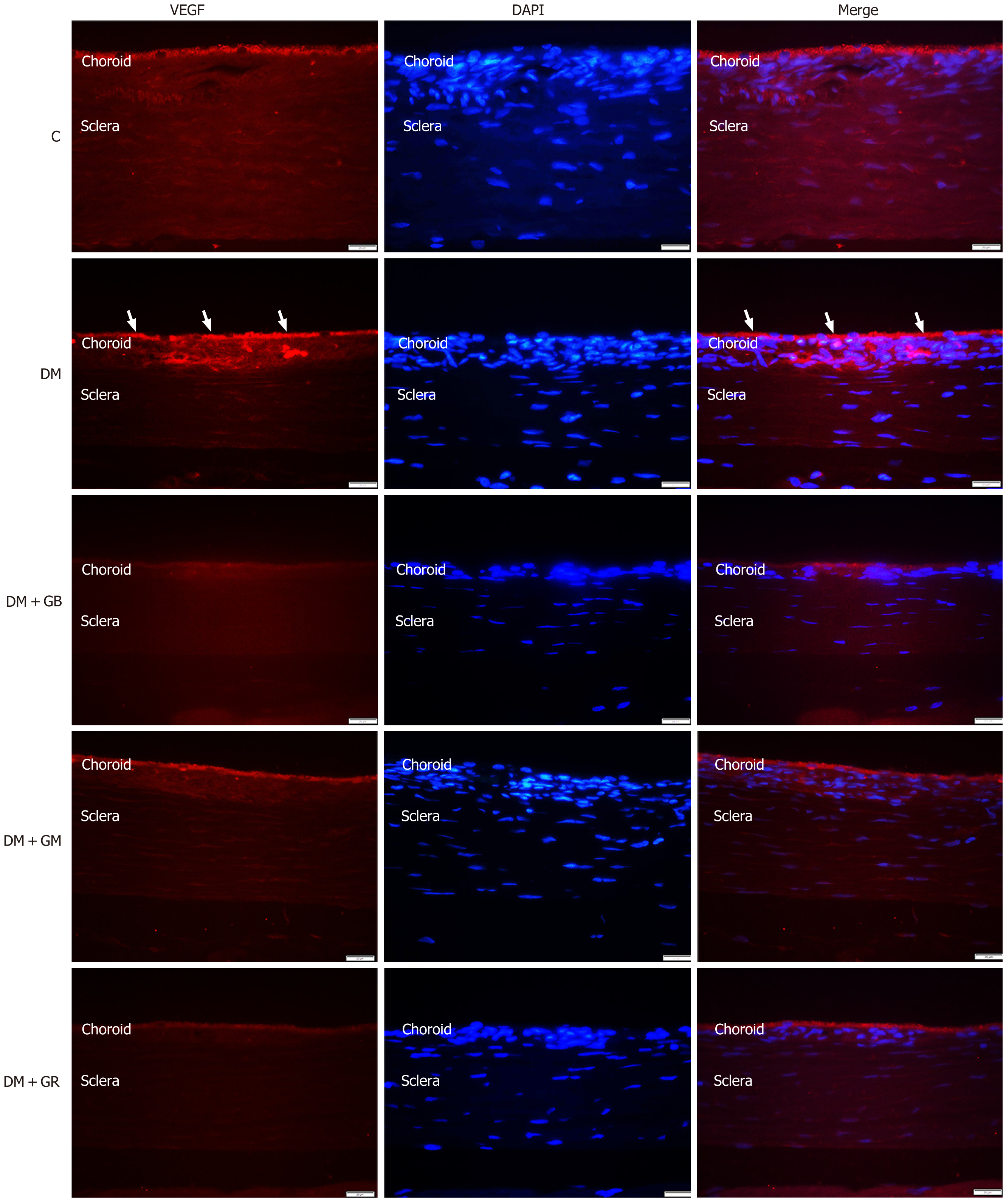
Figure 3 Glabridin and gymnemic acid can reduce a potent angiogenic cytokine.
Representative images of the immunohistochemical sections of the choroidal layer with specific vascular endothelial growth factor (VEGF) antibodies in rat eyes. Examples of immunoreactive VEGF, particularly in the choriocapillaris and the retinal pigment epithelium (white arrowheads), were present in the diabetic group at 600 magnifications. Scale bar = 20 μm. VEGF: Vascular endothelial growth factor; DAPI: 4’,6-diamidino-2-phenylindole; DM: Diabetic; DM + GB: Diabetic rats treated with glabridin 40 mg/kg body weight; DM + GM: Diabetic rats treated with gymnemic acid 400 mg/kg body weight; DM + GR: Diabetic rats treated with glyburide 4 mg/kg body weight.
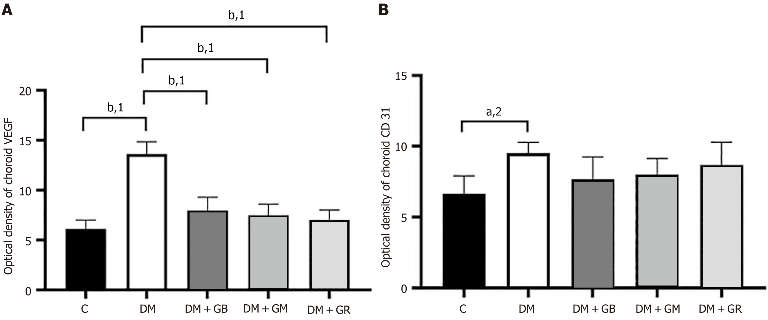
Figure 4 Optical density.
A: The effect of glabridin and gymnemic acid on the immunoreactive cytokines of the choroidal layer in five groups of rats at 8 weeks. A representative graph presents the relative vascular endothelial growth factor optical density in the choroidal layer; B: The relative cluster of differentiation 31 optical density in choroidal layer was analyzed. aP < 0.01. bP < 0.001. 1P calculated in the diabetic group when compared with the control, diabetic rats treated with glabridin 40 mg/kg body weight group, diabetic rats treated with gymnemic acid 400 mg/kg body weight group, and diabetic rats treated with glyburide 4 mg/kg body weight group. 2P calculated in the diabetic group when compared with the control group. VEGF: Vascular endothelial growth factor; CD: Cluster of differentiation; DM: Diabetic; DM + GB: Diabetic rats treated with glabridin 40 mg/kg body weight; DM + GM: Diabetic rats treated with gymnemic acid 400 mg/kg body weight; DM + GR: Diabetic rats treated with glyburide 4 mg/kg body weight.
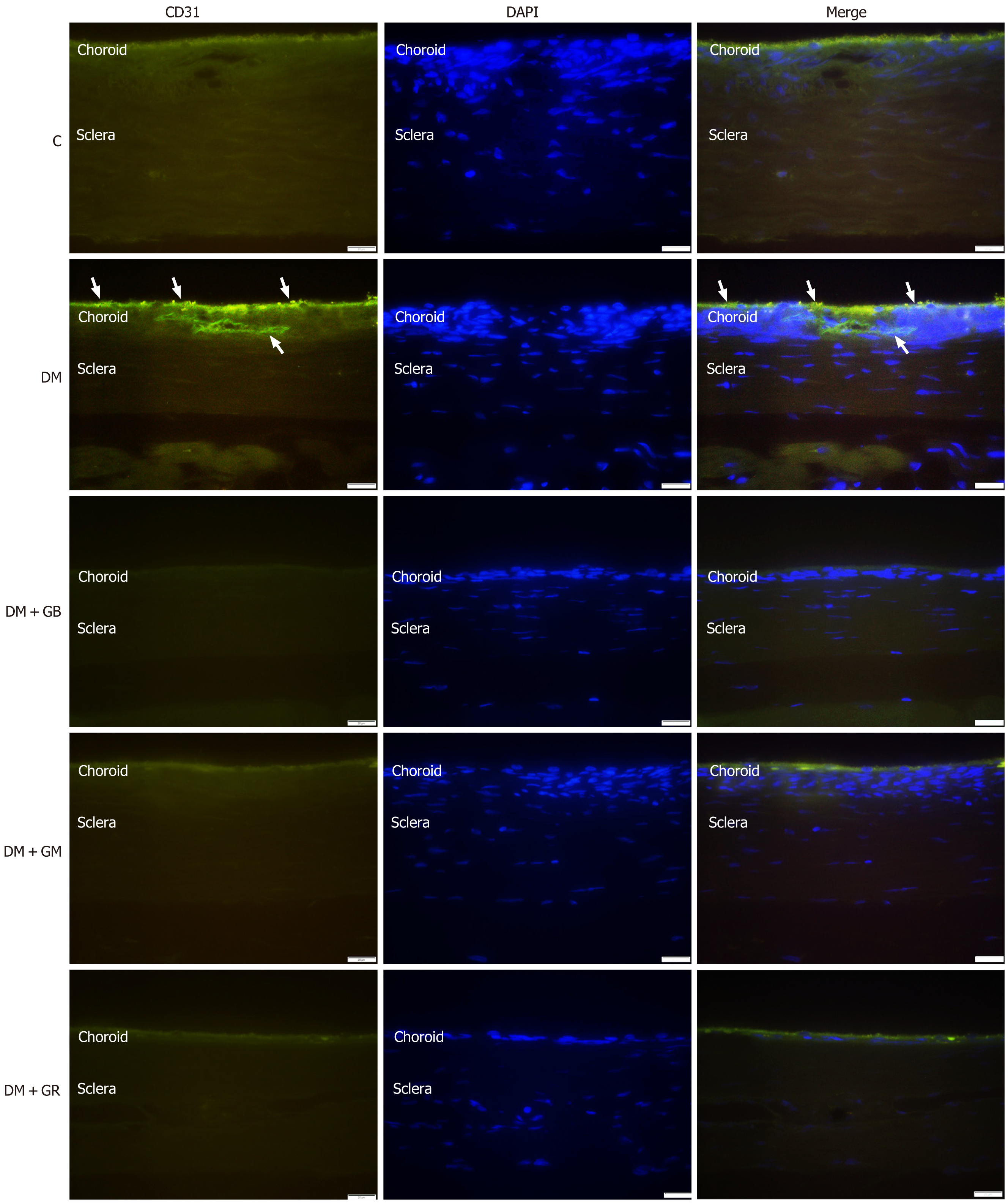
Figure 5 Glabridin and gymnemic acid have the ability to reduce the amount of angiogenic cytokine, which is essential for the process of survival.
Representative images of the immunohistochemical sections of the choroidal layer with specific cluster of differentiation (CD) 31 antibodies in rat eyes. Examples of immunoreactive CD31, particularly in the regions of the retinal pigment epithelial and choriocapillaris as well as in the walls of blood vessels (white arrowheads), were present in the diabetic group at 600 magnifications. Scale bar = 20 μm. DAPI: 4’,6-diamidino-2-phenylindole; CD: Cluster of differentiation; DM: Diabetic; DM + GB: Diabetic rats treated with glabridin 40 mg/kg body weight; DM + GM: Diabetic rats treated with gymnemic acid 400 mg/kg body weight; DM + GR: Diabetic rats treated with glyburide 4 mg/kg body weight.
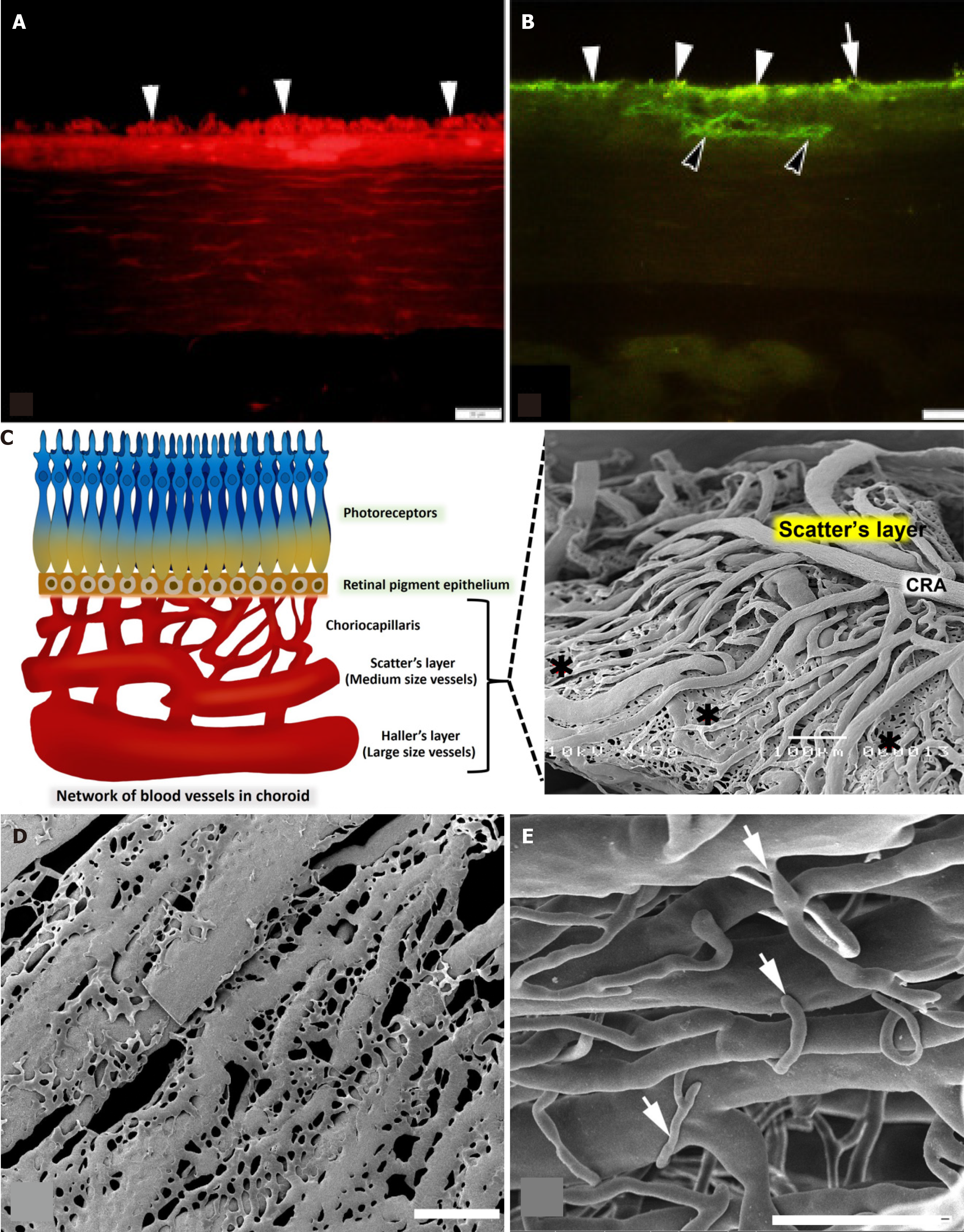
Figure 6 Schematic diagrams and the representation of results.
A: Photomicrographs of immunohistochemical sections of the choroidal layer with specific antibodies in the diabetic group. An example of immunoreactive vascular endothelial growth factor in the choriocapillaris and the retinal pigment epithelium (RPE) (white arrowheads); B: Immunoreactive cluster of differentiation 31 in the region of the RPE (white arrowheads), choriocapillaris (white arrow), and the wall of blood vessels (black arrowheads) was present in diabetic group. Scale bar = 20 μm; C: A comparison of the normal choroidal vascular structure and the vascular corrosion casting framework is indicated in this diagram. The outermost layer of the choroid, which is also known as Haller’s layer, is composed of vessels with a large vessel capacity. Sattler’s layer is the name given to the innermost layer of the choroid, which is composed of vessels that are significantly smaller in size than the vessels that make up the outermost layer such as choroidal artery. An extensive number of anastomotic capillaries are what constitute the choriocapillaris (black roundness) of the choroid; D: Scanning electron microscopy (SEM) micrographs using a vascular corrosion casting technique/SEM in the diabetic group. The arrangement of the choriocapillaris is not dense and untidy; they link to one another, resulting in the formation of a flat sheet; E: Furthermore, the sprouting of blood vessels (white arrows) was discovered in Sattler’s layer, which included choroidal arteries that were significantly more exposed. Scale bar = 300 μm. CRA: Choroid arteries.
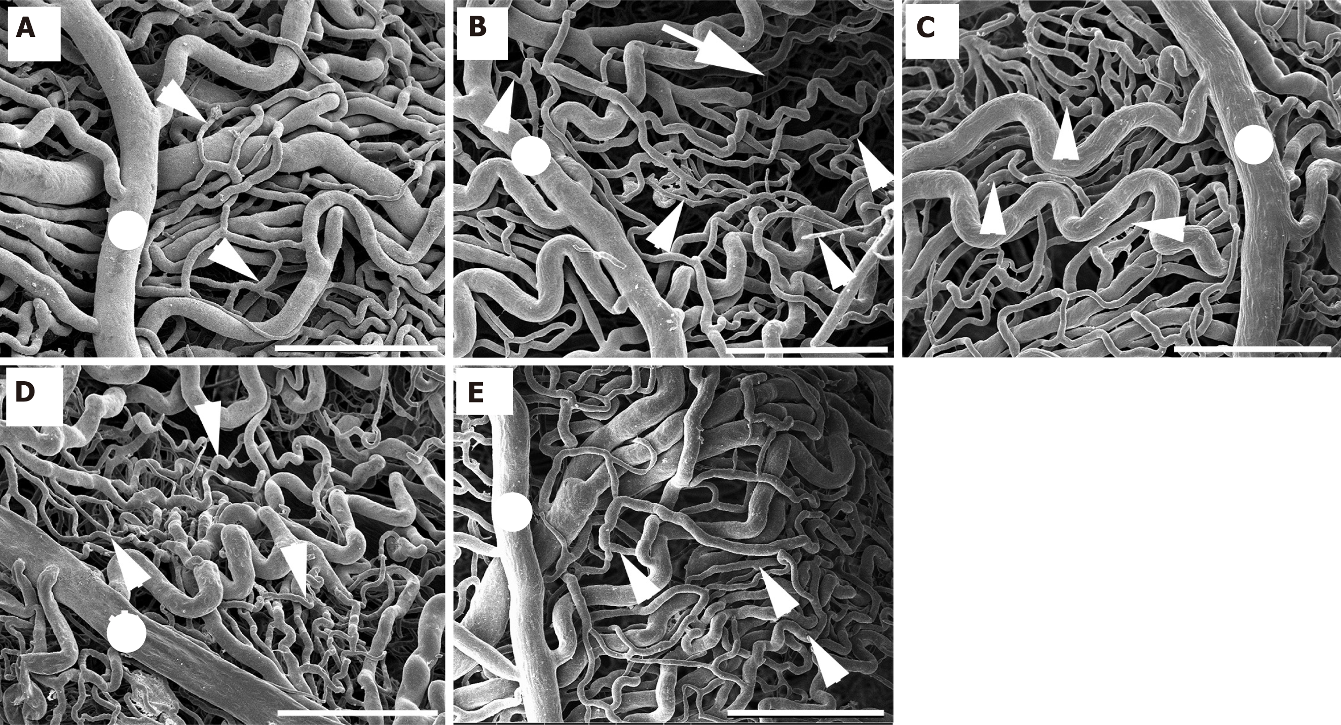
Figure 7 Glabridin and gymnemic acid have the capacity to enhance the choroid microvasculature feature.
Scanning electron microscopy (SEM) micrographs with vascular corrosion casting technique/SEM in all five groups of rats. A: Control group; B: Diabetic group; C: Diabetic rats treated with glabridin 40 mg/kg body weight group; D: Diabetic rats treated with gymnemic acid 400 mg/kg body weight group; E: Diabetic rats treated with glyburide 4 mg/kg body weight group. The choroid artery (white roundness) splits into branches to supply the choroidal layer. Many anastomotic capillaries are what make up the choriocapillaris of the choroid (white arrowheads). In the diabetic group, choroid artery had a small diameter and the choriocapillaris shrank. The capillaries dropping out of the choriocapillaris (white arrow) were also present in diabetic group. Scale bar = 300 μm.
- Citation: Matsathit U, Komolkriengkrai M, Khimmaktong W. Glabridin and gymnemic acid alleviates choroid structural change and choriocapillaris impairment in diabetic rat’s eyes. World J Diabetes 2025; 16(3): 97336
- URL: https://www.wjgnet.com/1948-9358/full/v16/i3/97336.htm
- DOI: https://dx.doi.org/10.4239/wjd.v16.i3.97336


