©The Author(s) 2025.
World J Diabetes. Mar 15, 2025; 16(3): 102126
Published online Mar 15, 2025. doi: 10.4239/wjd.v16.i3.102126
Published online Mar 15, 2025. doi: 10.4239/wjd.v16.i3.102126
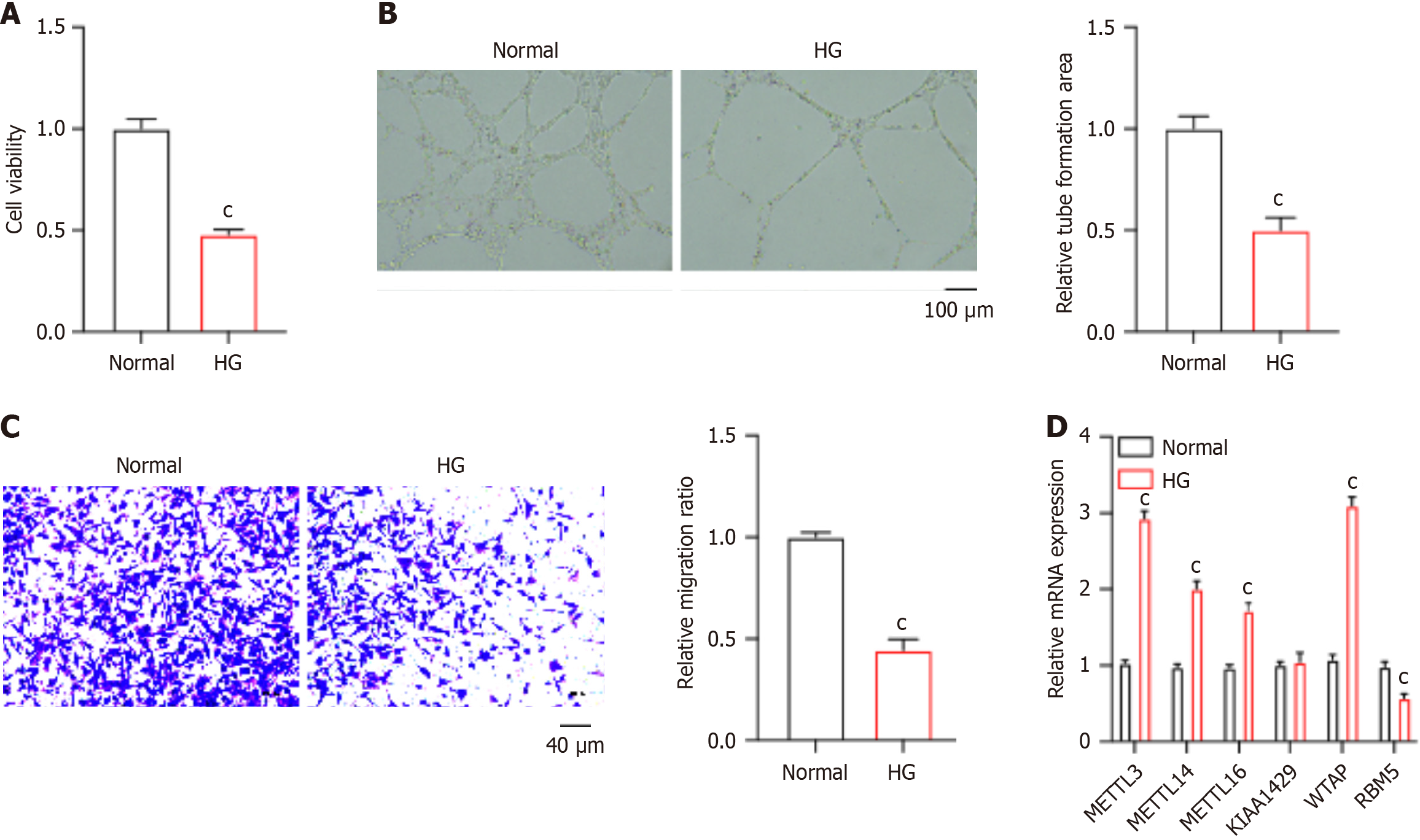
Figure 1 Wilms tumor 1-associated protein expression is elevated in high glucose-induced human umbilical vein endothelial cells.
Human umbilical vein endothelial cells were stimulated with high glucose. A: Cell viability was measured using cell counting kit 8 assay; B: The in vitro angiogenesis was measured by tube formation assay; C: Cell migration was analyzed by Transwell assay; D: The relative RNA levels of RNA methyltransferases were measured by quantitative PCR assay. cP < 0.001; HG: High glucose; METTL3: Methyltransferase-like 3; WTAP: Wilms tumor 1-associated protein.
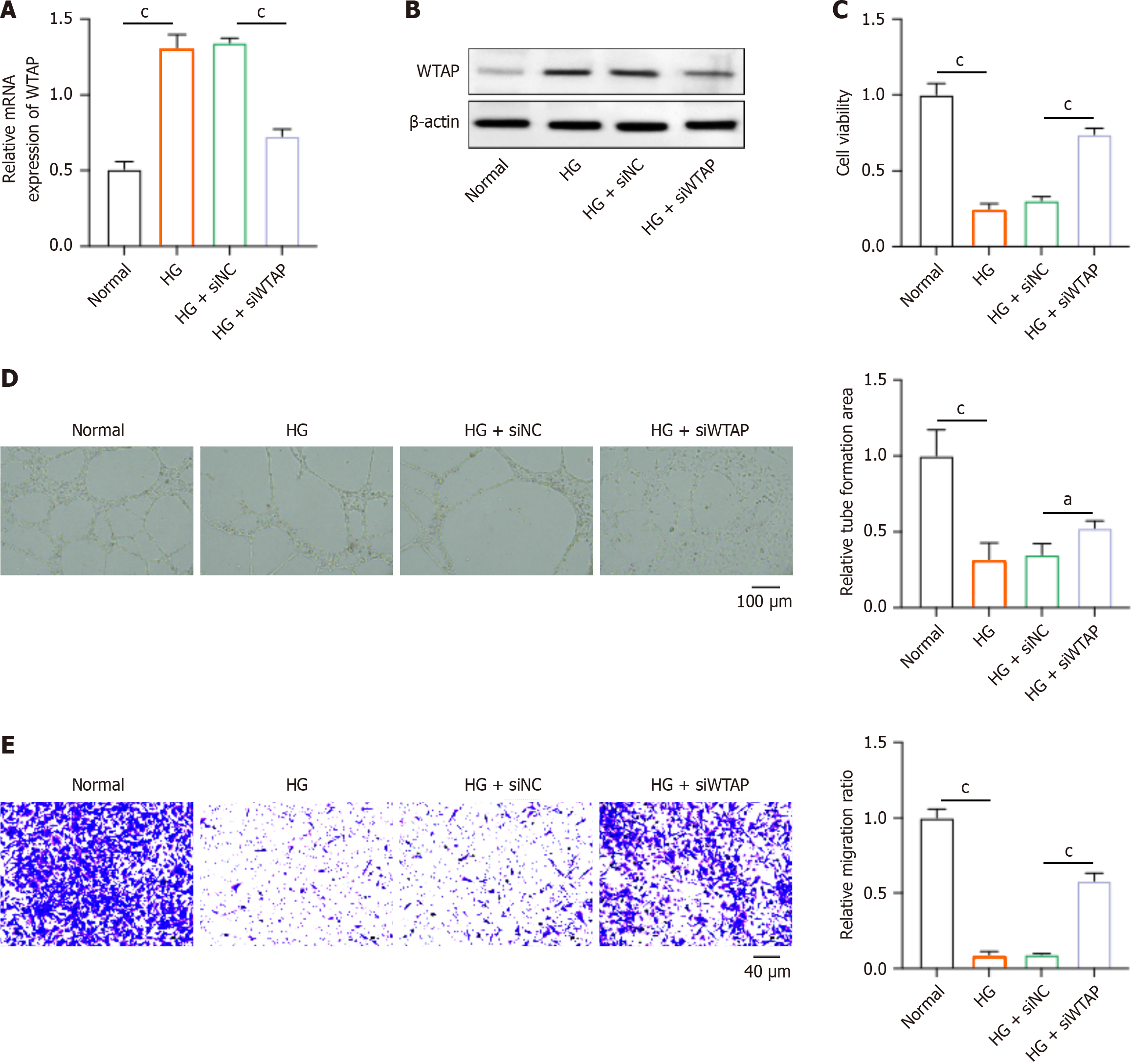
Figure 2 Knockdown of Wilms tumor 1-associated protein activates proliferation, invasion, and angiogenesis of human umbilical vein endothelial cells.
Human umbilical vein endothelial cells were stimulated with high glucose and transfected with siWilms tumor 1-associated protein. A: Cell viability was measured using cell counting kit 8 assay; B: The in vitro angiogenesis was measured by tube formation assay; C: Cell migration was analyzed by Transwell assay; D: The relative RNA levels of RNA methyltransferases were measured by quantitative PCR assay; E: Migration of high glucose-stimulated human umbilical vein endothelial cells. aP < 0.05; cP < 0.001; HG: High glucose; WTAP: Wilms tumor 1-associated protein.
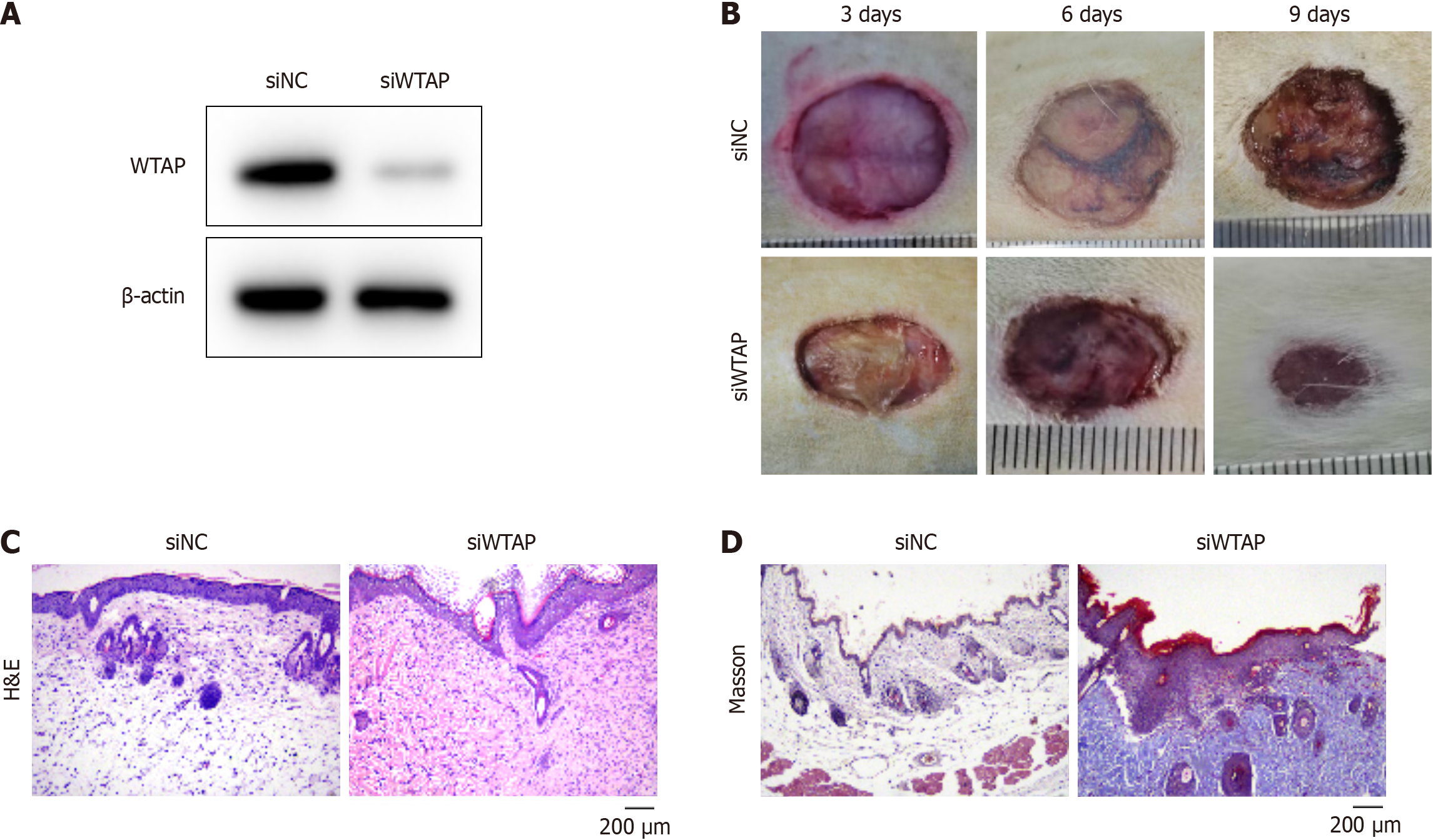
Figure 3 Knockdown of Wilms tumor 1-associated protein improves wound healing in diabetic mice.
A: Protein level of Wilms tumor 1-associated protein (WTAP) in skin tissues from mice treated with siNC or siWTAP; B: Images of sounds in dorsal skin at day 0, 6, and 9 post-wounding; C: Hematoxylin & eosin staining of skin tissues to determine epithelialization; D: Masson’s trichrome staining was conducted to measure collagen deposition in dorsal skin. WTAP: Wilms tumor 1-associated protein; H&E: Hematoxylin & eosin staining; NC: Negative controls.
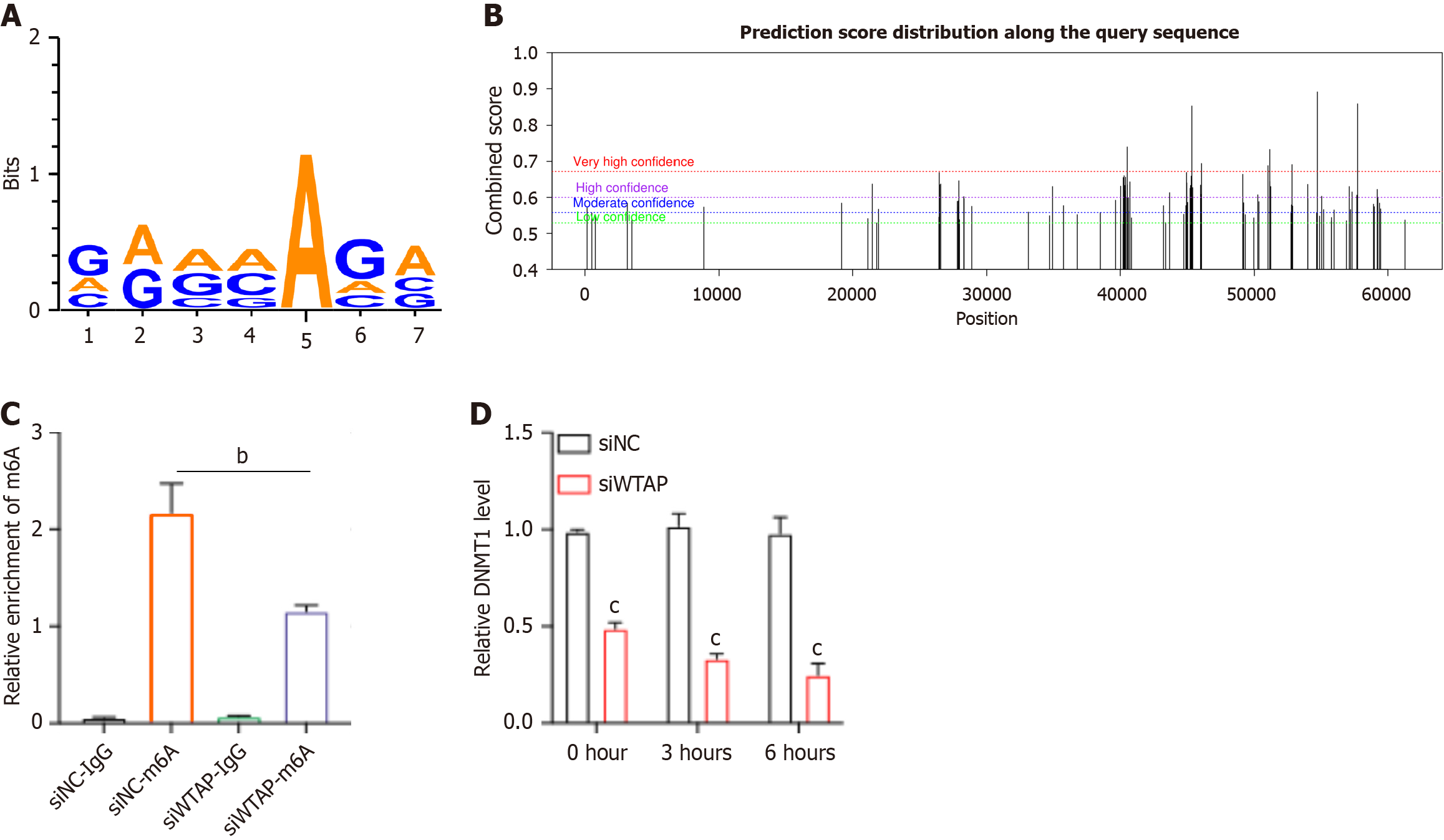
Figure 4 Wilms tumor 1-associated protein modulates the N6-methyladenosine modification of DNA methyltransferase 1.
A: The N6-methyladenosine (m6A)-modified motif on DNA methyltransferase 1 (DNMT1) mRNA towards Wilms tumor 1-associated protein (WTAP) was predicted; B: A sequence-based m6A modification site predictor SRAMP identified the m6A modification site on DNMT1 mRNA; C: MeRIP-PCR analysis was performed to measure the m6A enrichment in human umbilical vein endothelial cells treated with siNC or siWTAP; D: The stability of DNMT1 mRNA was analyzed by actinomycin D assay in human umbilical vein endothelial cells treated with siNC or siWTAP. bP < 0.01; cP < 0.001; WTAP: Wilms tumor 1-associated protein; DNMT1: DNA methyltransferase 1; m6A: N6-methyladenosine; NC: Negative controls.
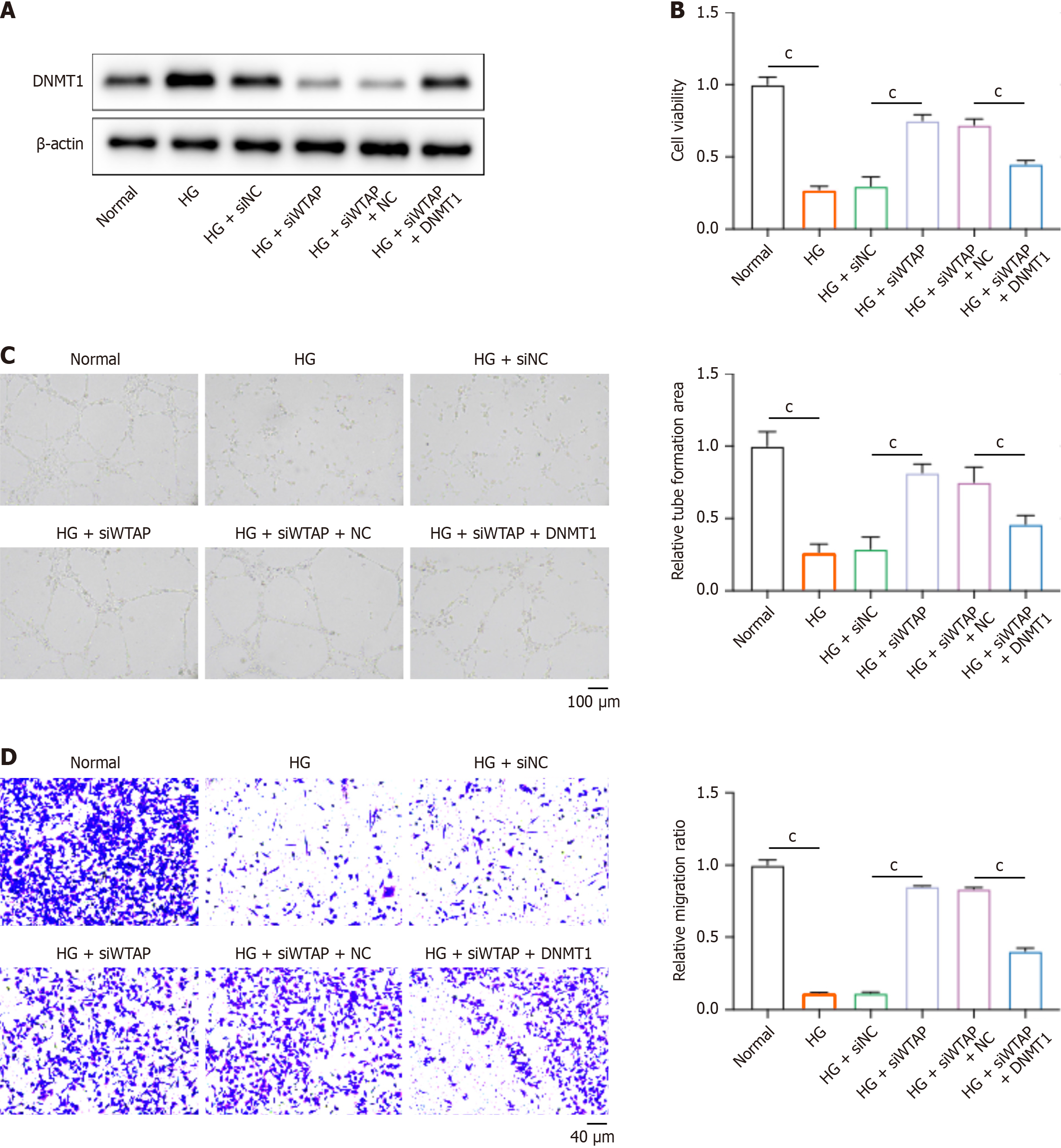
Figure 5 Wilms tumor 1-associated protein-DNA methyltransferase 1 axis regulates human umbilical vein endothelial cell function.
Human umbilical vein endothelial cells (HUVECs) were stimulated with high glucose and treated with siWilms tumor 1-associated protein and DNA methyltransferase 1 (DNMT1) overexpression vectors. A: The protein level of DNMT1 in the dorsal skin of mice was detected by western blotting; B: Cell viability was detected by cell counting kit 8 assay; C: The angiogenesis ability of HUVECs was measured by tube formation assay; D: Cell migration was analyzed by Transwell assay. cP < 0.001; HG: High glucose; WTAP: Wilms tumor 1-associated protein; DNMT1: DNA methyltransferase 1; m6A: N6-methyladenosine; NC: Negative controls.
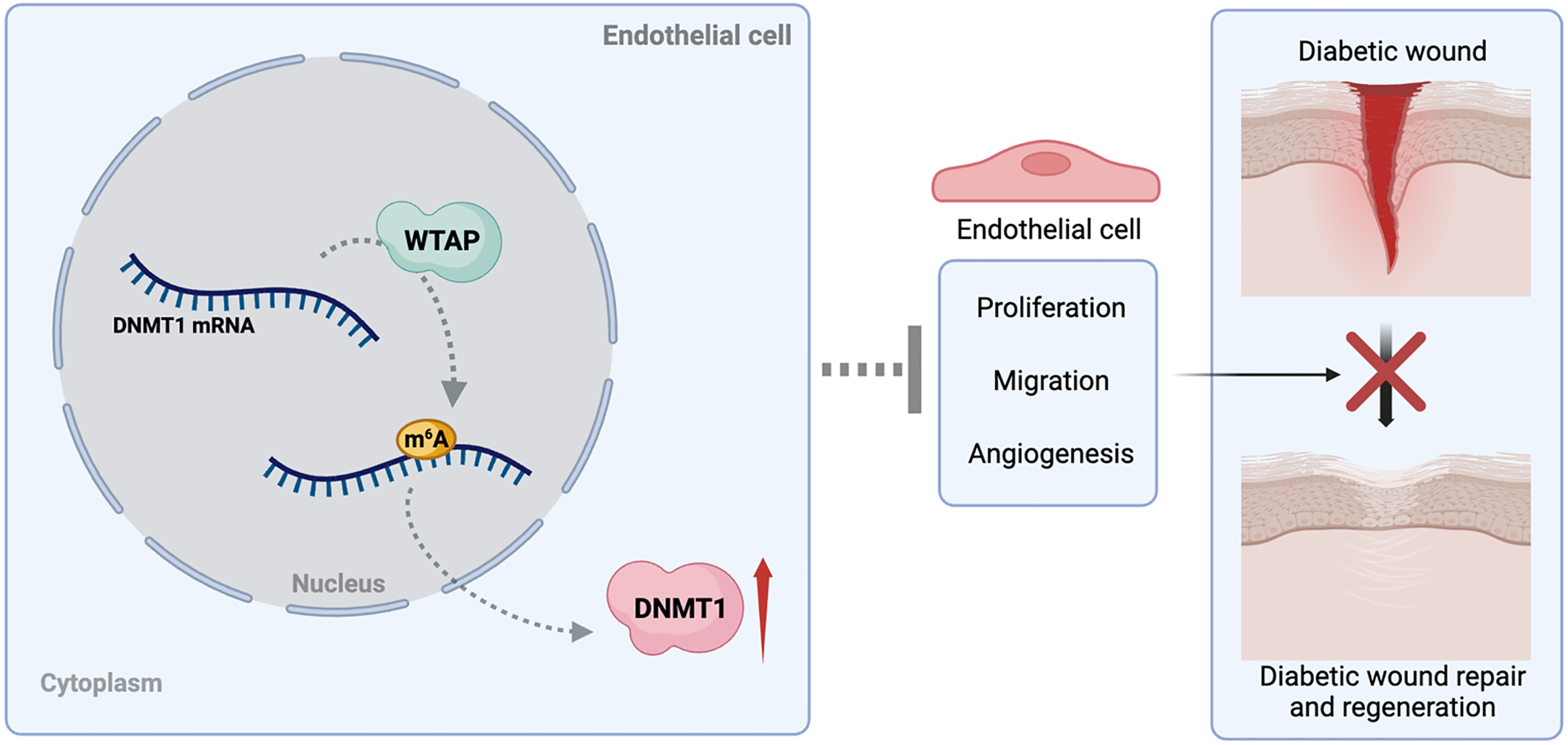
Figure 6 Work model illustrating that N6-methyladenosine methyltransferase Wilms tumor 1-associated protein impedes diabetic wound healing through epigenetically activating DNA methyltransferase 1.
DNMT1: DNA methyltransferase 1; m6A: N6-methyladenosine; WTAP: Wilms tumor 1-associated protein.
- Citation: Xiao RJ, Wang TJ, Wu DY, Yang SF, Gao H, Gan PD, Yi YY, Zhang YL. N6-methyladenosine methyltransferase Wilms tumor 1-associated protein impedes diabetic wound healing through epigenetically activating DNA methyltransferase 1. World J Diabetes 2025; 16(3): 102126
- URL: https://www.wjgnet.com/1948-9358/full/v16/i3/102126.htm
- DOI: https://dx.doi.org/10.4239/wjd.v16.i3.102126


