©The Author(s) 2025.
World J Diabetes. Mar 15, 2025; 16(3): 101328
Published online Mar 15, 2025. doi: 10.4239/wjd.v16.i3.101328
Published online Mar 15, 2025. doi: 10.4239/wjd.v16.i3.101328
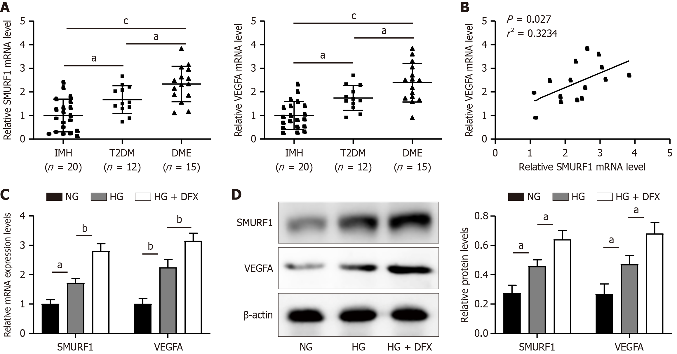
Figure 1 Expression of SMAD-specific E3 ubiquitin protein ligase 1 and vascular endothelial growth factor A was upregulated in diabetic macular edema.
A: The aqueous humor was collected from diabetic macular edema patients (n = 15), type 2 diabetes mellitus patients (n = 12), and idiopathic macular hole patients (n = 20), SMAD-specific E3 ubiquitin protein ligase (SMURF) 1 and vascular endothelial growth factor A (VEGFA) expression was detected by quantitative real-time PCR (qRT-PCR); B: Correlation analysis between SMURF1 expression and VEGFA expression; C: ARPE-19 cells were treated with normal concentration of glucose (NG), high concentration of glucose (HG) or desferrioxamine mesylate (DFX), and the mRNA levels of SMURF1 and VEGFA were then detected by qRT-PCR; D: NG, HG or DFX treated ARPE-19 cells were collected, and the protein levels of SMURF1 and VEGFA were detected by western blot and the gray scale of the bands was quantified. The experiments were repeated three times. aP < 0.05, bP < 0.01, cP < 0.001. VEGFA: Vascular endothelial growth factor A; SMURF 1: SMAD-specific E3 ubiquitin protein ligase 1; IMH: Idiopathic macular hole; HG: High glucose; DFX: Desferrioxamine mesylate.
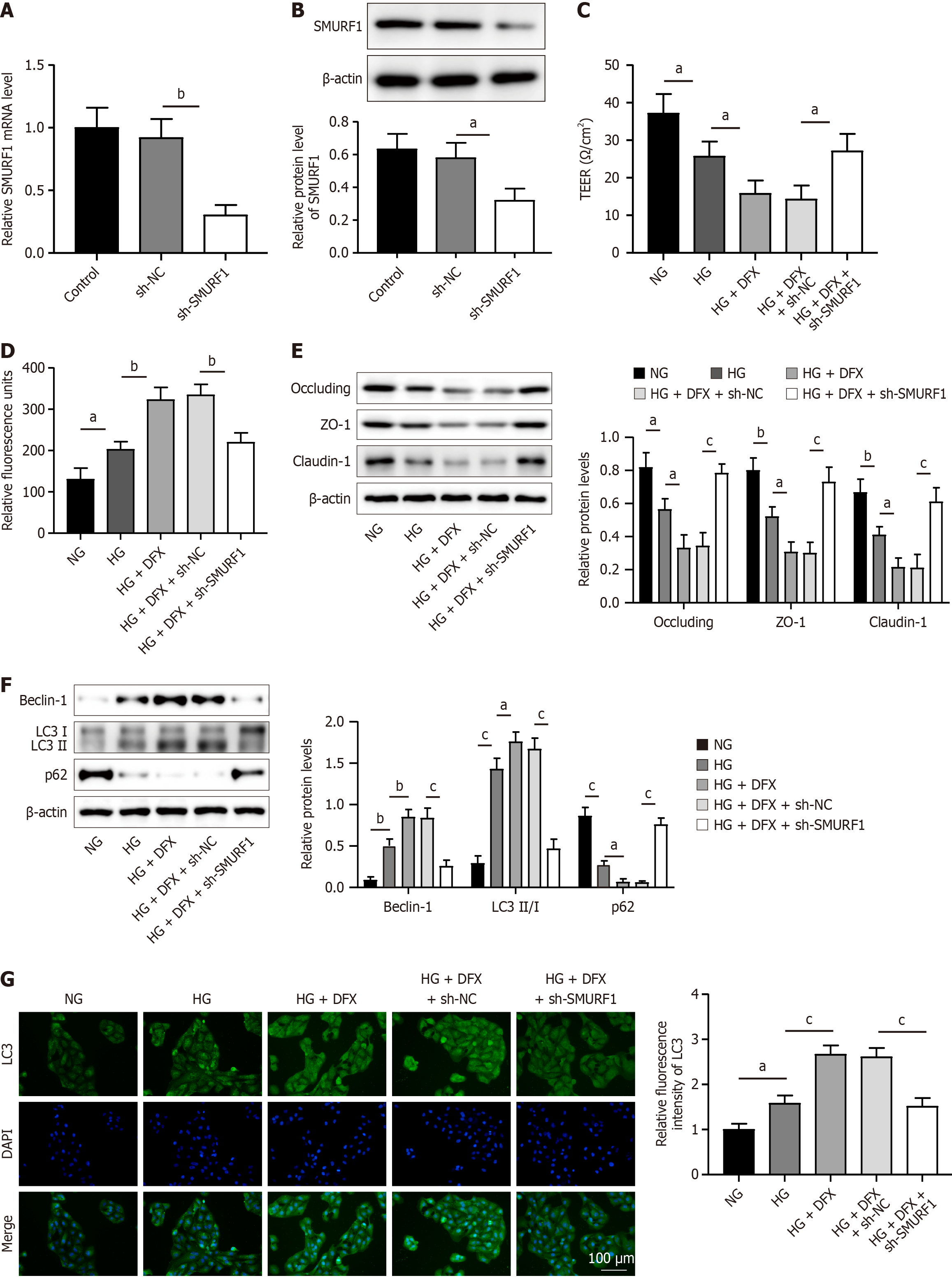
Figure 2 SMAD-specific E3 ubiquitin protein ligase 1 knockdown promoted retinal pigment epithelium cell tight junctions by inhibiting autophagy in diabetic macular edema.
A: The SMAD-specific E3 ubiquitin protein ligase (SMURF) 1 knockdown construct or vector were transfected in ARPE-19 cells, and the mRNA level of SMURF1 was then detected by quantitative real-time PCR; B: ARPE-19 cells transfected with the vector or SMURF1 construct were collected, followed by western blot to detect SMURF1 protein levels; C: Vector or SMURF1 knockdown ARPE-19 cells were treated with normal concentration of glucose (NG), high concentration of glucose (HG) or desferrioxamine mesylate (DFX), followed by determination of cellular trans-epithelial electrical resistance; D: Vector or SMURF1 knockdown ARPE-19 cells were treated with NG, HG or DFX, and the detection of cell permeability was performed; E: Vector or SMURF1 knockdown ARPE-19 cells were treated with NG, HG or DFX, and then the expression of occluding, ZO-1, and claudin-1 was detected by western blot; F: Vector or SMURF1 knockdown ARPE-19 cells were treated with NG, HG or DFX, and the expression of Beclin-1, LC3I, LC3II, and p62 was then detected by western blot; G: Vector or SMURF1 knockdown ARPE-19 cells were treated with NG, HG or DFX, followed by immunofluorescence analysis of LC3. The experiments were repeated three times. aP < 0.05, bP < 0.01, cP < 0.001. SMURF1: SMAD-specific E3 ubiquitin protein ligase 1; NG: Normal concentration of glucose; HG: High concentration of glucose; DFX: Desferrioxamine mesylate.
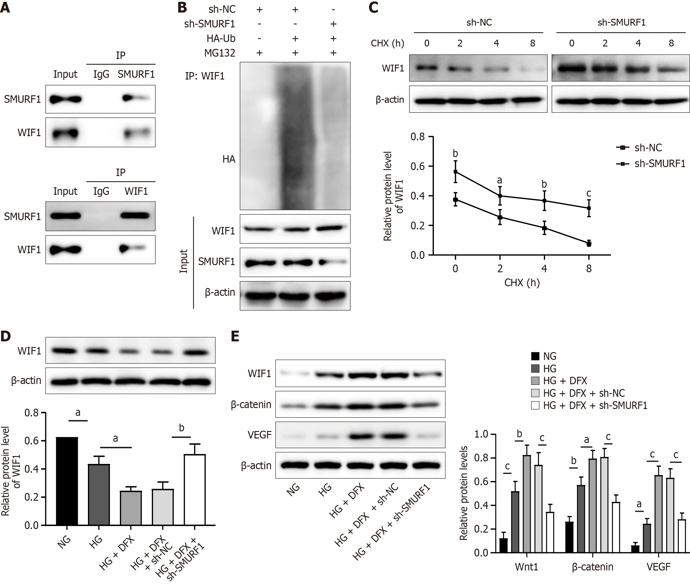
Figure 3 SMAD-specific E3 ubiquitin protein ligase 1-mediated ubiquitination degradation of WNT inhibitory factor 1 activated the Wnt/β-catenin-vascular endothelial growth factor signaling pathway.
A: ARPE-19 cells were collected, followed by the interaction between SMAD-specific E3 ubiquitin protein ligase (SMURF) 1 and WNT inhibitory factor 1 (WIF1) was verified by the Co-immunoprecipitation (Co-IP) method; B: The SMURF1 knockdown construct or vector was transfected into ARPE-19 cells, followed by the Co-IP method for detecting the ubiquitination level of WIF1; C: The SMURF1 knockdown construct or vector was transfected into ARPE-19 cells, and then cells were treated with Cycloheximide (50 ng/mL) for 0, 2, 4, and 8 hours, followed by the protein level detection of WIF1 by western blot; D: The SMURF1 knockdown construct or vector was transfected into ARPE-19 cells, and cells were then treated with normal concentration of glucose (NG), high concentration of glucose (HG) or desferrioxamine mesylate (DFX), followed by western blot for the detection of WIF1 expression; E: The SMURF1 knockdown construct or vector was transfected into ARPE-19 cells, and cells were then treated with NG, HG, or DFX, followed by western blot for the detection of Wnt1, β-tcatenin, and vascular endothelial growth factor. The experiments were repeated three times. aP < 0.05, bP < 0.01, cP < 0.001. SMURF1: SMAD-specific E3 ubiquitin protein ligase 1; WIF1: WNT inhibitory factor 1; NG: Normal concentration of glucose; HG: High concentration of glucose; DFX: Desferrioxamine mesylate.
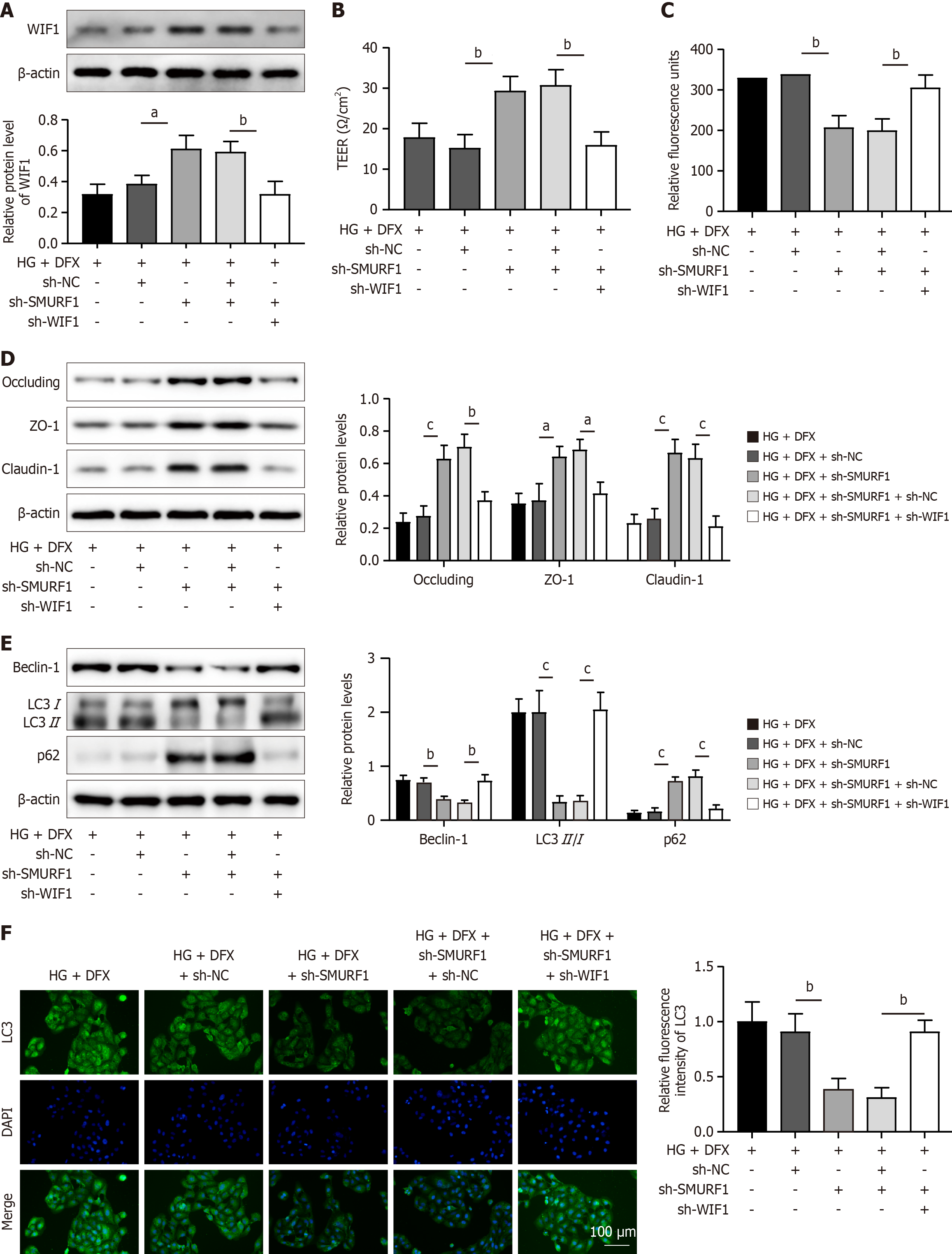
Figure 4 SMAD-specific E3 ubiquitin protein ligase 1 regulated high concentration of glucose/desferrioxamine mesylate-induced tight junction inhibition and autophagy in retinal pigment epithelium cells via WNT inhibitory factor 1.
A: ARPE-19 cells were transfected with the SMAD-specific E3 ubiquitin protein ligase (SMURF) 1 or WNT inhibitory factor (WIF) 1 knockdown construct or vector, followed by treatment of the cells with high concentration of glucose or desferrioxamine mesylate, and then the protein level of WIF1 was detected by western blot; B: ARPE-19 cells were transfected with the SMURF1 or WIF1 knockdown construct or vector, followed by the measurement of cellular trans-epithelial electrical resistance; C: ARPE-19 cells were transfected with the SMURF1 or WIF1 knockdown construct or vector, followed by the detection of cell permeability; D: SMURF1, WIF1 or vector knockdown ARPE-19 cells were collected, and the expression of occluding, ZO-1 and claudin-1 was detected by western blot; E: SMURF1, WIF1 or vector knockdown ARPE-19 cells were collected, and the expression of Beclin-1, LC3I, LC3II and p62 was detected by western blot; F: ARPE-19 cells were transfected with the SMURF1 or WIF1 knockdown construct or vector, followed by the immunofluorescence analysis of LC3. The experiments were repeated three times. aP < 0.05, bP < 0.01, cP < 0.001. SMURF1: SMAD-specific E3 ubiquitin protein ligase 1; WIF1: WNT inhibitory factor 1; HG: High concentration of glucose; DFX: Desferrioxamine mesylate.
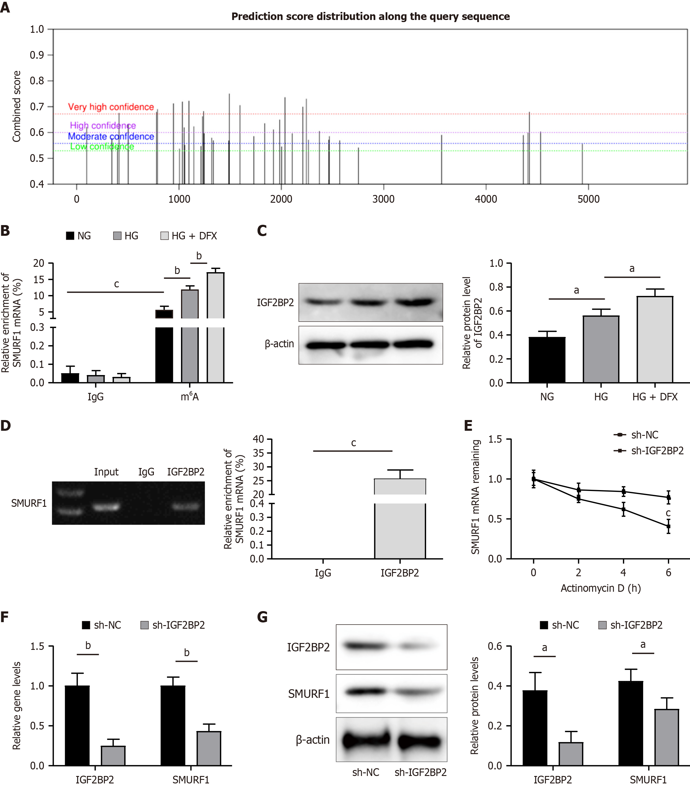
Figure 5 Insulin like growth factor 2 mRNA binding protein 2 upregulated SMAD-specific E3 ubiquitin protein ligase 1 expression in an N6-methyladenosine modification-dependent manner.
A: The N6-methyladenosine (m6A) modification site of SMAD-specific E3 ubiquitin protein ligase (SMURF) 1 was predicted by the SRAMP database; B: ARPE-19 cells were treated with normal concentration of glucose (NG) or high concentration of glucose (HG), and the m6A level of SMURF1 was then detected by methylated RNA immunoprecipitation (RIP); C: ARPE-19 cells were treated with NG or HG, and then the expression of insulin like growth factor 2 mRNA binding protein 2 (IGF2BP2) was detected by western blot; D: The interaction between IGF2BP2 protein and SMURF1 mRNA was verified by RIP method; E: ARPE-19 cells were transfected with IGF2BP2 knockdown construct or vector, and then cells were treated with dactinomycin (10 µg/mL) for 0, 2, 4, or 6 hours, followed by quantitative real-time PCR (qRT-PCR) to detect the stability of SMURF1 mRNA; F: IGF2BP2 or vector knockdown ARPE-19 cells were collected, and then the mRNA levels of IGF2BP2 and SMURF1 were detected by qRT-PCR; G: ARPE-19 cells were transfected with IGF2BP2 knockdown construct or vector, followed by western blot to detect the protein levels of IGF2BP2 and SMURF1. The experiments were repeated three times. aP < 0.05, bP < 0.01, cP < 0.001. SMURF1: SMAD-specific E3 ubiquitin protein ligase 1; IGF2BP2: Insulin like growth factor 2 mRNA binding protein 2; HG: High concentration of glucose; DFX: Desferrioxamine mesylate; NG: Normal concentration of glucose.
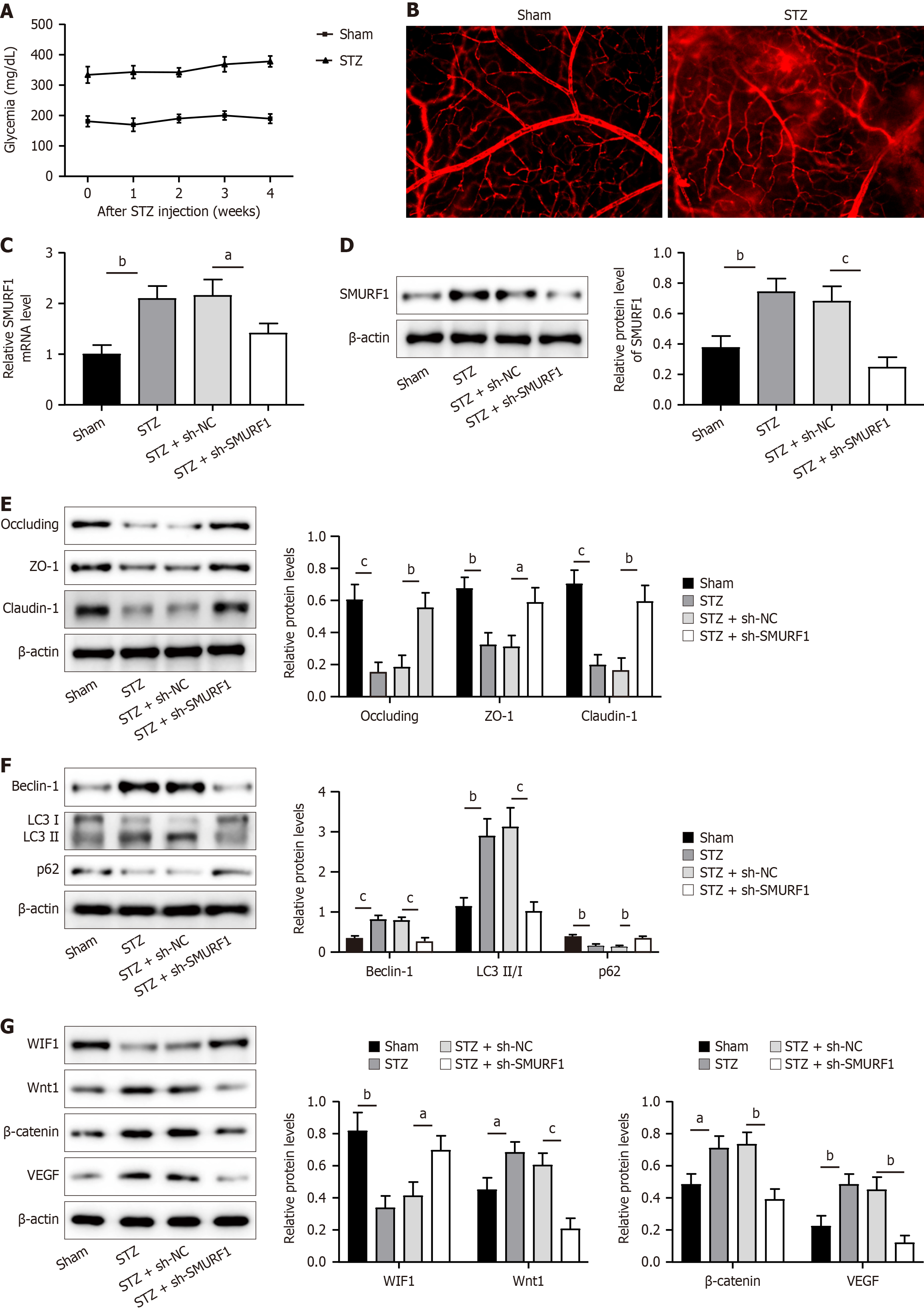
Figure 6 SMAD-specific E3 ubiquitin protein ligase 1 knockdown improved retinal damage in diabetic macular edema mice.
A: The Streptozotocin (STZ) mouse model was constructed, and then the blood glucose concentration of the mice was examined; B: The STZ mouse model was constructed, and the permeability of mouse retinal blood vessels was then examined by Evans Blue assay; C: STZ model mice were injected intraocularly with adenoviruses of sh-NC or sh-SMAD-specific E3 ubiquitin protein ligase (SMURF) 1, and then the expression of SMURF1 was detected by quantitative real-time PCR; D: STZ model mice were injected intraocularly with adenoviruses of sh-NC or sh-SMURF1, and the expression of SMURF1 was then detected by western blot; E: STZ model mice were injected intraocularly with adenoviruses of sh-NC or sh-SMURF1, followed by western blot to detect the expression of occluding, ZO-1, and claudin-1; F: STZ model mice were injected intraocularly with adenoviruses of sh-NC or sh-SMURF1, and then the expression of Beclin-1, LC3I, LC3II, and p62 was detected by western blot; G: STZ model mice were injected intraocularly with adenoviruses of sh-NC or sh-SMURF1, followed by western blot to detect the expression of WIF1, Wnt1, β-catenin, and vascular endothelial growth factor was detected by western blot. n = 6. aP < 0.05, bP < 0.01, cP < 0.001. SMURF1: SMAD-specific E3 ubiquitin protein ligase 1; VEGF: Vascular endothelial growth factor; STZ: Streptozotocin.
- Citation: Liang LF, Zhao JQ, Wu YF, Chen HJ, Huang T, Lu XH. SMAD specific E3 ubiquitin protein ligase 1 accelerates diabetic macular edema progression by WNT inhibitory factor 1. World J Diabetes 2025; 16(3): 101328
- URL: https://www.wjgnet.com/1948-9358/full/v16/i3/101328.htm
- DOI: https://dx.doi.org/10.4239/wjd.v16.i3.101328


