©The Author(s) 2025.
World J Diabetes. Feb 15, 2025; 16(2): 99053
Published online Feb 15, 2025. doi: 10.4239/wjd.v16.i2.99053
Published online Feb 15, 2025. doi: 10.4239/wjd.v16.i2.99053
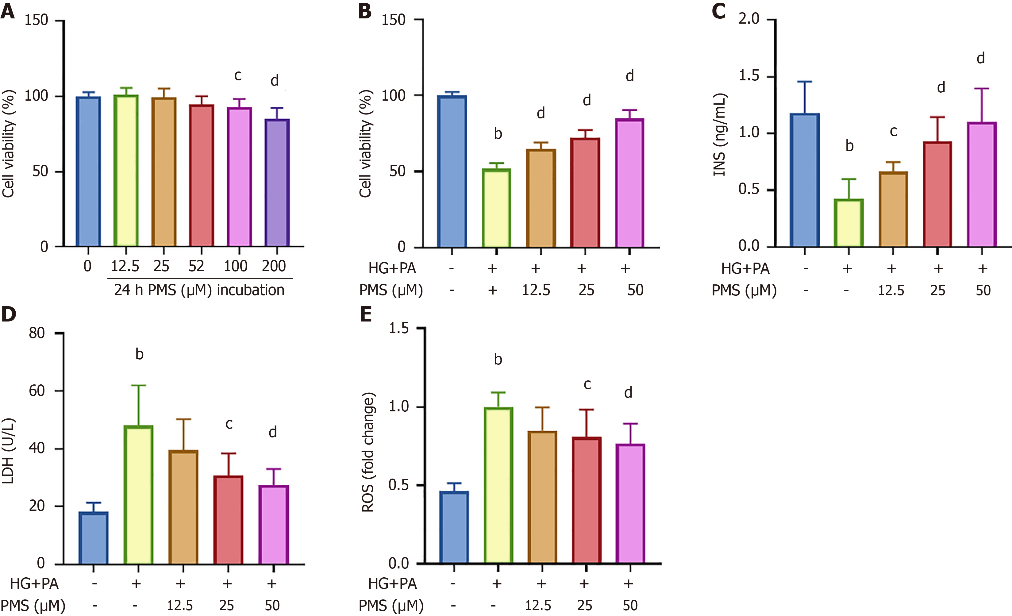
Figure 1 Plantamajoside intervention improves high glucose + palmitic acid-induced injury in pancreatic β-cells.
A: MIN6 cells were first treated with plantamajoside (PMS) and then subjected to MTT assay to measure cell viability; B: Subsequently, cells were treated with 40 mmol/L high glucose + 0.4 mmol/L palmitic acid, and concurrently treated with different concentrations of PMS. MTT results indicate that 12.5 μM, 25 μM, and 50 μM PMS did not significantly affect MIN6 cell viability; C: ELISA results showing that PMS intervention increases insulin (INS) levels in the supernatant; D: PMS intervention decreases the activity of lactate dehydrogenase (LDH); E: In the supernatant and reactive oxygen species (ROS) levels within the cells. Data are presented as mean ± SD, n = 6 for A-D. aP < 0.05, bP < 0.01 vs the normal control group; cP < 0.05, dP < 0.01 vs the high glucose + palmitic acid group.
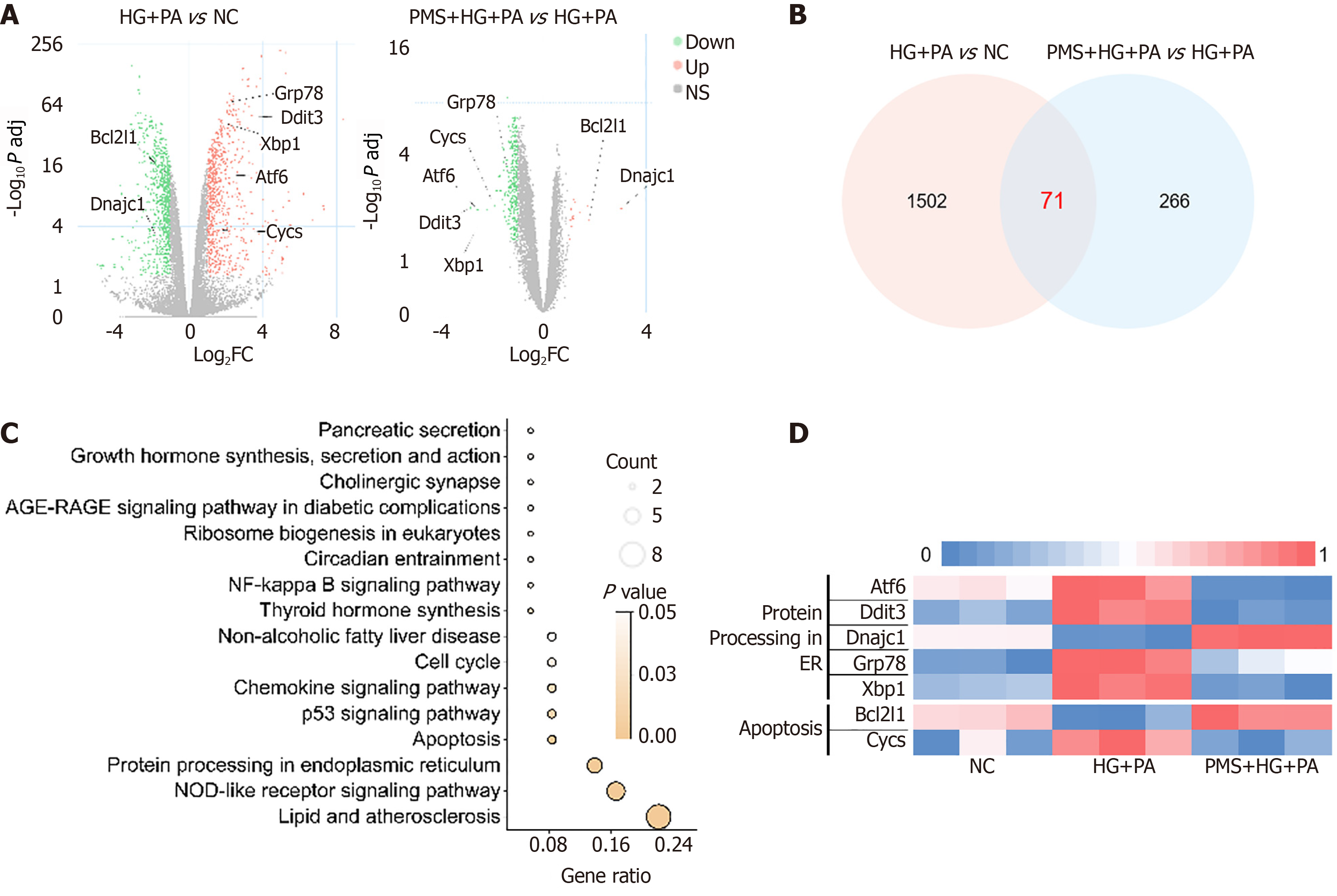
Figure 2 Impact of plantamajoside intervention on gene expression in high glucose + palmitic acid-induced pancreatic β-cells using transcriptomics.
Differentially expressed genes between high glucose (HG) + palmitic acid (PA) vs normal control (NC) groups, and plantamajoside (PMS) + HG + PA vs HG + PA groups, filtered based on P adj < 0.05 and |Log2 (Fold change)| > 1. A: Genes were visualized using volcano plots; B: With the intersecting genes depicted in Venn diagrams; C: KEGG pathway enrichment analysis of intersecting genes revealing that protein processing in the endoplasmic reticulum and apoptosis pathways are enriched following PMS intervention; D: Expression of apoptosis pathway-related genes visualized using a heatmap, showing that PMS intervention upregulated Bcl-2 and downregulated Bax and CytC. Expression of genes related to protein processing in the endoplasmic reticulum pathway visualized using a heatmap, demonstrating that PMS intervention upregulated Dnajc1 and downregulated ATF6, XBP1, Ddit3 (CHOP)and GRP78. n = 6 per group.
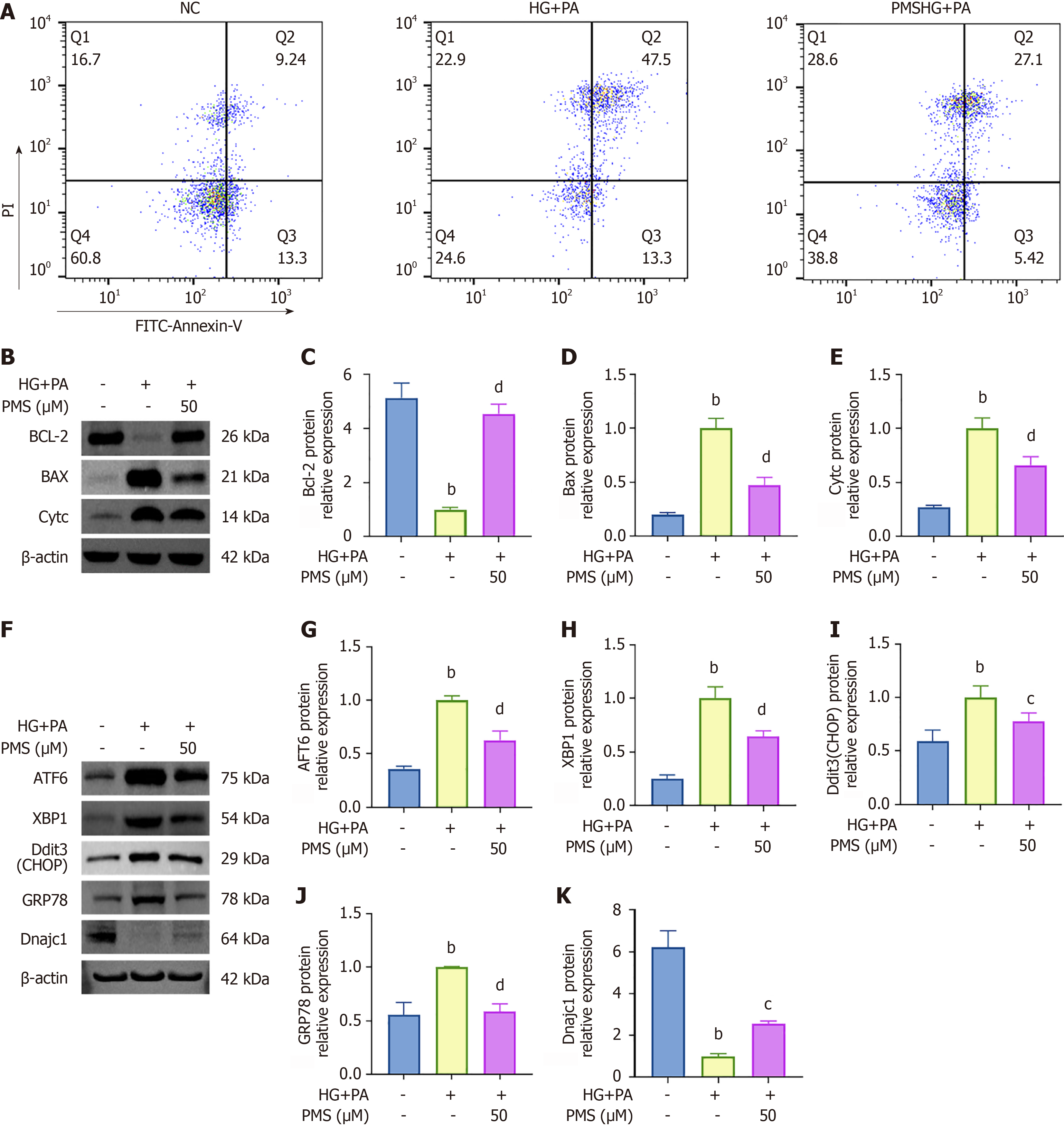
Figure 3 Validation of the impact of plantamajoside intervention on apoptosis and endoplasmic reticulum stress in high glucose + palmitic acid-induced pancreatic β-cells.
A: Flow cytometry results indicate that plantamajoside (PMS) intervention reduced the rate of apoptosis; B-E: Western blot results showing that PMS intervention upregulated the expression of Bcl-2 and downregulated the expression of Bax and CytC; F-K: Western blot results revealing that PMS intervention upregulated the expression of Dnajc1 and downregulated the expression of ATF6, XBP1, Ddit3 (CHOP) and GRP78. n = 3 per group. aP < 0.05, bP < 0.01 vs the normal control group; cP < 0.05, dP < 0.01 vs the high glucose (HG) + palmitic acid (PA) group.
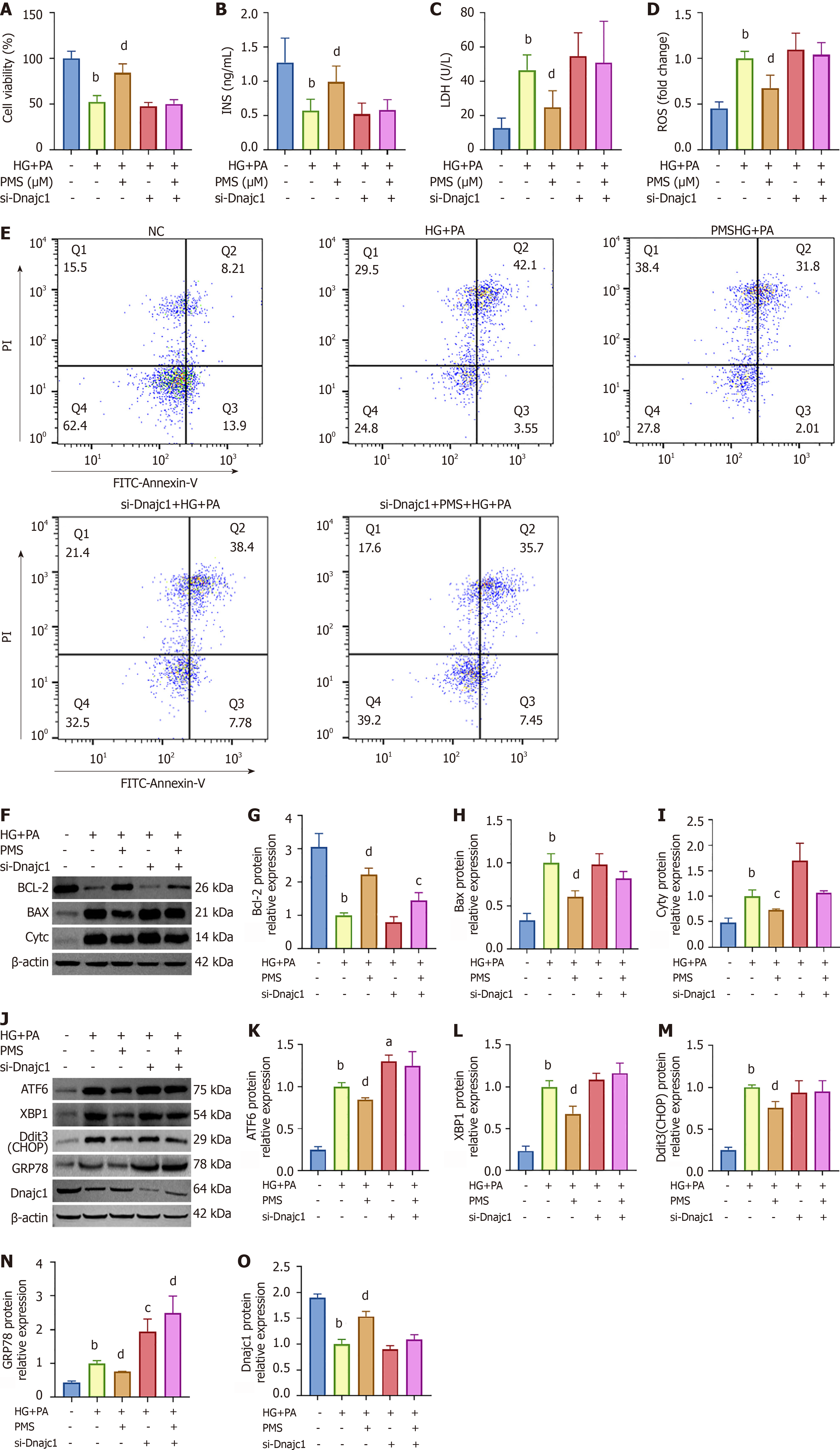
Figure 4 Protective effects of plantamajoside on high glucose + palmitic acid -induced pancreatic β-cell damage are attenuated after Dnajc1 silencing.
A: Cell viability was assessed using MTT assay. Dnajc1 was silenced using a transfection reagent, and the cells were divided into five groups: Normal control (NC) group, high glucose (HG) + palmitic acid (PA) group, plantamajoside (PMS) high-dose group (PMS-H) + HG + PA group, si-Dnajc1 + HG + PA group, and si-Dnajc1 + PMS-H + HG + PA group; B-D: The protective effect of PMS on HG + PA-induced pancreatic β-cell damage was weakened after Dnajc1 silencing. Silencing Dnajc1 weakened or eliminated the effects of PMS on insulin levels B), lactate dehydrogenase activity in the supernatant, and reactive oxygen species levels within the cells; E-I: Silencing Dnajc1 weakened or eliminated the impact of PMS intervention on the apoptosis rate and the expression of apoptosis-related proteins (Bcl-2, Bax, and CytC) in HG + PA-induced MIN6 cells; J-O: The regulatory effect of PMS on endoplasmic reticulum stress-related proteins [ATF6, XBP1, Ddit3 (CHOP), GRP78, Dnajc1] disappeared after silencing Dnajc1. A-D: n = 6 per group; E-O: n = 3 per group. aP < 0.05, bP < 0.01 vs the NC group; cP < 0.05, dP < 0.01 vs the HG + PA group.
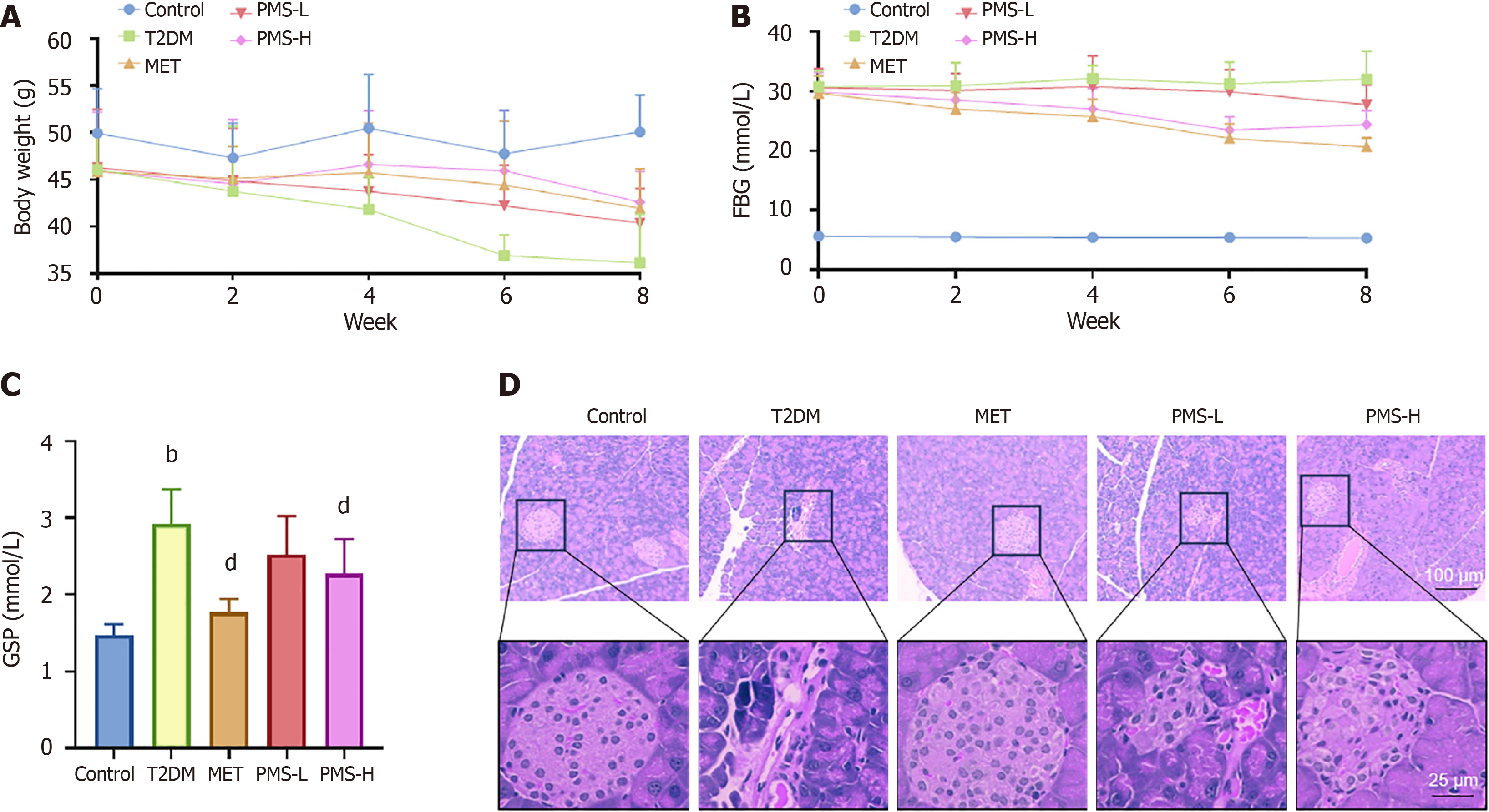
Figure 5 Therapeutic effects of plantamajoside on type 2 diabetes mellitus mice.
A: After establishing a type 2 diabetes mellitus (T2DM) mouse model with a high-fat diet, different doses of plantamajoside (PMS) were administered for 8 weeks to evaluate the therapeutic effects of PMS on T2DM mice. PMS intervention significantly increased mouse body weight; B: Improved fasting blood glucose (FBG); C: Glycated serum protein (GSP) levels; D: Pancreatic hematoxylin and eosin staining showing a reduction in pancreatic tissue damage in T2DM mice after PMS intervention. A-C: n = 10 per group; D: n = 3 per group. bP < 0.01 vs normal control group; dP < 0.01 vs T2DM group. PMS-L: Plantamajoside low-dose group; PMS-H: Plantamajoside high-dose group.
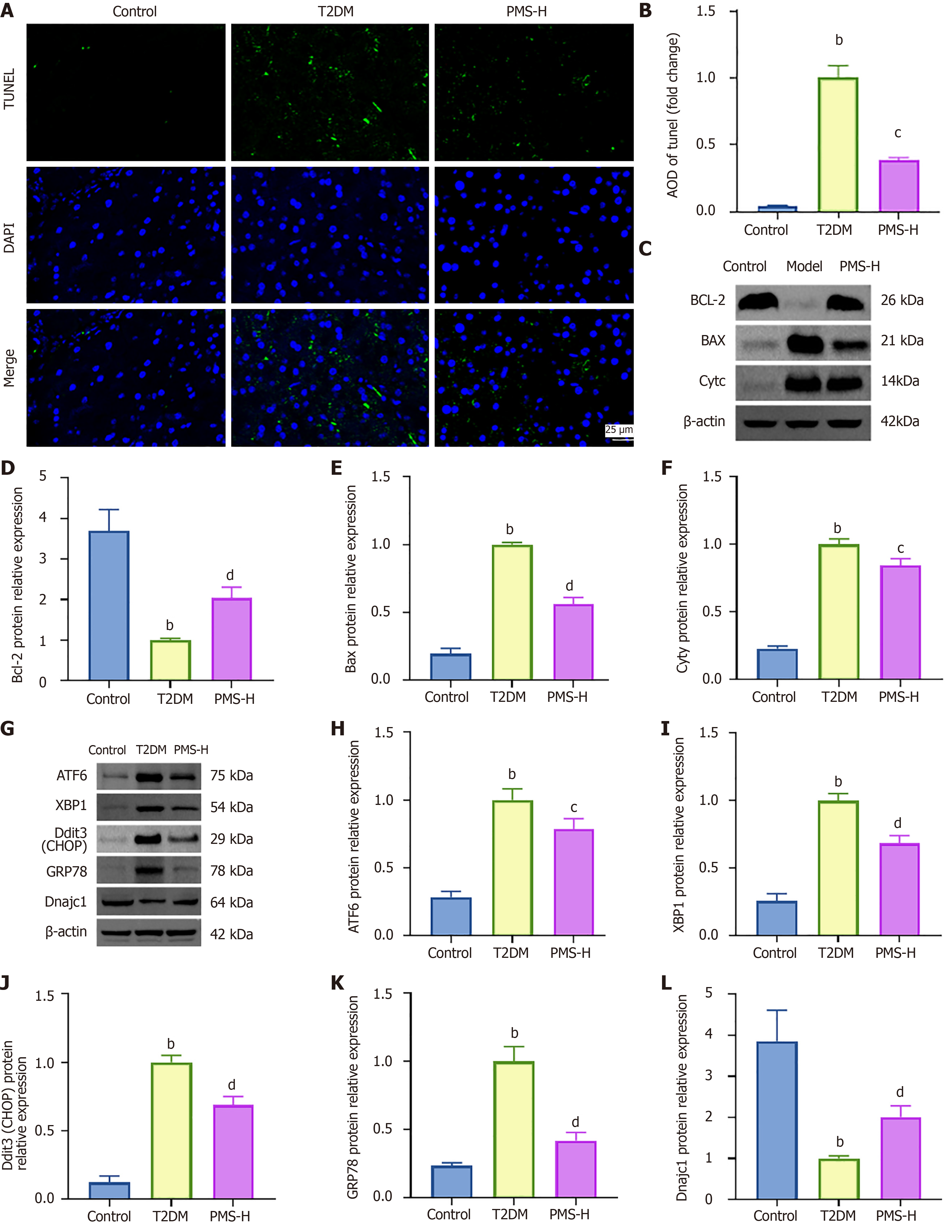
Figure 6 Plantamajoside downregulates the expression of apoptosis and endoplasmic reticulum stress-related factors in pancreatic tissue of type 2 diabetes mellitus mice and upregulates Dnajc1 expression.
A and B: Impact of plantamajoside (PMS) on apoptosis and the expression of endoplasmic reticulum stress-related proteins in the pancreatic tissue of type 2 diabetes mellitus (T2DM) mice was evaluated using TUNEL staining and Western Blot. After PMS intervention, the TUNEL-positive area in the pancreatic tissue of T2DM mice decreased; C-F: Western blot results showing that expression levels of Bcl-2 increased, while expression levels of Bax and CytC decreased after PMS intervention; G-L: Western blot results showing that expressions of ATF6, XBP1, Ddit3 (CHOP), and GRP78 were downregulated, and expression of Dnajc1 was upregulated after PMS intervention. n = 3 per group. bP < 0.01 vs the normal control group. cP < 0.05, dP < 0.01 vs the T2DM group. PMS-H: Plantamajoside high-dose group.
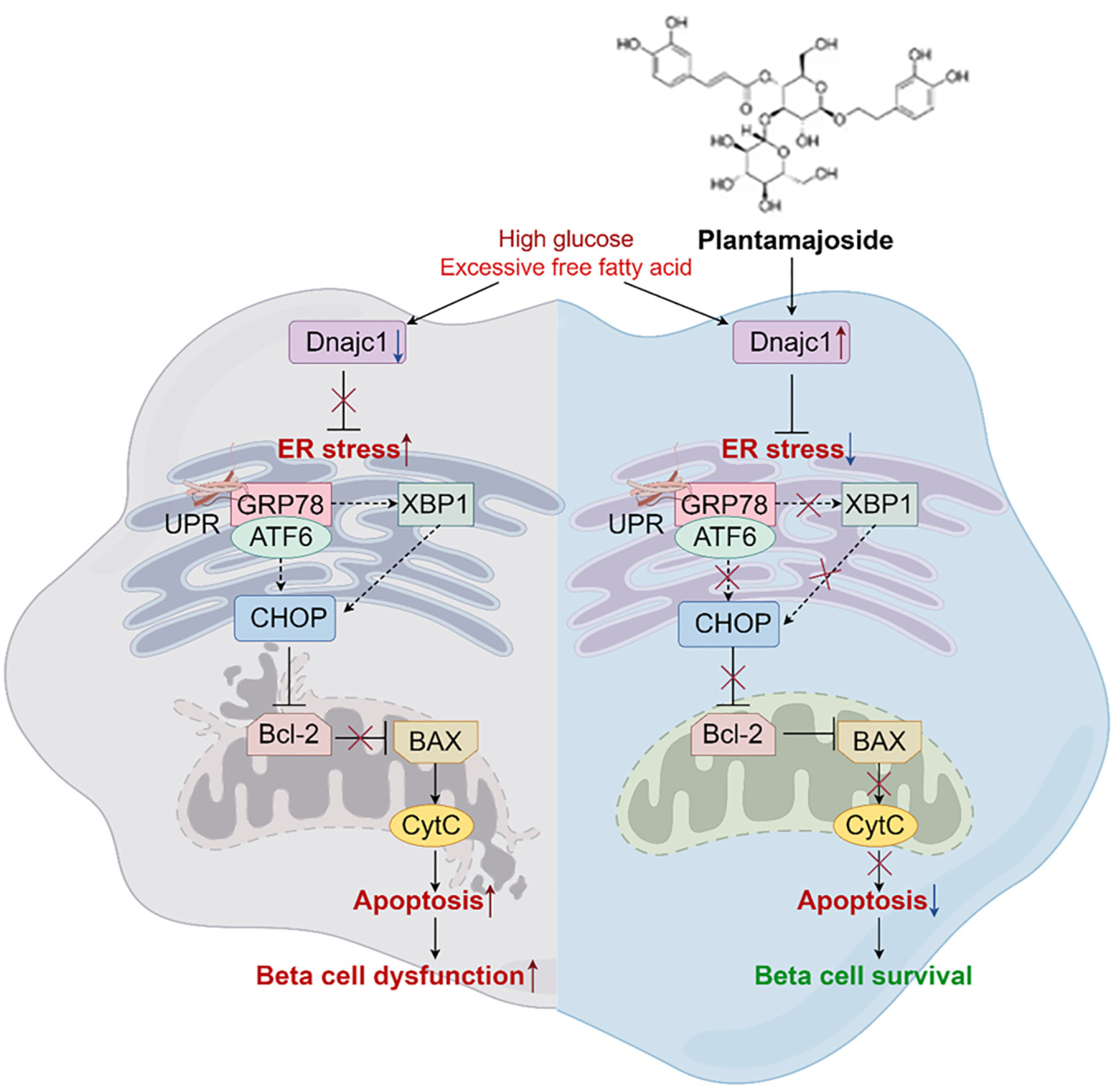
Figure 7 Plantamajoside alleviates pancreatic β-cell damage in type 2 diabetes mellitus mice by promoting the expression of Dnajc1, which inhibits endoplasmic reticulum stress and apoptosis.
- Citation: Wang D, Wang YS, Zhao HM, Lu P, Li M, Li W, Cui HT, Zhang ZY, Lv SQ. Plantamajoside improves type 2 diabetes mellitus pancreatic β-cell damage by inhibiting endoplasmic reticulum stress through Dnajc1 up-regulation. World J Diabetes 2025; 16(2): 99053
- URL: https://www.wjgnet.com/1948-9358/full/v16/i2/99053.htm
- DOI: https://dx.doi.org/10.4239/wjd.v16.i2.99053


