©The Author(s) 2025.
World J Diabetes. Jan 15, 2025; 16(1): 99745
Published online Jan 15, 2025. doi: 10.4239/wjd.v16.i1.99745
Published online Jan 15, 2025. doi: 10.4239/wjd.v16.i1.99745
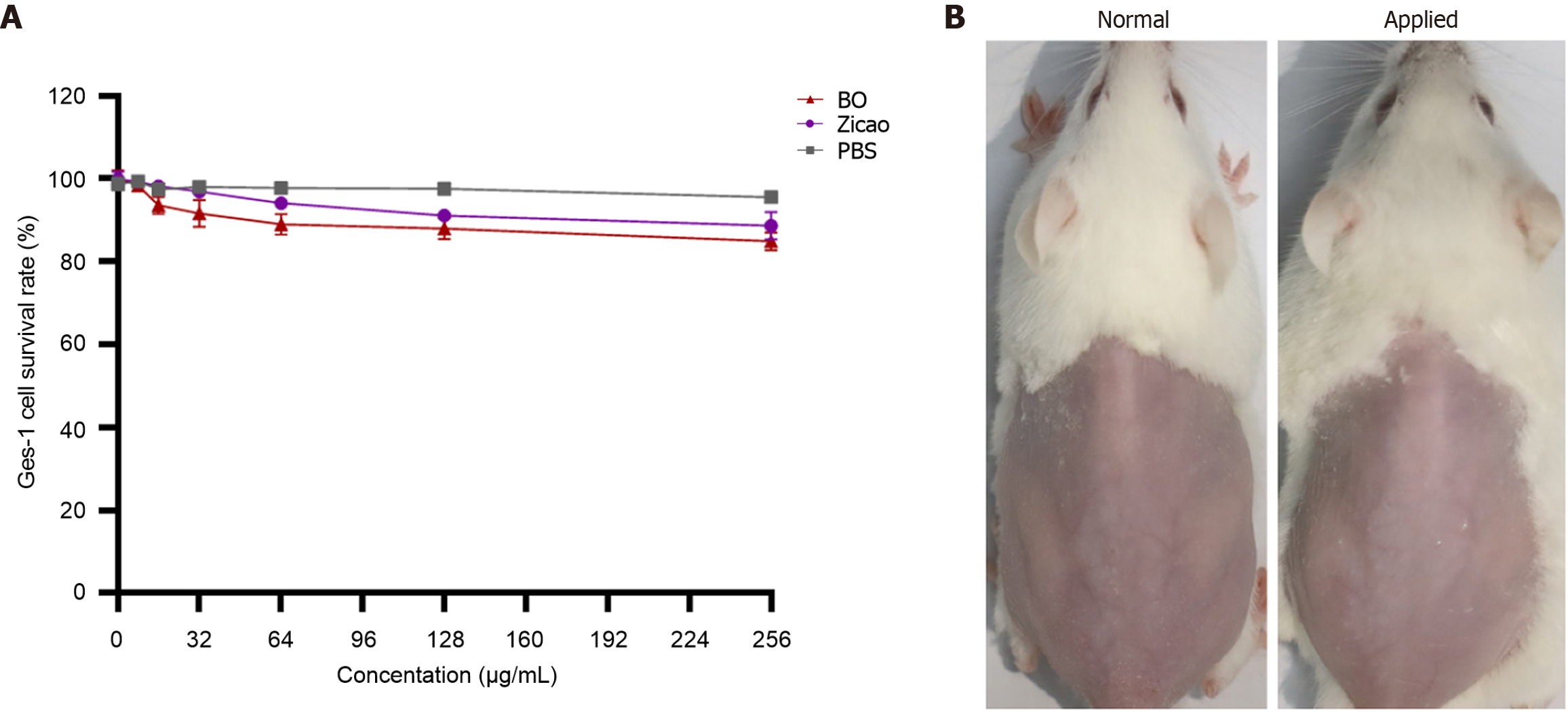
Figure 1 Safety evaluation of betaine ointment.
A: Toxicity to normal cells; B: Picture of mice after application of the ointment. BO: Betaine ointment; PBS: Phosphate buffer solution.
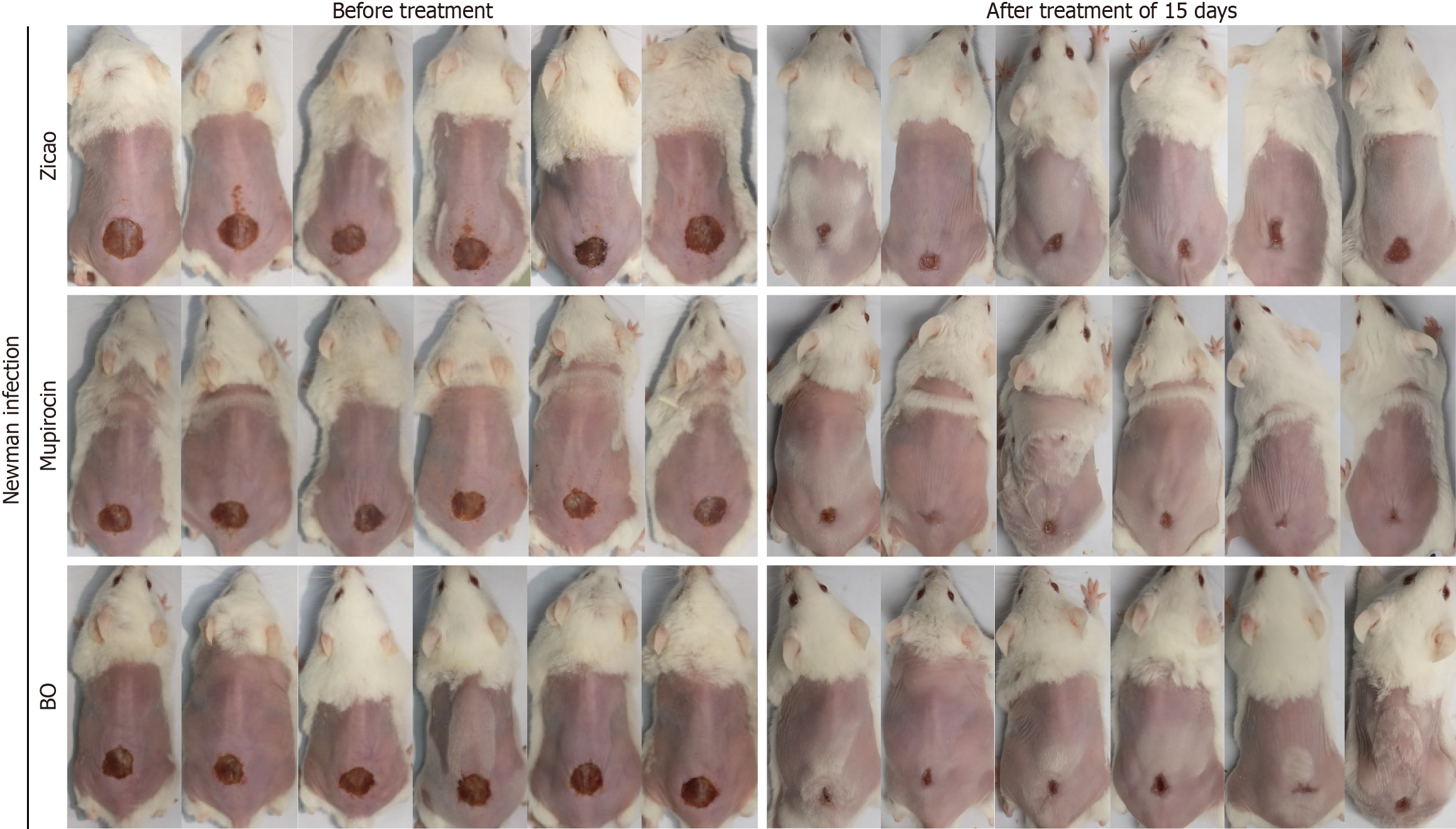
Figure 2 Wound healing of mice in the in vivo antibacterial experiment.
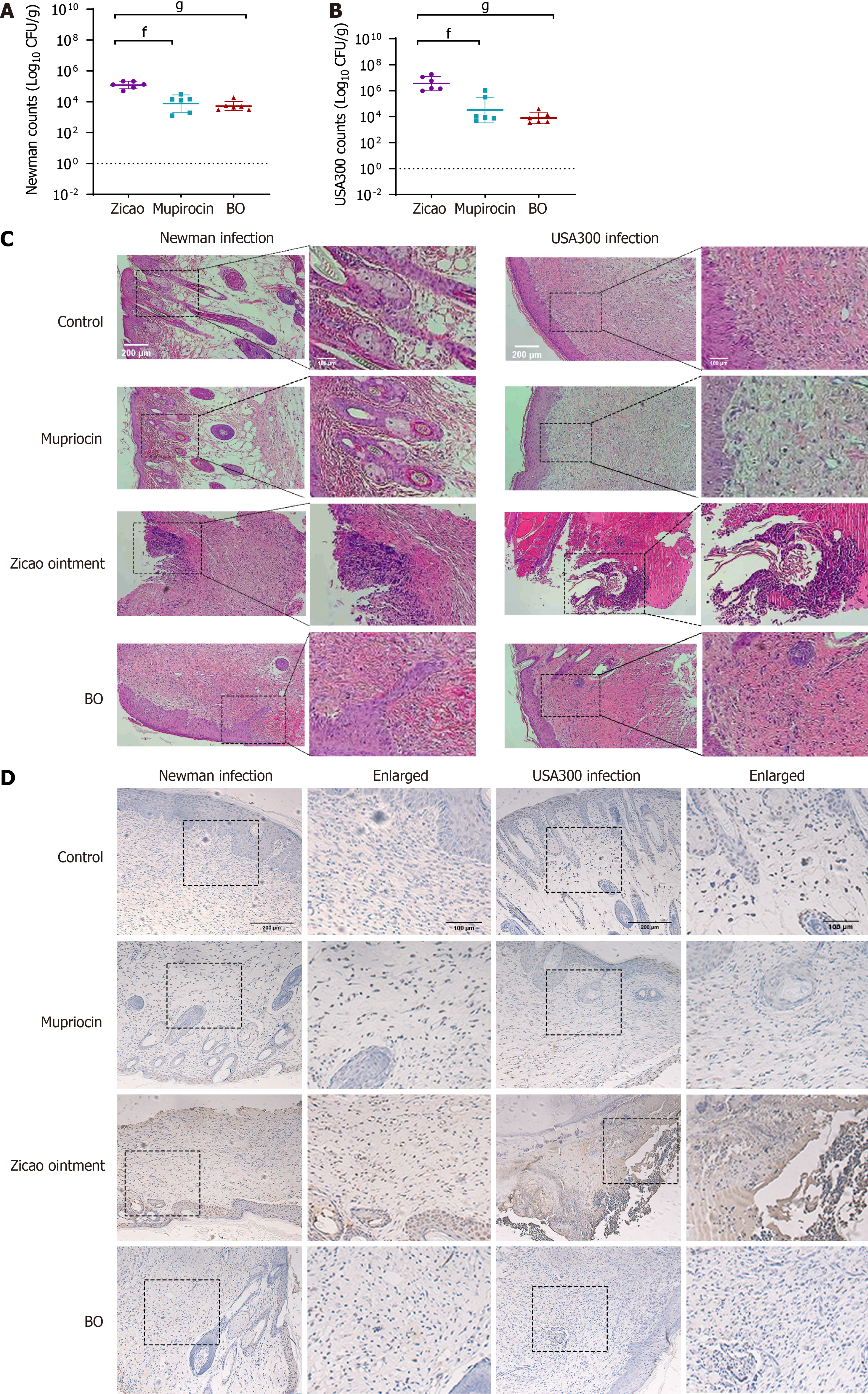
Figure 3 Detection results of wound-infected mouse skin tissue.
A: Newman strain colonization; B: USA300 strain colonization; C: HE staining; D: TUNEL staining. fP < 0.05; gP < 0.01. BO: Betaine ointment.
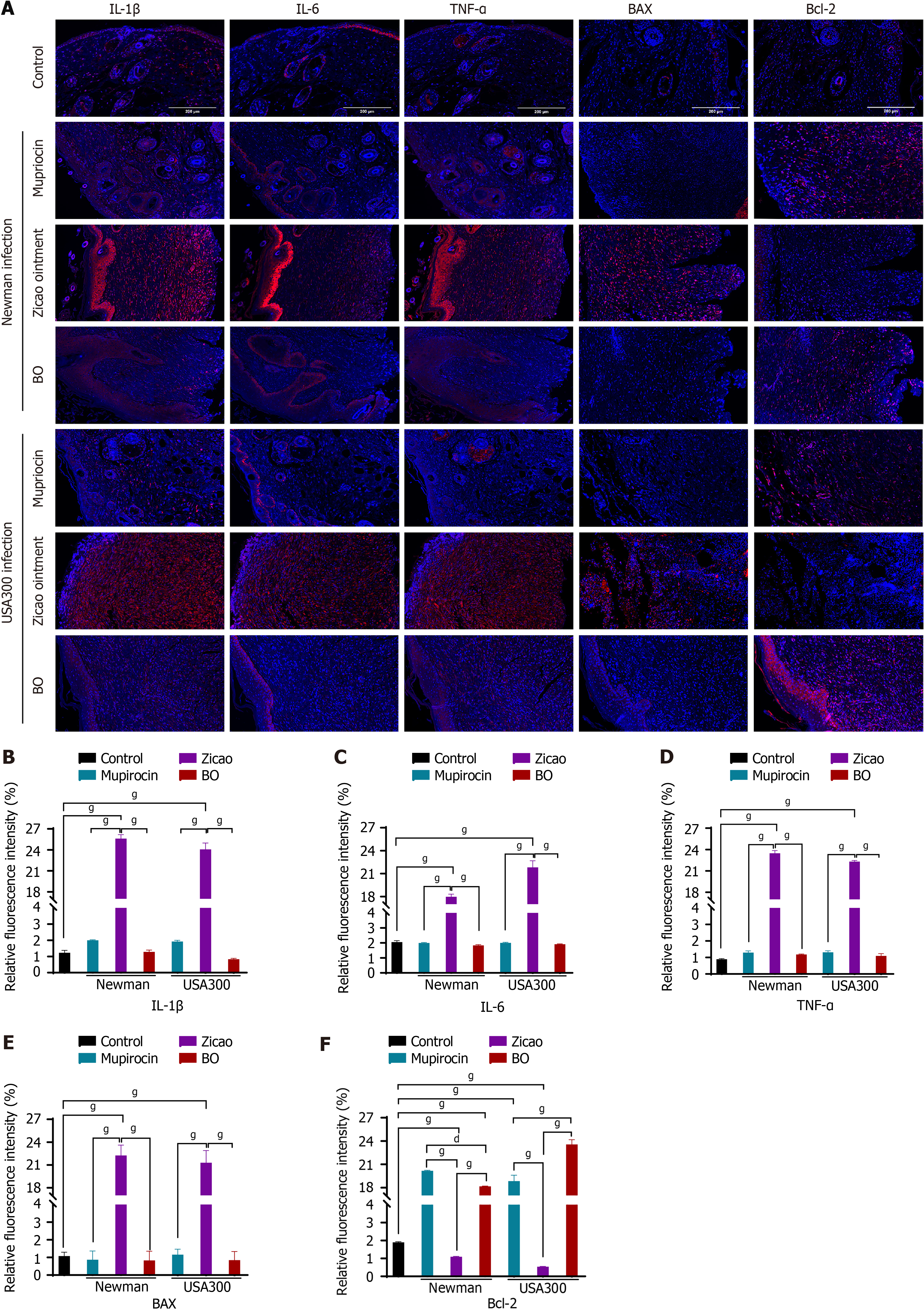
Figure 4 Fluorescence staining of inflammatory factors and apoptotic factors in the skin wound tissue of mice in the in vivo antibacterial experiment.
A: Fluorescence microscopy view; B-F: Fluorescence quantification. gP < 0.01. BO: Betaine ointment.
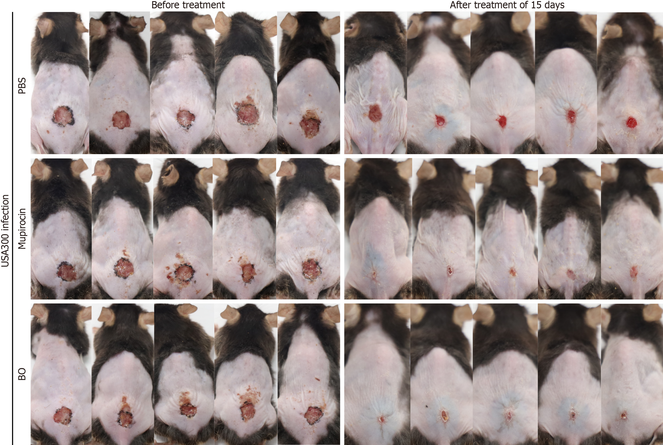
Figure 5 Wound healing of mice in the in vivo antibacterial experiment.
PBS: Phosphate buffer solution; BO: Betaine ointment.
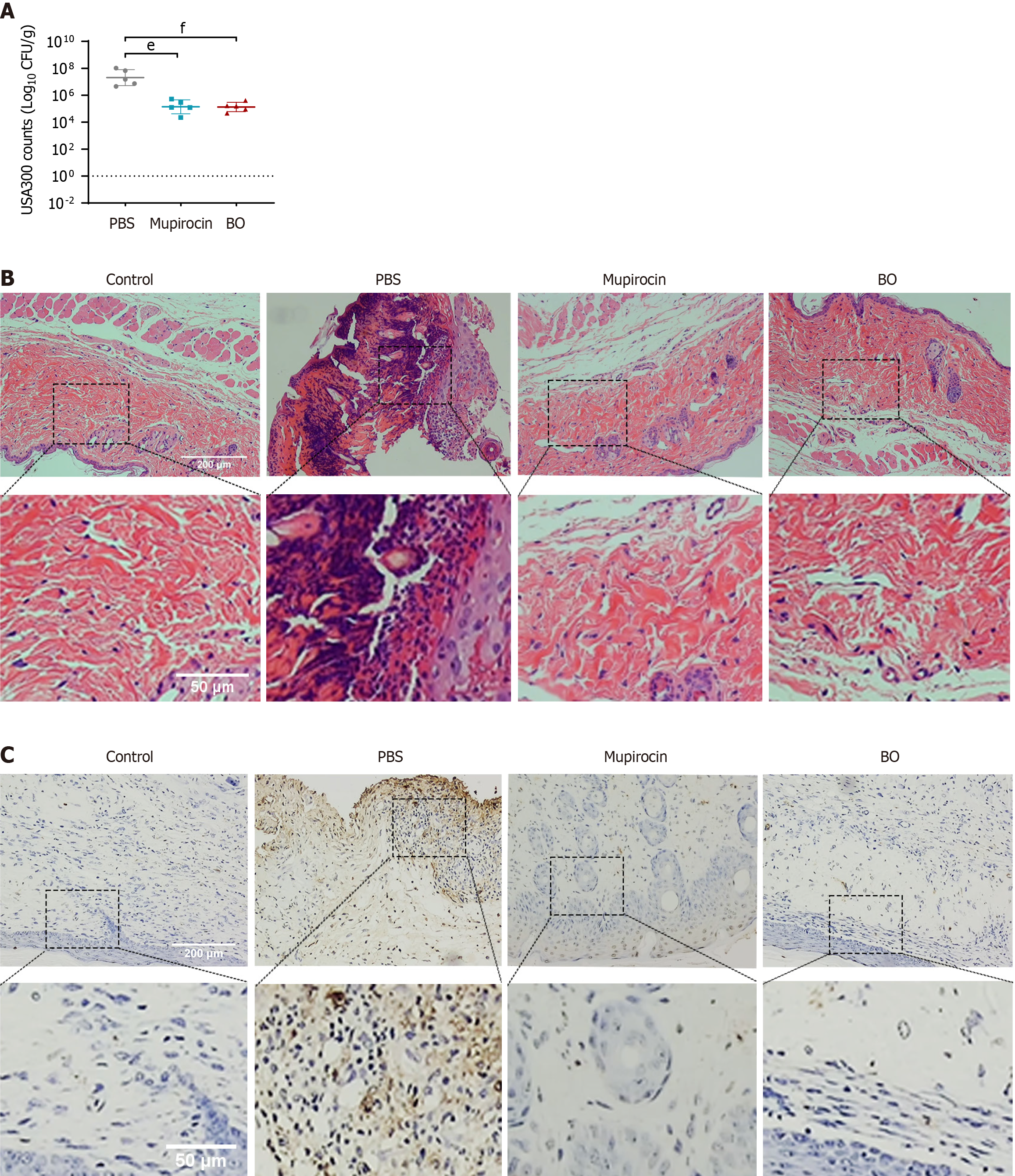
Figure 6 Colonization of bacterial strains, inflammation, and apoptosis in the skin tissues of diabetic mice with traumatic infection.
A: Colonization of the USA300 strain; B: HE staining; C: TUNEL staining. eP < 0.01; fP < 0.05. PBS: Phosphate buffer solution; BO: Betaine ointment.
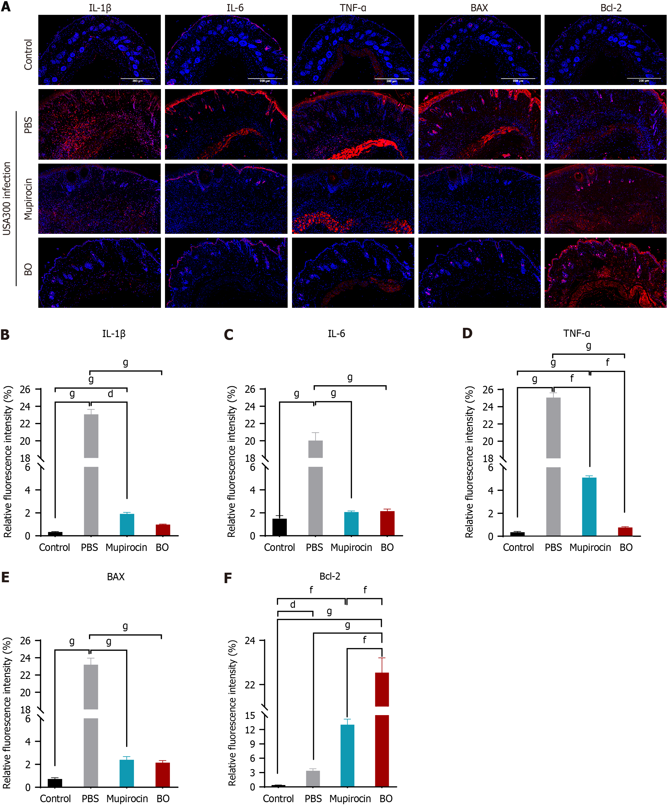
Figure 7 Fluorescence staining of inflammatory factors and apoptotic factors in skin wound tissues from diabetic mice.
A: Fluorescence microscopy view; B-F: Fluorescence quantification. dP < 0.05; fP < 0.05; gP < 0.01. PBS: Phosphate buffer solution; BO: Betaine ointment.
- Citation: Xu WY, Dai YY, Yang SX, Chen H, Huang YQ, Luo PP, Wei ZH. Betaine combined with traditional Chinese medicine ointment to treat skin wounds in microbially infected diabetic mice. World J Diabetes 2025; 16(1): 99745
- URL: https://www.wjgnet.com/1948-9358/full/v16/i1/99745.htm
- DOI: https://dx.doi.org/10.4239/wjd.v16.i1.99745


