©The Author(s) 2023.
World J Diabetes. Jun 15, 2023; 14(6): 724-740
Published online Jun 15, 2023. doi: 10.4239/wjd.v14.i6.724
Published online Jun 15, 2023. doi: 10.4239/wjd.v14.i6.724
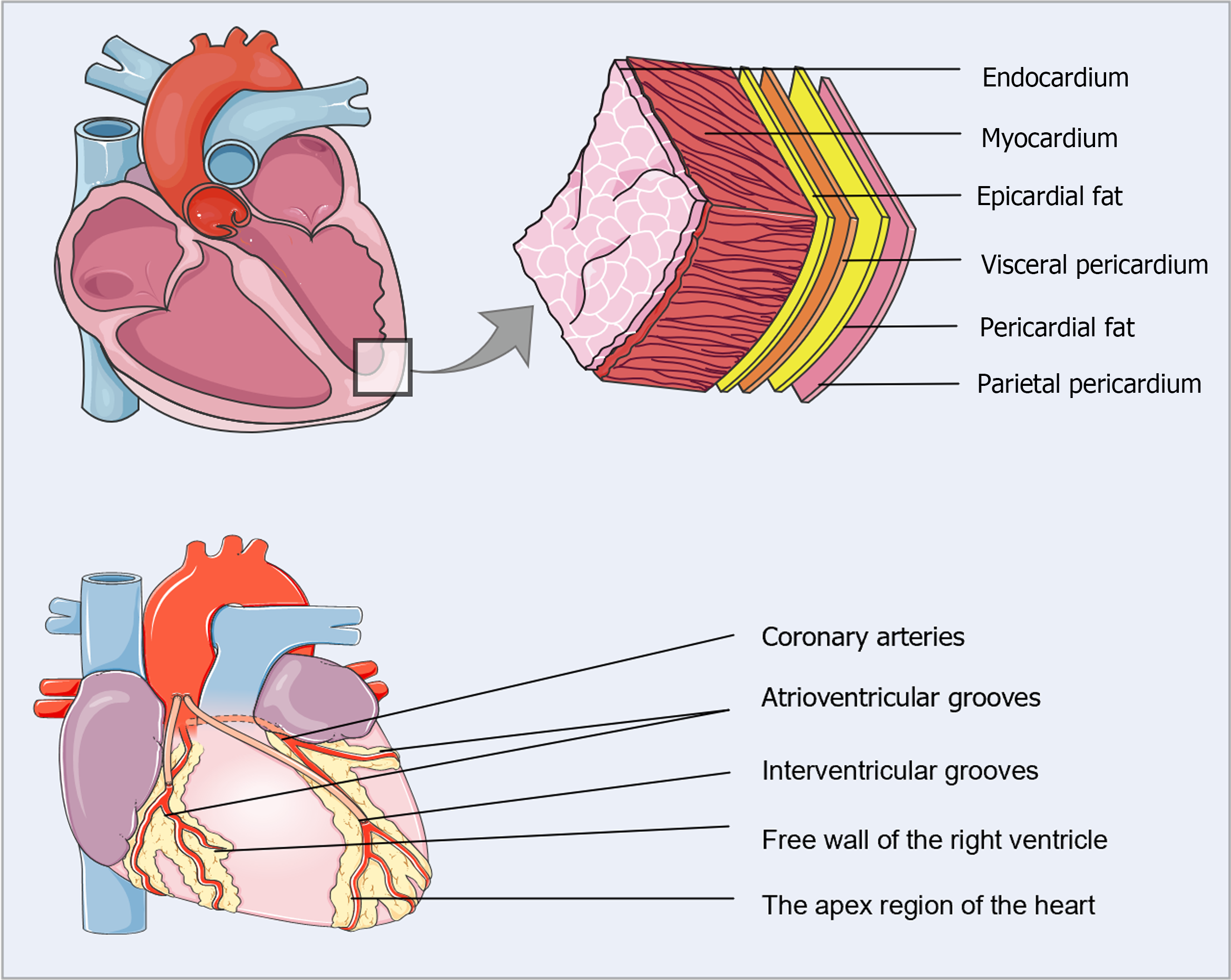
Figure 1 Anatomical location of epicardial adipose tissue.
Epicardial adipose tissue (EAT) is situated between the myocardium and the visceral pericardium. In normal adults, EAT usually accompanies the coronary arteries and their major epicardial branches, mainly concentrated in the interventricular and atrioventricular grooves, with lesser amounts covering the atria, the free wall of the right ventricle, and the apex.
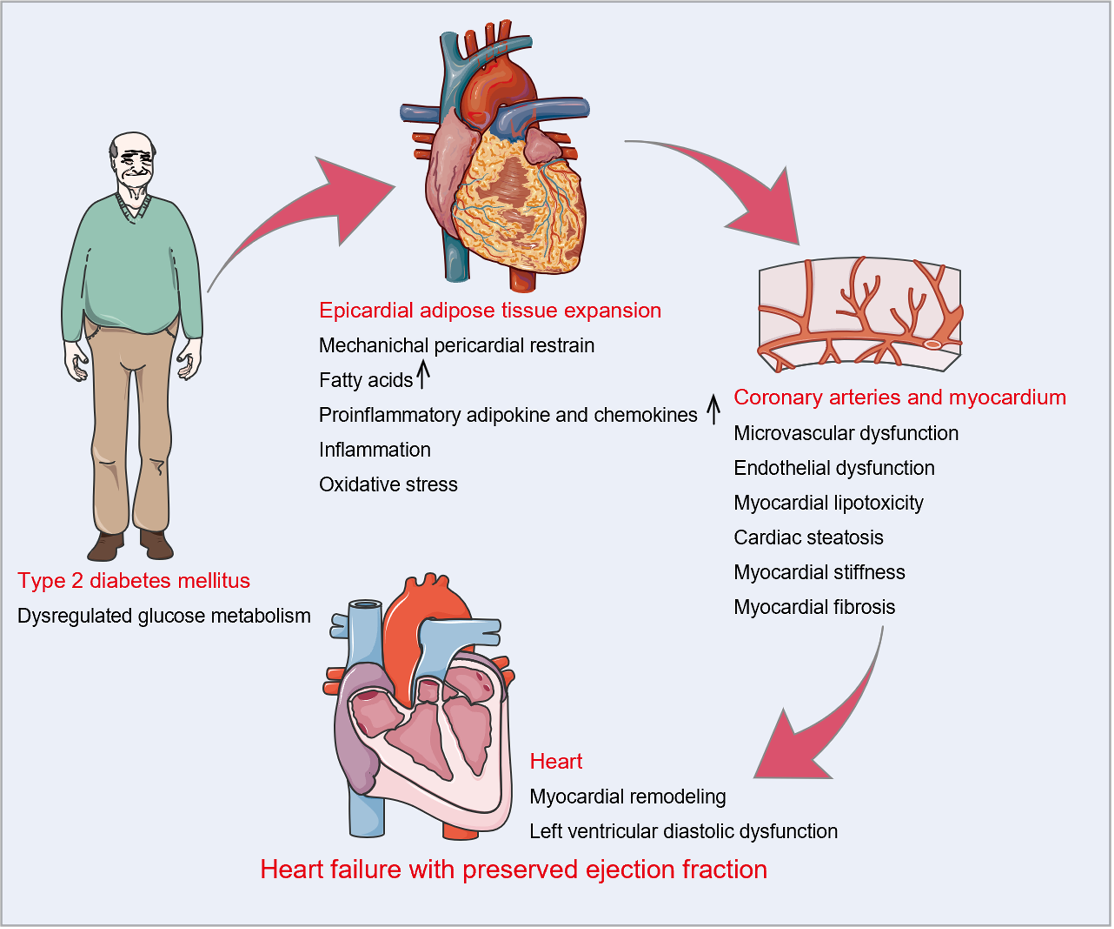
Figure 2 Epicardial adipose tissue in the pathophysiology of heart failure with preserved ejection fraction with type 2 diabetes mellitus.
Dysregulated glucose metabolism is intimately related to the expansion of epicardial adipose tissue (EAT). Increased EAT deposition interacts directly with the heart through mechanical and metabolic mechanisms. Mechanically, EAT expansion may directly contribute to pericardial restrain, resulting in left ventricular (LV) diastolic dysfunction. Metabolically, EAT enlargement is linked to the buildup of free fatty acids, which may induce myocardial lipotoxicity and cardiac steatosis. Simultaneously, hypertrophic adipocytes and activated macrophages secrete numerous proinflammatory adipocytokines and chemokines in EAT. Subsequent local inflammation, excessive oxidative stress, microvascular and endothelial dysfunction, and myocardial stiffness and fibrosis ultimately lead to LV remodeling and diastolic dysfunction.
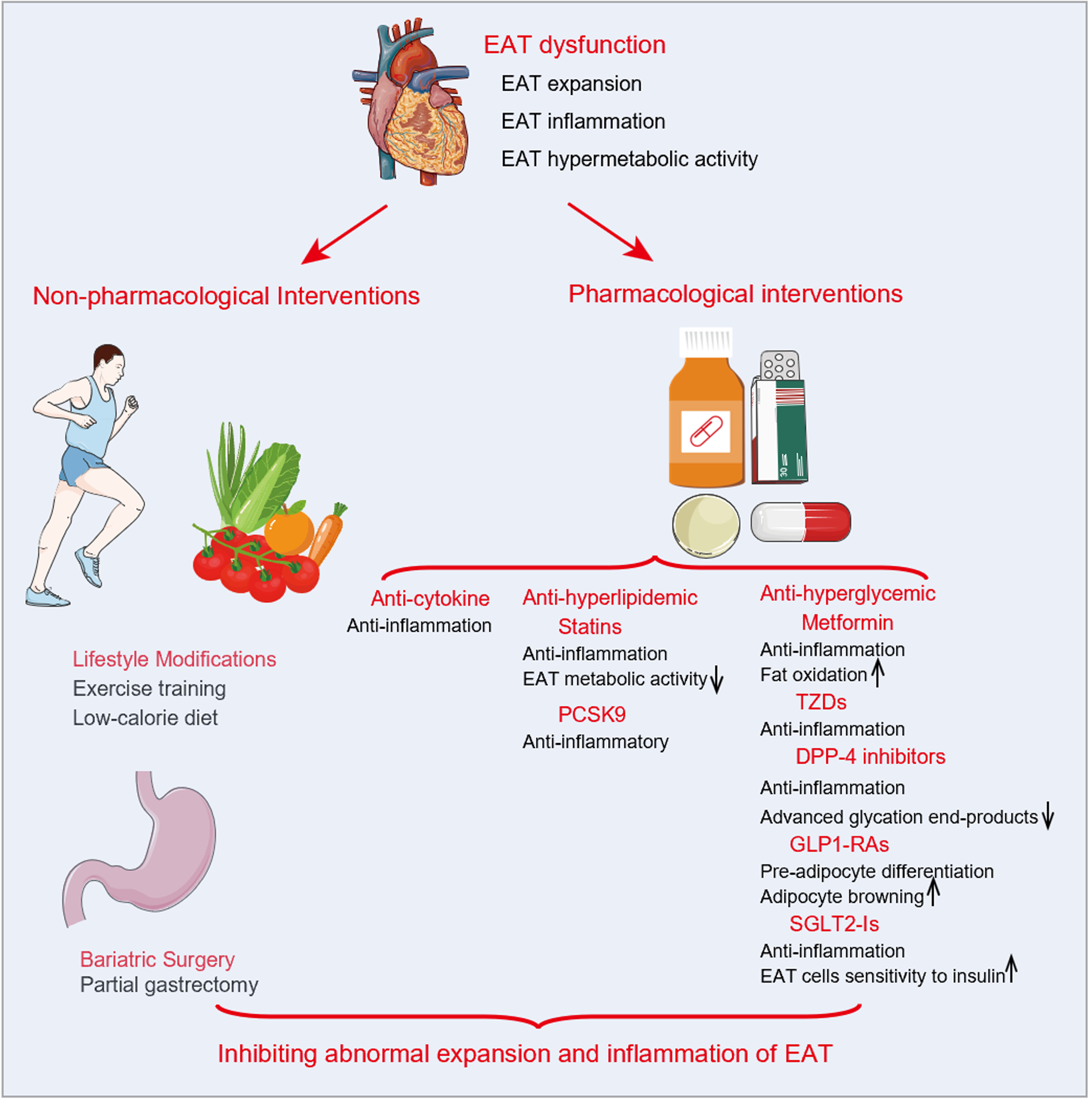
Figure 3 Current interventions targeting epicardial adipose tissue and possible mechanisms.
Current interventions targeting epicardial adipose tissue (EAT) reported in the literature include non-pharmacological interventions (lifestyle management and bariatric surgery) and pharmacological interventions related to anti-cytokines, anti-hyperlipidemia, and anti-hyperglycemia. By increasing fat oxidation or sensitivity to insulin and inhibiting inflammation or hypermetabolic activity, these interventions may prevent abnormal expansion and inflammation of EAT. EAT: Epicardial adipose tissue; DPP-4: Dipeptidyl peptidase 4; GLP1-RAs: Glucagon-like peptide-1 receptor agonists; PCSK9: Proprotein convertase subtilisin/kexin type 9; SGLT2-Is: Sodium-glucose cotransporter 2 inhibitors; TZDs: Thiazolidinediones.
- Citation: Shi YJ, Dong GJ, Guo M. Targeting epicardial adipose tissue: A potential therapeutic strategy for heart failure with preserved ejection fraction with type 2 diabetes mellitus. World J Diabetes 2023; 14(6): 724-740
- URL: https://www.wjgnet.com/1948-9358/full/v14/i6/724.htm
- DOI: https://dx.doi.org/10.4239/wjd.v14.i6.724


