Copyright
©The Author(s) 2022.
World J Diabetes. Jul 15, 2022; 13(7): 498-520
Published online Jul 15, 2022. doi: 10.4239/wjd.v13.i7.498
Published online Jul 15, 2022. doi: 10.4239/wjd.v13.i7.498
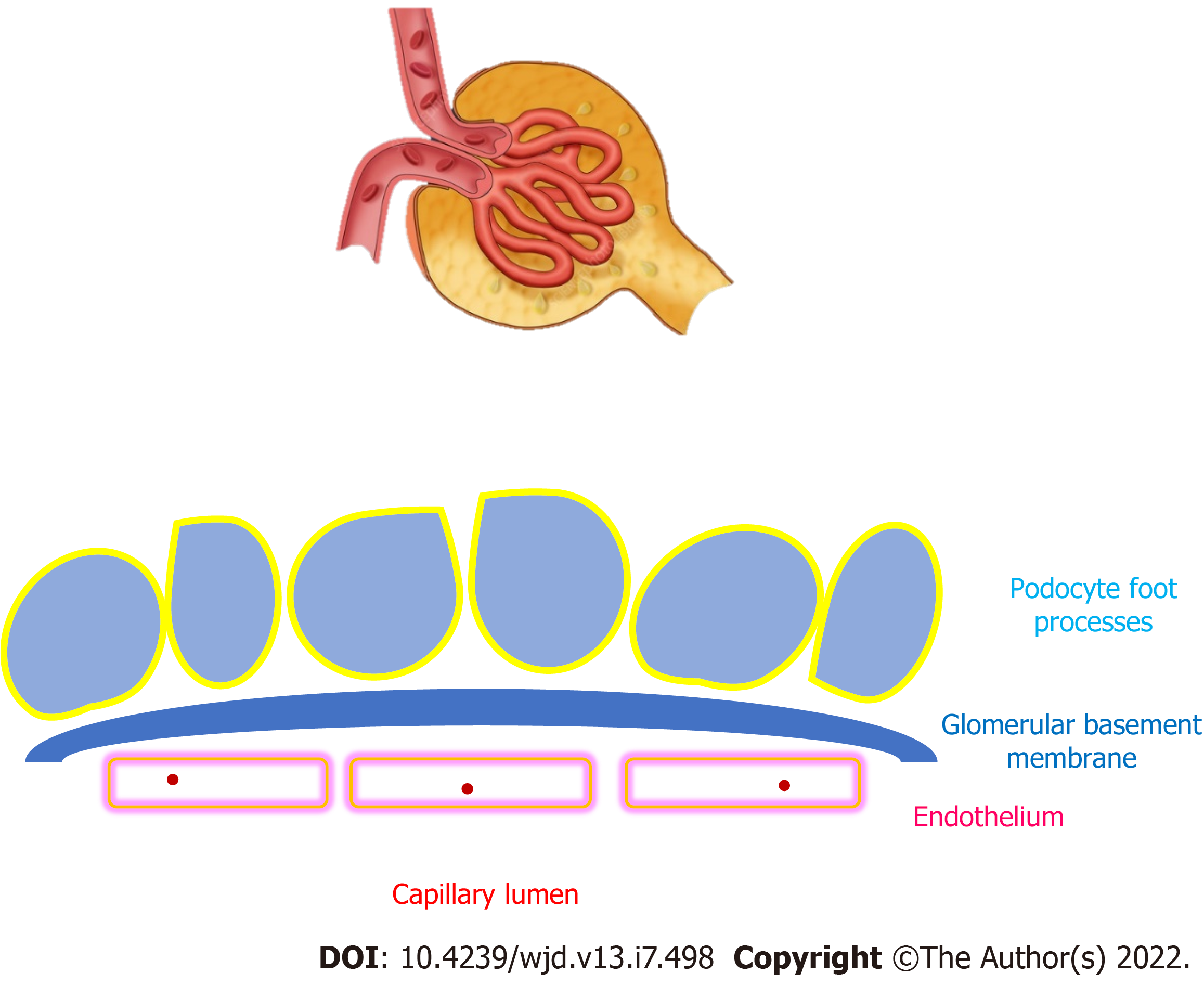
Figure 1 Glomerular capillaries and glomerular basement membrane.
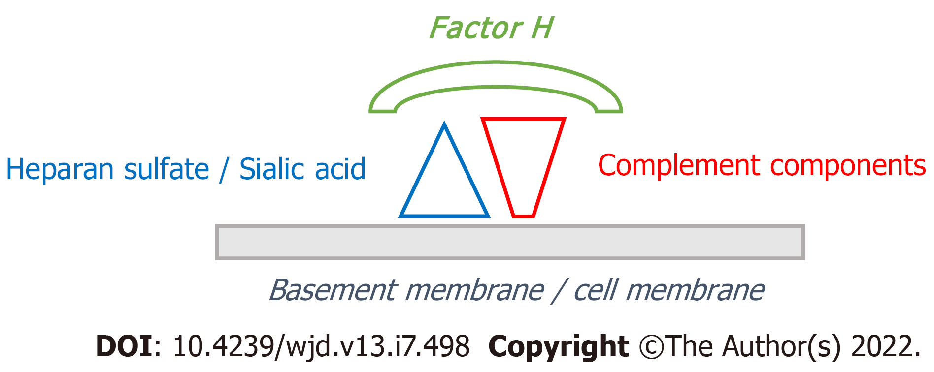
Figure 2 Binding of complement factor H to components of the alternative pathway of complement (C3b) and heparan sulfate/sialic acid on “self” structures (cell membranes or basement membranes).
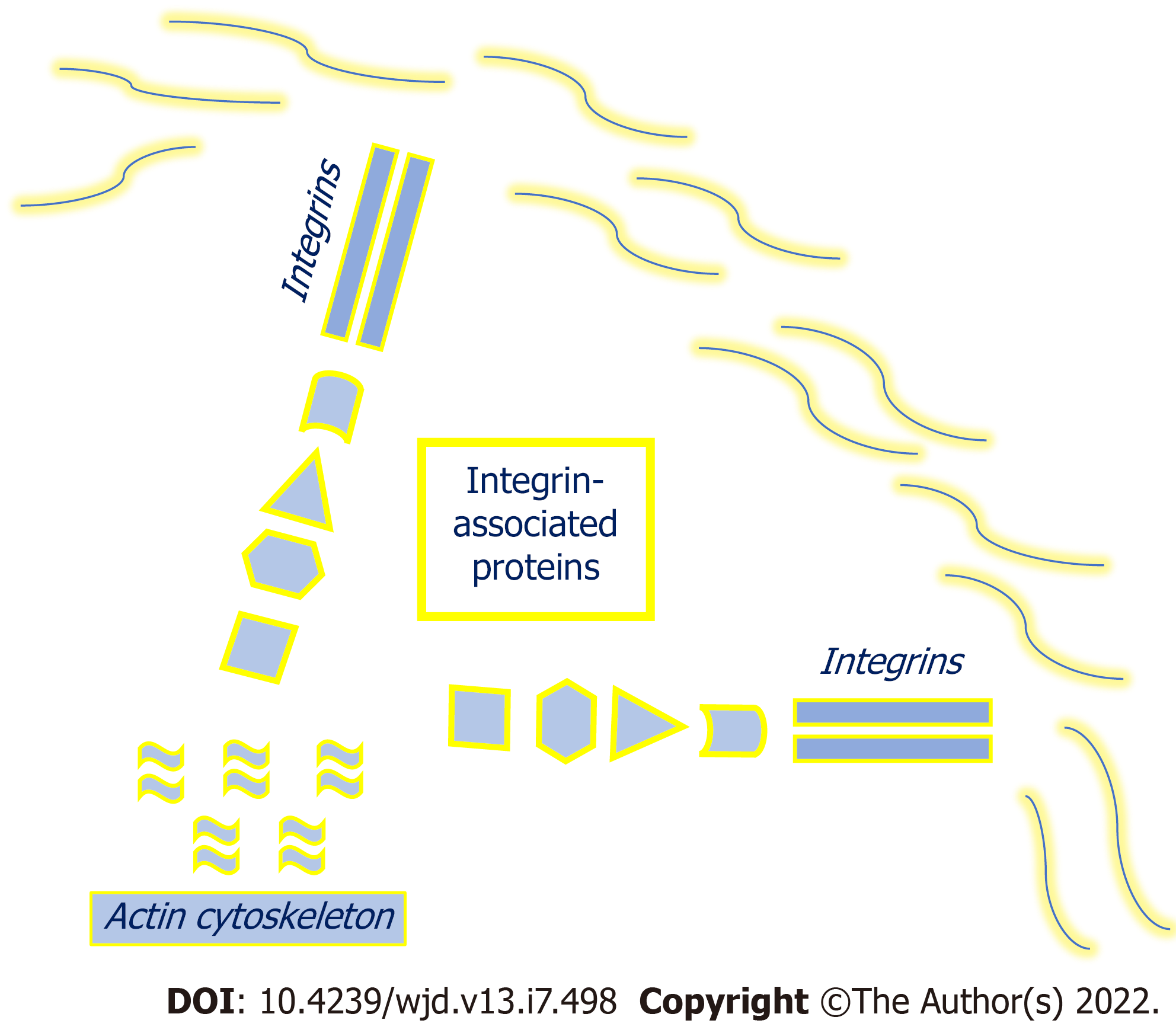
Figure 3 Integrins and integrin-associated proteins.
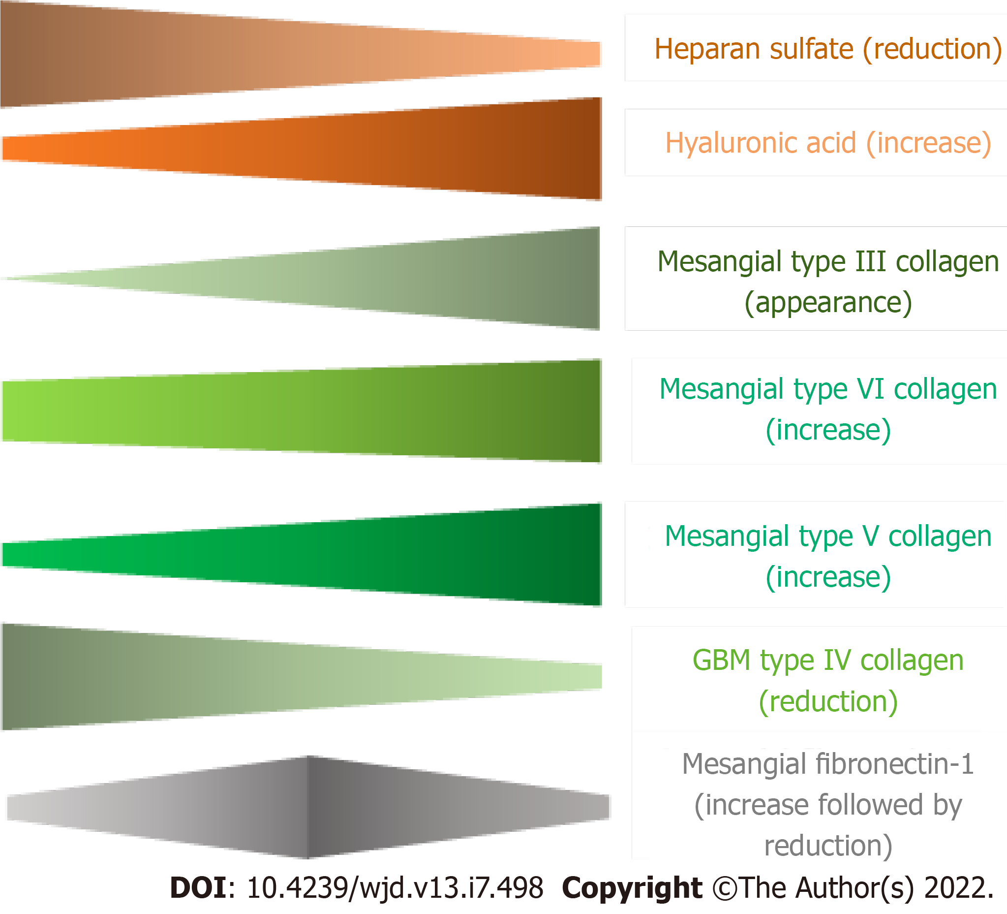
Figure 4 Schematic variation in some components of the glomerular extracellular matrix according to the progression of diabetic kidney disease.
GBM: Glomerular basement membrane.
- Citation: Adeva-Andany MM, Carneiro-Freire N. Biochemical composition of the glomerular extracellular matrix in patients with diabetic kidney disease. World J Diabetes 2022; 13(7): 498-520
- URL: https://www.wjgnet.com/1948-9358/full/v13/i7/498.htm
- DOI: https://dx.doi.org/10.4239/wjd.v13.i7.498


