©The Author(s) 2022.
World J Diabetes. Nov 15, 2022; 13(11): 972-985
Published online Nov 15, 2022. doi: 10.4239/wjd.v13.i11.972
Published online Nov 15, 2022. doi: 10.4239/wjd.v13.i11.972
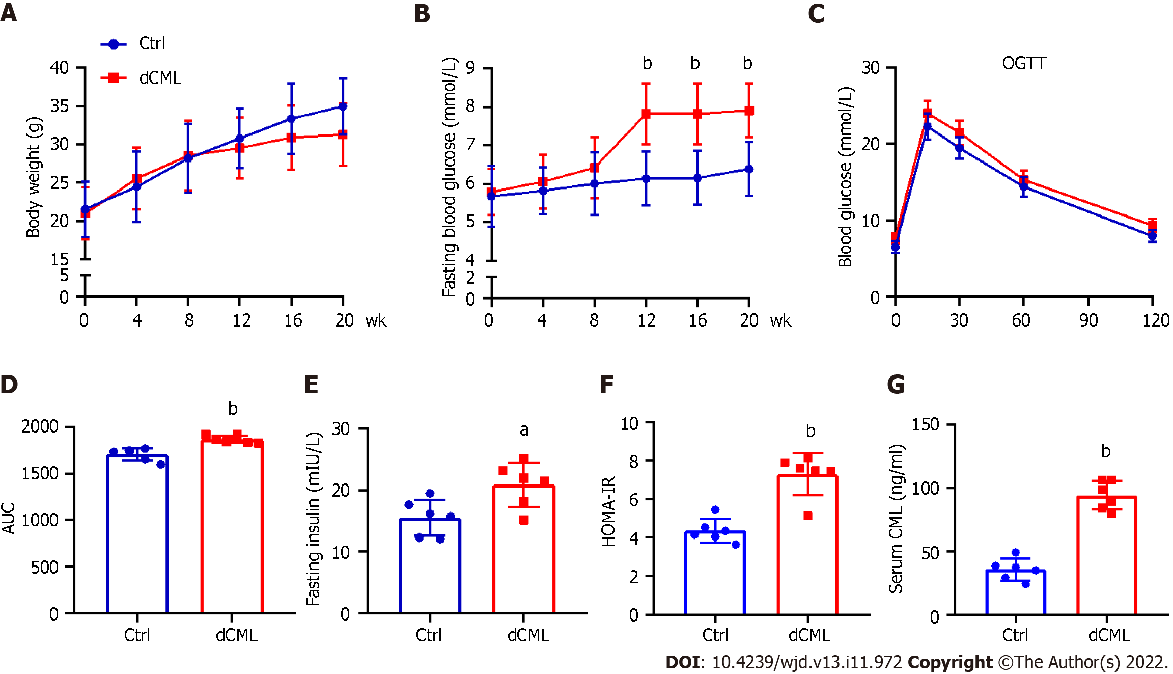
Figure 1 Dietary Nε-(carboxymethyl)lysine increases blood glucose and induces insulin resistance in mice.
A: Body weight of mice; B: Fasting blood glucose of mice; C: Oral glucose tolerance test (OGTT) test of mice; D: Area under the curve (AUC) of OGTT test; E: Fasting insulin of mice; F: Mouse homeostatic model assessment insulin resistance; G: Mouse serum Nε-(carboxymethyl)lysine (CML) level. Ctrl: Control; dCML: Dietary CML; n = 6. aP < 0.05, bP < 0.01, compared with the Ctrl group.
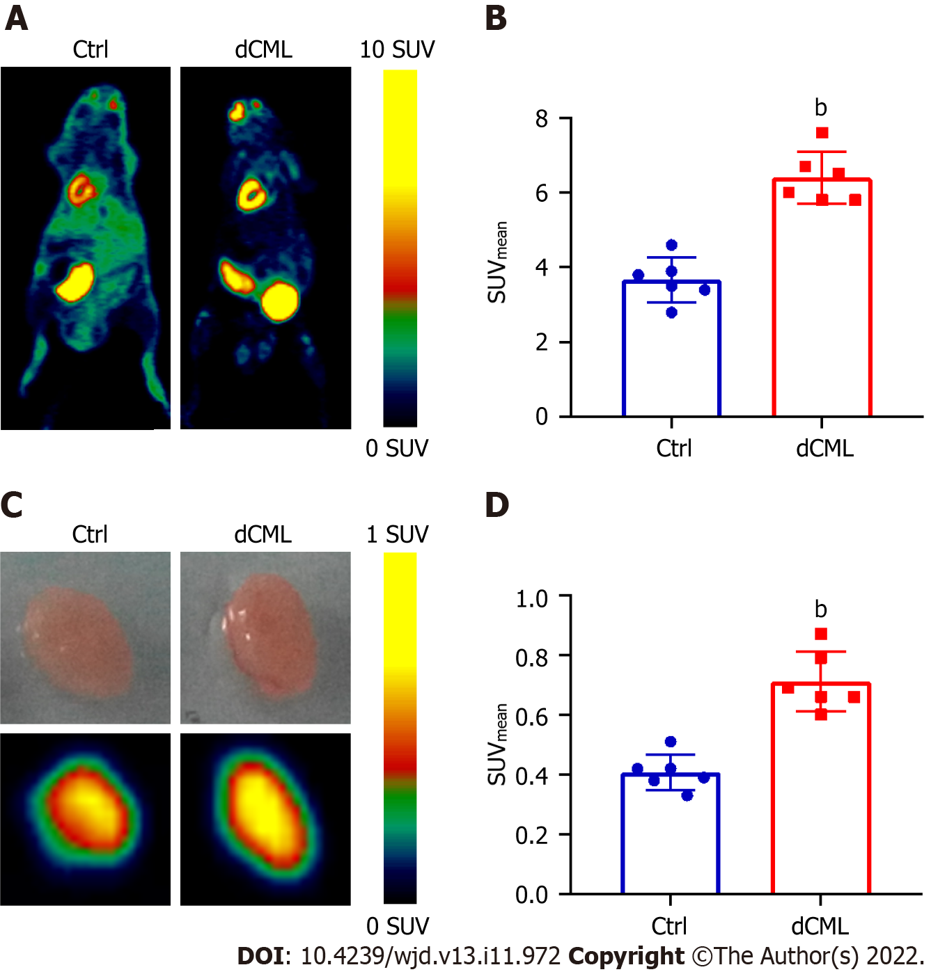
Figure 2 Myocardial glucose uptake is increased after dietary Nε-(carboxymethyl)lysine.
A and B: Micro-positron emission tomography scanning of 18F-fluorodeoxyglucose (FDG) accumulation in mouse myocardium; C and D: Uptake of 18F-FDG by isolated mouse hearts of the control (Ctrl) group and dietary Nε-(carboxymethyl)lysine (dCML) group. SUV: Standard uptake value. n = 6. bP < 0.01, compared with the Ctrl group.
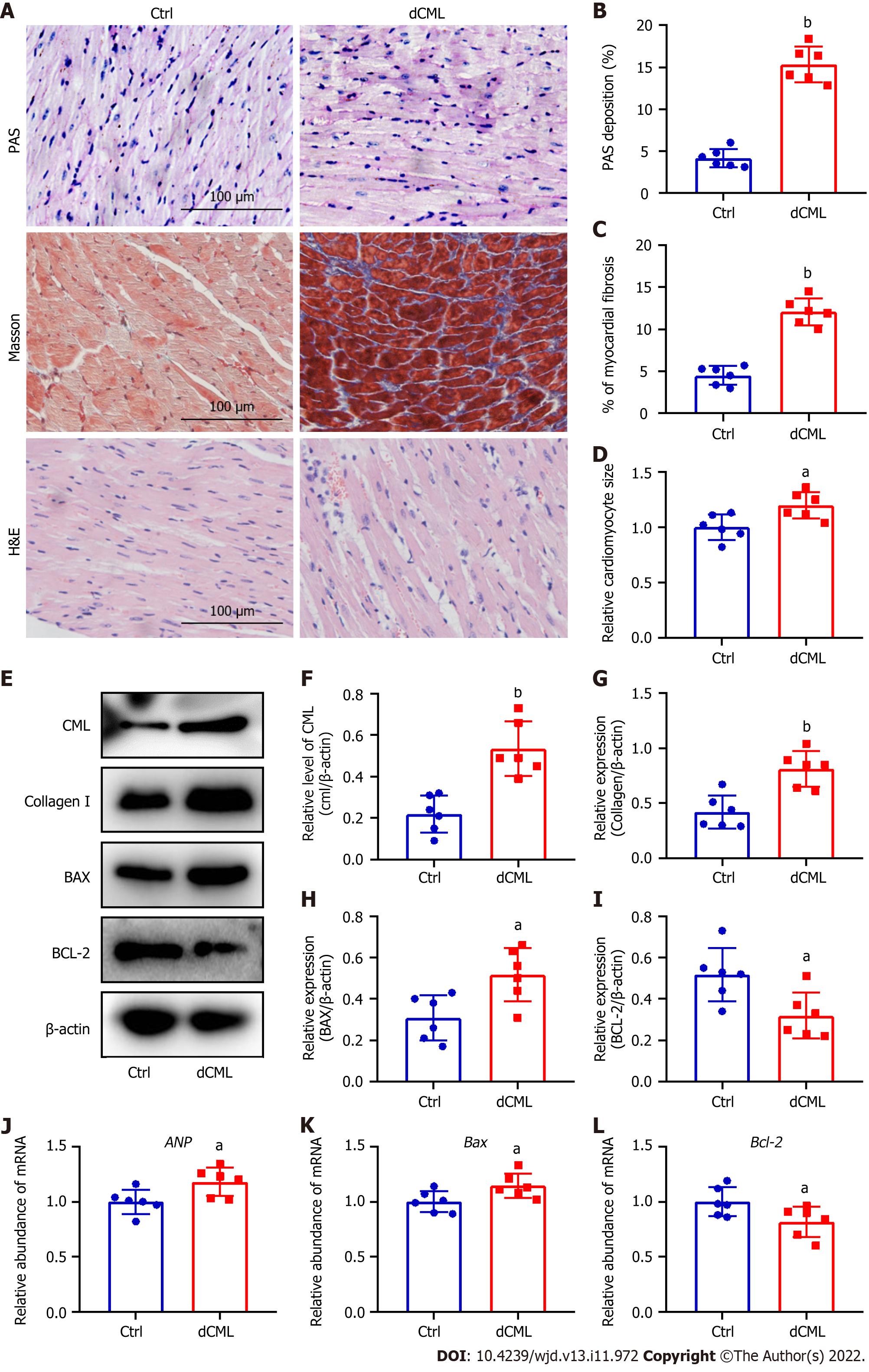
Figure 3 Dietary Nε-(carboxymethyl)lysine increases myocardial fibrosis, hypertrophy and apoptosis in mice.
A: Mouse myocardial glycogen Periodic Acid Schiff (PAS) staining, Masson’s trichrome staining, and hematoxylin and eosin staining; Scale 100 μm; B and C: Percentage of PAS-positive and fibrotic areas in the mouse myocardium; D: Relative area of myocardial cells in the myocardium; E-I: Western blotting and its relative level of Nε-(carboxymethyl)lysine (CML), collagen I, B-cell leukemia/lymphoma 2 (Bcl-2) and Bcl-2-associated X (BAX) in the mouse myocardium; J-L: Atrial natriuretic peptide (ANP), Bax, and Bcl-2 mRNA levels in the mouse myocardium. dCML: Dietary CML. n = 6. aP < 0.05, bP < 0.01, compared with the control (Ctrl) group.
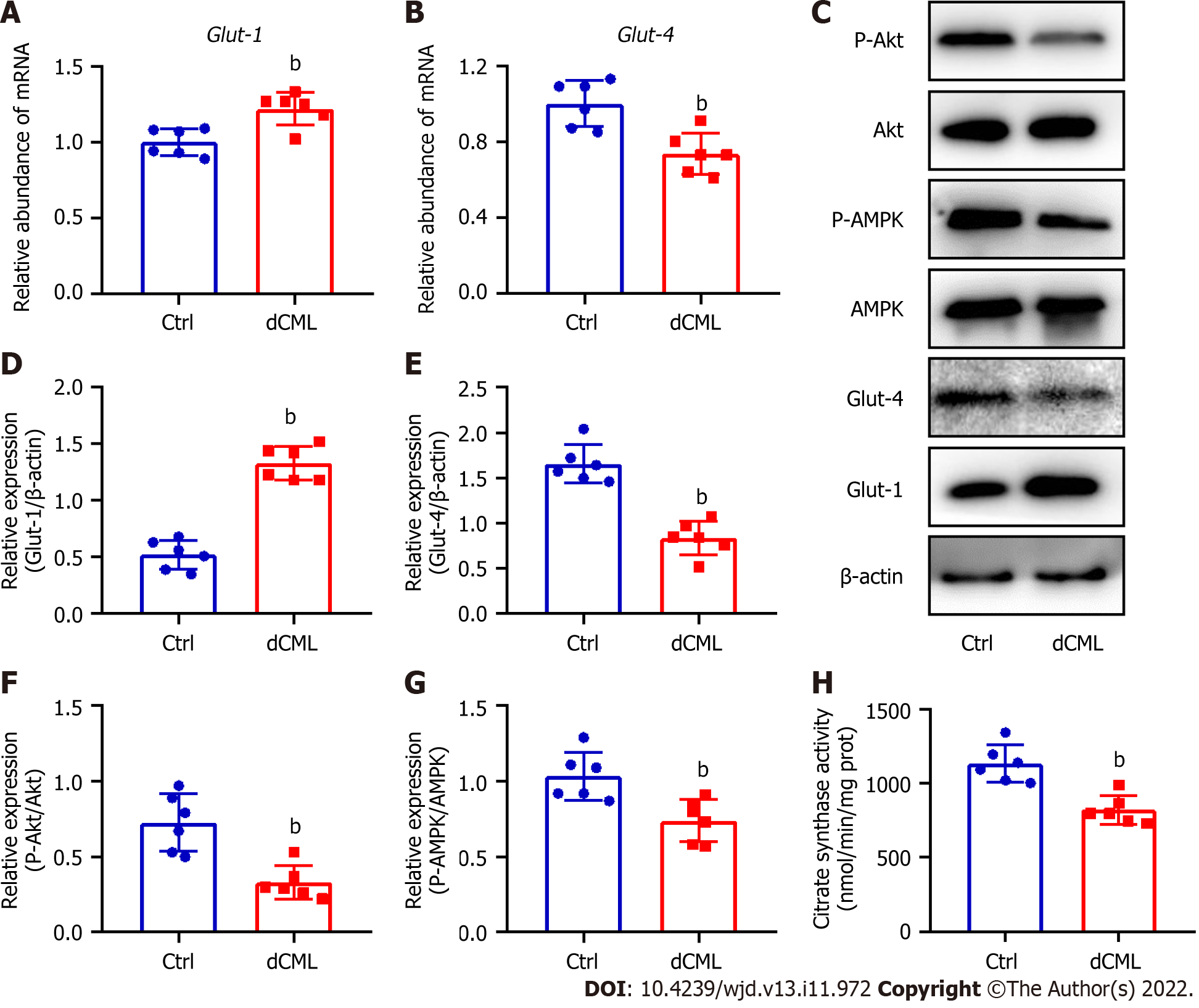
Figure 4 Dietary Nε-(carboxymethyl)lysine impairs glucose metabolism in mouse myocardium.
A and B: mRNA levels of glucose transporter (Glut)-1 and Glut-4 in the mouse myocardium; C-G: Western blotting and its relative quantification of Glut-1, Glut-4, phospho-Akt, and phospho-AMP-activated protein kinase (AMPK) in the mouse myocardium; H: Citrate synthase (CS) activity in the mouse myocardium. dCML: Dietary Nε-(carboxymethyl)lysine; Bcl-2: B-cell leukemia/lymphoma 2; BAX: Bcl-2-associated X; ANP: Atrial natriuretic peptide. n = 6. bP < 0.01, compared with the control (Ctrl) group.
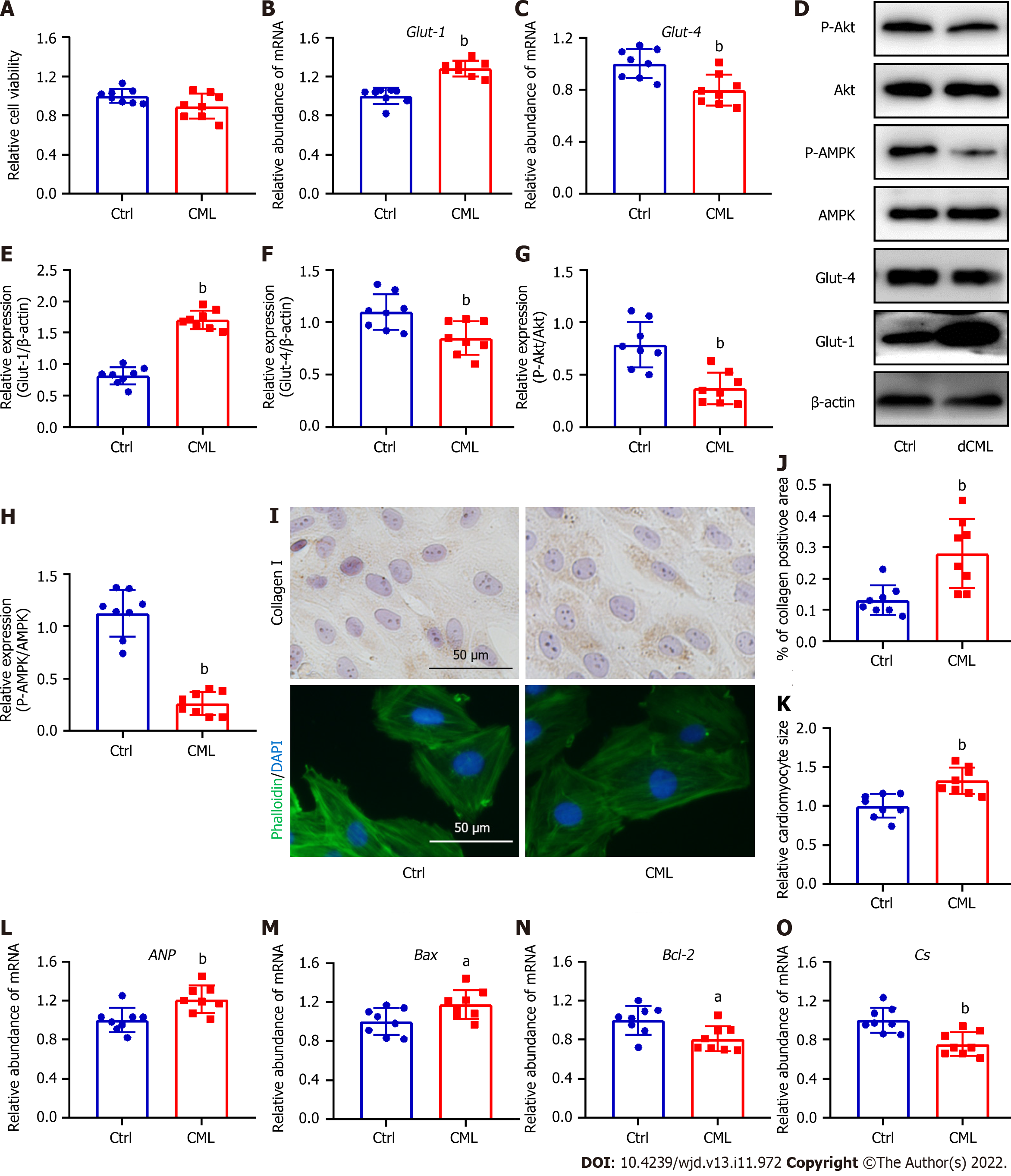
Figure 5 Exogenous Nε-(carboxymethyl)lysine inhibits the glucose metabolism and promotes collagen I expression, hypertrophy and apoptosis in H9C2 cells.
A: Cell viability after the simulation of Nε-(carboxymethyl)lysine (CML); B and C: Quantitative PCR detection of glucose transporter (Glut)-1 and Glut-4 mRNA; D-H: Relative expression of Glut-1, Glut-4, phospho-Akt, and phospho-AMP-activated protein kinase (AMPK) in H9C2 cells; I: Upper: Detection of collagen I content with immunocytochemical staining; bottom: Phalloidin-labeled H9C2 cardiomyocytes; J: Quantification of collagen I-positive areas; K: Quantitative analysis of cardiomyocyte area; L-O: Atrial natriuretic peptide (ANP), Bcl-2-associated X (Bax), B-cell leukemia/lymphoma 2 (Bcl-2), and citrate synthase (CS) mRNA levels in H9C2 cardiomyocytes. dCML: Dietary CML. n = 8 independent experiments. aP < 0.05, bP < 0.01, compared with the control (Ctrl) group.
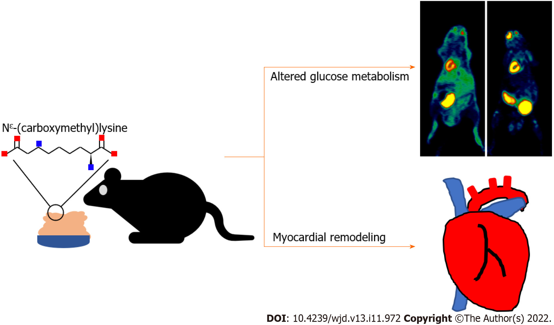
Figure 6 Dietary Nε-(carboxymethyl)lysine alters myocardial glucose metabolism and promotes myocardial remodeling.
- Citation: Wang ZQ, Sun Z. Dietary Nε-(carboxymethyl) lysine affects cardiac glucose metabolism and myocardial remodeling in mice. World J Diabetes 2022; 13(11): 972-985
- URL: https://www.wjgnet.com/1948-9358/full/v13/i11/972.htm
- DOI: https://dx.doi.org/10.4239/wjd.v13.i11.972


