Published online Nov 15, 2013. doi: 10.4251/wjgo.v5.i11.204
Revised: September 30, 2013
Accepted: October 11, 2013
Published online: November 15, 2013
Processing time: 72 Days and 4.5 Hours
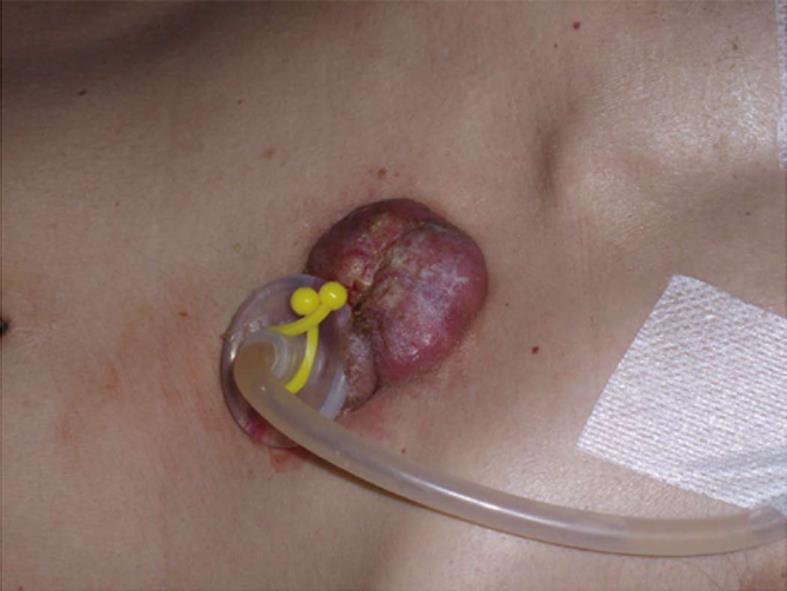
The authors present the case of a 55-year-old male with a stage III (T4N1M0) squamous-cell esophageal carcinoma, who underwent percutaneous endoscopic gastrostomy (PEG). The pull method of tube placement was used. Five months after the procedure, the patient was referred to the hospital with a hard palpable tumour at the ostomy site. The histologic exam revealed an abdominal wall metastasis of the esophageal cancer. The authors present this case because of the rarity of metastasis in ostomy after placement of PEG in patients with tumours located in the head and neck. In this particular context and judging by the rarity of situation, the clinical impact of this phenomenon is limited. Nevertheless, metastasis in ostomy site could be prevented by the push method, laparoscopy or laparotomy.
Core tip: We present this case to alert endoscopists to the possibility of metastatic cells in the ostomy when the percutaneous endoscopic gastrostomy is the chosen method for the nutrition support of the patient with head and neck cancer. This is a very rare but a possible complication so, in these situations, probably we must think about other possibilities of tube placement namely using an overtube, the introducer method or performing a surgical gastrostomy.
- Citation: Sousa AL, Sousa D, Velasco F, Açucena F, Lopes A, Guerreiro H. Rare complication of percutaneous endoscopic gastrostomy: Ostomy metastasis of esophageal carcinoma. World J Gastrointest Oncol 2013; 5(11): 204-206
- URL: https://www.wjgnet.com/1948-5204/full/v5/i11/204.htm
- DOI: https://dx.doi.org/10.4251/wjgo.v5.i11.204
A percutaneous endoscopic gastrostomy (PEG) is a way of long-term enteral nutrition, simple to perform, safe and highly accepted in patients with head and neck tumours[1,2]. The pull method of the tube placement, described by Gauderer et al[1] in 1980, is the most commonly used in clinical practice. A potential, but very rare complication is the metastasis of malignant cells from the primary tumour to the ostomy[3,4]. It is believed that direct implantation of the tumour by instrumentation is the explanation for this phenomenon; however, hematogenous metastasis is also a possible mechanism[5].
The authors describe the case of a 55-year-old man that presented to the emergency room with dysphagia to solids, with progressive worsening in the last week to semi-liquids, late regurgitation of undigested food, non-selective anorexia and weight loss. Relevant social history included heavy smoking and chronic alcoholism.
He underwent upper endoscopy which revealed an infiltrating neoplasm with almost complete stenosis of the upper third of the esophagus (21-26 cm), with poorly differentiated squamous cell carcinoma histology.
The computerized tomography showed neoplastic involvement of a higher level, occupying the left vallecula and the left piriform sinus with extrinsic anterioposterior compression of the trachea and presence of ipsilateral internal jugular lymphadenopathies.
The bronchoscopy showed extrinsic compression of the upper third posterior wall of the trachea with intact mucosa. The throat examination revealed neoformation at the left piriform sinus with decreased hemilarynx motility, without affecting the airway.
It is, therefore, a case of a man with a stage III (T4N1M0) squamous cell carcinoma of the esophagus with indication for chemotherapy (cisplatin + 5 - fluorouracil) and palliative radiotherapy.
As the patient presented with significant symptoms, several dilations with through-the-scope balloons and Savary dilators were attempted and the proximal area of the tumour was destroyed with argon-plasma coagulation. The PEG insertion was selected as a long-term pathway for nutritional support. The pull method is the most frequently used for PEG insertion both generally as in our institution, thus being the selected method.
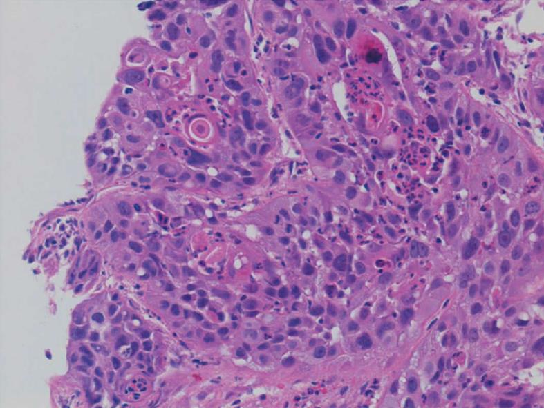
Five months after the placement of PEG, the patient was referred to the Gastroenterology Department after presenting with a hard palpable tumor at the ostomy site, with approximately 10 cm in diameter (Figure 1). Biopsy of the mass confirmed identical histology to the primary carcinoma (Figure 2). The patient died approximately 1 mo after this finding.
The use of PEG as a means of nutritional support for patients with head and neck tumors is one of the clearest indications for this procedure. This is a safe technique however, complications may occur, ranging between 6% and 30% in different series, being skin infections the most common[2,6,7].
We report a very rare but potential complication: metastasis of malignant cells from the primary tumour site to the ostomy. In the literature review about 50 cases of metastasis in ostomy are described, after placement of PEG in patients with head and neck tumours[8]. The pull method was used in our patient, as in most other cases reported in literature[5,8,9]. The time from tube placement to ostomy metastasis range between 3 and 18 mo[10]. However, when head and neck tumours metastasize to the site of ostomy, even with extensive tumor resection, patient survival is low[11]. Since the implantation of the tumor by direct instrumentation is the more likely explanation of metastasis, this complication might possibly be prevented by alternative PEG methods, such as introducer method, laparoscopy or laparotomy, or by using an overtube[4,5,10]. However, due to the rarity and clinical impact of this situation we consider that the practical relevance of the using of these methods will be low.
| 1. | Gauderer MW, Ponsky JL, Izant RJ. Gastrostomy without laparotomy: a percutaneous endoscopic technique. J Pediatr Surg. 1980;15:872-875. [RCA] [PubMed] [DOI] [Full Text] [Cited by in Crossref: 1408] [Cited by in RCA: 1311] [Article Influence: 28.5] [Reference Citation Analysis (1)] |
| 2. | Löser C, Aschl G, Hébuterne X, Mathus-Vliegen EM, Muscaritoli M, Niv Y, Rollins H, Singer P, Skelly RH. ESPEN guidelines on artificial enteral nutrition--percutaneous endoscopic gastrostomy (PEG). Clin Nutr. 2005;24:848-861. [RCA] [PubMed] [DOI] [Full Text] [Cited by in Crossref: 360] [Cited by in RCA: 370] [Article Influence: 17.6] [Reference Citation Analysis (0)] |
| 3. | Ponsky JL, Gauderer MW. Percutaneous endoscopic gastrostomy: a nonoperative technique for feeding gastrostomy. Gastrointest Endosc. 1981;27:9-11. [PubMed] |
| 4. | Adelson RT, Ducic Y. Metastatic head and neck carcinoma to a percutaneous endoscopic gastrostomy site. Head Neck. 2005;27:339-343. [RCA] [PubMed] [DOI] [Full Text] [Cited by in Crossref: 52] [Cited by in RCA: 47] [Article Influence: 2.2] [Reference Citation Analysis (0)] |
| 5. | Mincheff TV. Metastatic spread to a percutaneous gastrostomy site from head and neck cancer: case report and literature review. JSLS. 2005;9:466-471. [PubMed] |
| 6. | McClave SA, Chang WK. Complications of enteral access. Gastrointest Endosc. 2003;58:739-751. [RCA] [PubMed] [DOI] [Full Text] [Cited by in Crossref: 146] [Cited by in RCA: 140] [Article Influence: 6.1] [Reference Citation Analysis (0)] |
| 7. | Lockett MA, Templeton ML, Byrne TK, Norcross ED. Percutaneous endoscopic gastrostomy complications in a tertiary-care center. Am Surg. 2002;68:117-120. [PubMed] |
| 8. | Volkmer K, Meyer T, Sailer M, Fein M. Metastasis of an esophageal carcinoma at a PEG site--case report and review of the literature. Zentralbl Chir. 2009;134:481-485. [RCA] [PubMed] [DOI] [Full Text] [Cited by in Crossref: 7] [Cited by in RCA: 10] [Article Influence: 0.6] [Reference Citation Analysis (0)] |
| 9. | Sinclair JJ, Scolapio JS, Stark ME, Hinder RA. Metastasis of head and neck carcinoma to the site of percutaneous endoscopic gastrostomy: case report and literature review. JPEN J Parenter Enteral Nutr. 2001;25:282-285. [RCA] [PubMed] [DOI] [Full Text] [Cited by in Crossref: 52] [Cited by in RCA: 53] [Article Influence: 2.1] [Reference Citation Analysis (0)] |
| 10. | Volkmer K, Meyer T, Sailer M, Fein M. Implantation of an esophageal squamous cell carcinoma at the site of a percutaneous endoscopic gastrostomy. Endoscopy. 2007;39 Suppl 1:E240-E241. [RCA] [PubMed] [DOI] [Full Text] [Cited by in Crossref: 1] [Cited by in RCA: 2] [Article Influence: 0.1] [Reference Citation Analysis (0)] |
| 11. | Cruz I, Mamel JJ, Brady PG, Cass-Garcia M. Incidence of abdominal wall metastasis complicating PEG tube placement in untreated head and neck cancer. Gastrointest Endosc. 2005;62:708-711; quiz 752, 753. [RCA] [PubMed] [DOI] [Full Text] [Cited by in Crossref: 73] [Cited by in RCA: 70] [Article Influence: 3.3] [Reference Citation Analysis (0)] |
P- Reviewer: Korpanty G S- Editor: Cui XM L- Editor: A E- Editor: Wu HL


