Published online Apr 15, 2011. doi: 10.4251/wjgo.v3.i4.67
Revised: March 3, 2011
Accepted: March 10, 2011
Published online: April 15, 2011
Primary gastric signet ring cell carcinoma presenting as cardiac tamponade is difficult to diagnosis early. Patients are generally asymptomatic until the disease is advanced. General practitioners usually focus on the initial symptoms related to pericarditis and pericardial effusion. We report a case of signet-ring cell carcinoma of the stomach presenting as cardiac tamponade with pericarditis and pericardial effusion but without any gastrointestinal symptoms. A 49-year old woman was admitted because of progressive dyspnea and cough. Chest X-ray revealed an increased cardiothoracic ratio and a small amount of bilateral pleural effusion. Two dimensional ultrasonographic echocardiography pericardial effusions with atrial and right ventricular early diastolic collapse were found, establishing the diagnosis of cardiac tamponade. Pericardiocentesis was performed and 420 mL of bloody fluid was taken. The patient died of respiratory failure and cardiac arrest on October 28, 2009. Post-mortem examination revealed diffuse gastric mucosa erosion and edema with stomach mucosa incrassation in the greater curvature. The primary lesion was histopathologically diagnosed as signet-ring cell carcinoma of the stomach.
- Citation: Huang JY, Jiang HP, Chen D, Tang HL. Primary gastric signet ring cell carcinoma presenting as cardiac tamponade. World J Gastrointest Oncol 2011; 3(4): 67-70
- URL: https://www.wjgnet.com/1948-5204/full/v3/i4/67.htm
- DOI: https://dx.doi.org/10.4251/wjgo.v3.i4.67
Signet-ring cell carcinoma of the stomach tends to spread transversely in early gastric cancer but once it invades the submucosal region, it shows high potential to metastasize. We report a patient with cardiac tamponade due to pericarditis and pericardial effusion, originating from gastric signet-ring cell carcinoma, a rare condition with few reports, mostly case reports, in the literature. It is usually detected during the terminal stages of gastric carcinoma and therefore the prognosis is poor. Here we discuss the clinicopathological study of cardiac tamponade due to pericardial metastasis originating from gastric cancer and the results of the postmortem examination.
A 49-year old woman presented with a 36-h history of fever and dry cough on October 15, 2009. She reported a worsening cough, especially from September to November with cold air and gas as the stimulation. She noted that the cough did not vary from day to day and there was no productive sputum. Her symptoms became worse so that she could not lie flat at night, had progressive dyspnea after exercise and an underlying xiphoid process pain. She had a 10-year history of paroxysmal cough variant asthma (CVA). The earlier diagnosis by a general practitioner was cough finding: CVA? She was then admitted to our hospital with progressive dyspnea and cough on October 26, 2009. Physical examination revealed that she was febrile, with heart rate 98 bpm and blood pressure 120/78 mmHg. She had distant heart sounds, normal lung sounds, pulsus paradoxus and jugular venous distension.
Chest X-ray revealed an increased cardiothoracic ratio and a small amount bilateral pleural effusion. Two dimensional ultrasonographic echocardiography pericardial effusions with atrial and right ventricular early diastolic collapse were found, establishing the diagnosis of cardiac tamponade. Laboratory studies showed lactate dehydrogenase (LDH) 590 U/L (Reference value: 109-245 U/L), creatine kinase (CK) 301 U/L (Reference value: 26-174 U/L) and creatine kinase cardiac isoenzyme (CKMB) 97 U/L (Reference value: 3-25 U/L). Pericardiocentesis was then performed and 220 mL of bloody fluid was taken. After that, the patient still complained of dyspnea, malaise, sweating and all four limbs had cold extremities. Pericardiocentesis was performed again and 200 mL of bloody fluid was taken, relieving her symptoms slightly. However, she developed pallor, had difficulty breathing and bradycardia on October 28. She died two days after admission to our hospital.
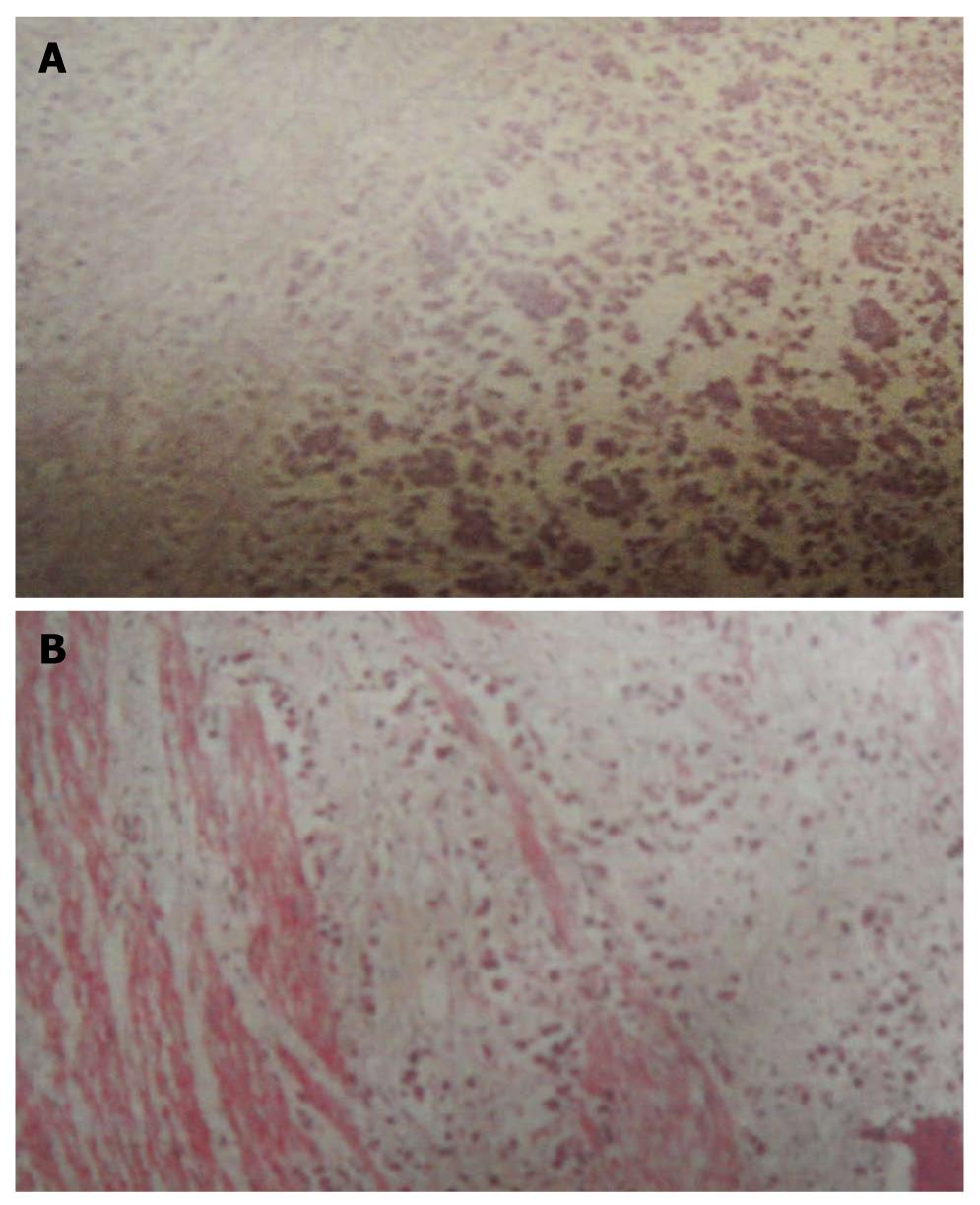
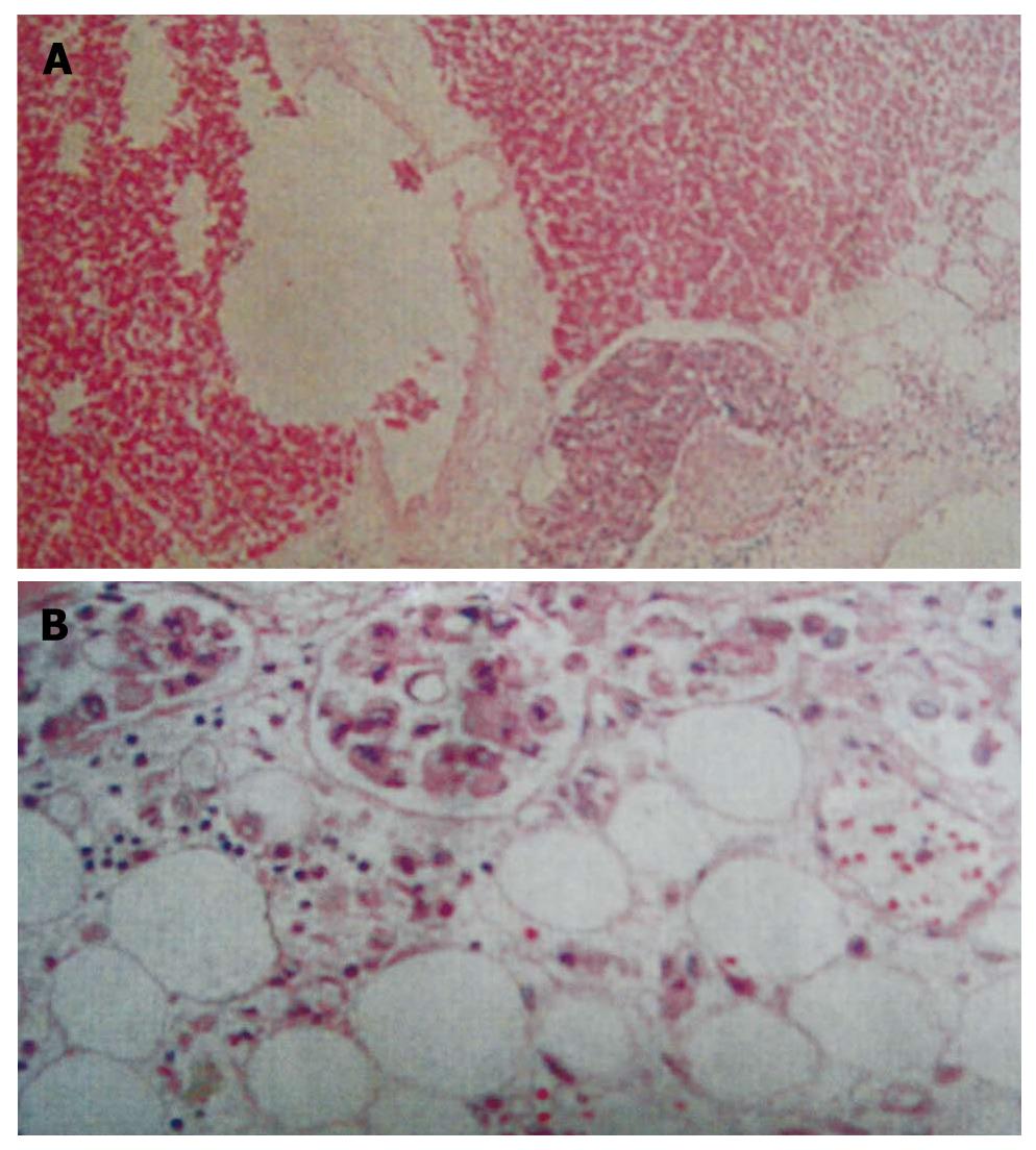
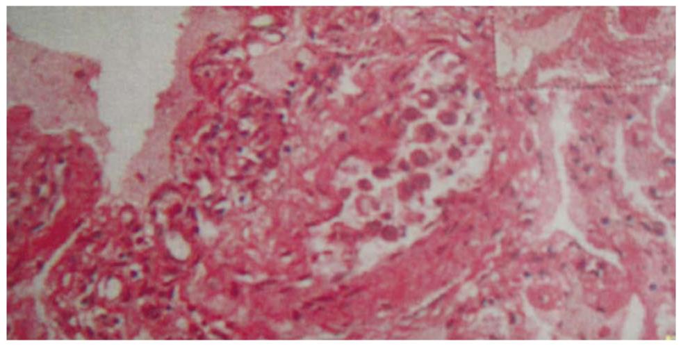
Postmortem examination revealed diffuse gastric mucosa erosion and edema with stomach mucosa incrassation in the greater curvature. The primary lesion was histopathologically diagnosed as signet-ring cell carcinoma of the stomach. The main tumor spread through the mucosa, submucosa, muscularis externa and adventitia (Figure 1A and B). The signet-ring cell carcinoma had metastasized to the para aortic and mediastinal lymph nodes. The pericardium was thickened and signet-ring cell carcinoma was also seen in the lymphatic vessels in both the pericardium and epicardium (Figure 2A and B). Malignant pulmonary embolus was also confirmed (Figure 3). Therefore, we conclude that the main diagnosis which led to the patient’s death was primary gastric signet ring cell carcinoma with multiple distance organ metastases.
There are three interesting aspects of this case: primary gastric carcinoma presenting as cardiac tamponade, the metastatic behavior of gastric signet ring cell carcinoma and the sudden unexpected death of a patient with malignancy.
Secondary tumors of the heart and/or pericardium are rarely diagnosed in clinical practice. Secondary cardiac metastases most frequently arise from primary lung tumors[1]. The most commonly reported of these include bronchial carcinoma, breast cancer, leukemia, Hodgkin’s disease, non-Hodgkin’s lymphoma, melanoma and sarcomas[2]. Primary gastric cancer rarely metastasizes to the heart. Abrams et al[3] reported metastases of 119 consecutive autopsied cases of stomach carcinoma. The incidence rates of primary gastric carcinoma metastasized to different organs are: abdominal lymph nodes 79.8% (95/119), liver 44.5% (53/119), lung 32.8% (39/119), ovary 15.1% (18/119) and only 4.2% (5/119) for pericardium and 2.5% (3/119) for heart. Some more autopsy investigations also summarized the pericardial metastasis incidence rate from stomach as 4.3%-7.7%[4-7]. Therefore, the probability of the original gastric cancer metastasis to heart is rare compared with the other systemic organs.
A search of the MEDLINE database revealed 8 cases of primary gastric carcinoma presenting as cardiac tamponade reported in the literature between 1982 and 2010[8-15]. In these reports, 3 out of 8 cases had signet-ring carcinoma and 1 case in which the tumor was confirmed by postmortem examination[13]. The case that reported the primary signet-ring cell carcinoma of the stomach was situated mostly in the mucosa without direct destruction of the mucosa muscularis[13]. Since then, there have been no cases in which the primary gastric signet-ring cell tumor invaded all the stomach layers from mucosa, submucosa, muscularis externa and to adventitia except the present study.
In our patient, the post-mortem examination revealed the late gastric carcinoma with multiple distance organs metastases (e.g. epicardium and lung), secondary to pericarditis, pericardial effusion and pulmonary effusion. This showed that the patient had a poorly differentiated and highly malignant carcinoma. The progression of the primary tumor was so fast that it had already metastasised before it was large enough to form a primary mass. Histopathological findings showed signet ring cell carcinoma and it met the morphologically poorly differentiated adenocarcinoma.
We concluded that the patient’s death was due to late stage primary gastric signet ring cell carcinoma from the postmortem examination, despite the fact that there were no gastrointestinal symptoms and signs such as abdominal pain, weight loss (> 10%), dysphagia, obstructive symptoms, upper gastrointestinal (UGI) hemorrhage, abdominal mass and adenopathy[16]. The patient was a sudden unexpected death from malignancy and the cause of death was only discovered from the postmortem examination. Since the patient had symptoms of cardiac tamponade more obviously than gastrointestinal symptoms, we focused on heart examinations and did not do tests such as an endoscopic examination of the stomach for the patient. Moreover, due to the rapid progression of the disease, serum levels of tumor markers, including carcinoembryonic antigen (CEA), carbohydrate antigen (CA) 19-9 and a-fetoprotein (AFP), and cytological diagnosis tests of pericardial effusion were not taken which would have shown the patient’s malignancy.
A study of 127 patients with a diagnosis of pericardial effusion with underlying malignancies states that the diagnosis of pericardial disease in patients with cancer is often extremely difficult because patients present with nonspecific symptoms and physical findings. Therefore, clinicians must consider pericardial disease in any patient with a known malignancy who develop unexplained dyspnea, tachycardia, arrhythmia or symptoms of heart failure[17]. Varvarigos et al[12], in common with this study, found a patient with no gastrointestinal symptoms until the onset of cardiac tamponade, the first sign of the disease. This shows primary gastric carcinoma presenting as cardiac tamponade is rare and medical practitioners should be aware of malignancy of the stomach when patients present with unexplained cardiac manifestations.
The routes by which the cardiac metastases commonly travel from primary tumors are believed to be: (1) by direct extension; (2) by lymphatic spread; (3) by hematogenous spread; and (4) by combinations of two or all three of the above[14]. A study has shown that the greater proportion of signet ring cancer cells, a cell type expressing nonspecific glycoproteins, express specific general neuroendocrine markers, indicating a neuroendocrine origin, and that at least a proportion of these tumors are derived from enterochromaffin-like (ECL) cells based on histidine decarboxylase (HDC) positivity[18]. In the present case, lymphatic spread with neuroendocrine origin is suggested since the main tumor spread through mucosa, submucosa, muscularis externa and adventitia and had metastasized to the para aortic and mediastinal lymph nodes.
In our case, the patient was a sudden unexpected death with malignancy. Kobayashi et al[19] reported cases that developed cardiac tamponade due to pericarditis carcinomatosa (PC) arising from primary gastric cancer (GC) more than 2 years after the diagnosis. The cases in which the levels of CEA and CA 19-9 were not elevated may have survived for a longer period of time. However, they were unable to reveal which therapy was the best for extending the life expectancy in cases of PC originating from GC[19]. Therefore, the period and the duration that the disease progresses highly affect the prognosis and survival of patients. Unfortunately, our patient died 2 d after admission. Her disease progression was so rapid that our team did not have enough time to treat her complications and she had already entered into an irreversible stage.
From this patient, we took a valuable lesson. The experience taught us the need for overall systematic physical examinations and laboratory examinations for general practitioners. At the time that the patient presented with cardiac complications, practitioners should have thought that there might be a tumor in the stomach. If our patient had had a regular gastroendoscopy, the gastric signet ring cell carcinoma would have been discovered earlier and there would have been the opportunity for surgical intervention. Therapy for cardiac metastases was given to reduce the patient’s discomfort. Pericardiocentesis, radiotherapy, local or systemic chemotherapy and the creation of a pericardiac window have been used to control cardiac metastases. The use of paclitaxel for systemic and cisplatin for local chemotherapy may be promising for the treatment of cardiac tamponade caused by PC arising from GC. As a result, the patient would have had an early diagnosis, faster treatments, better prognosis and longer survival.
In conclusion, we report a case of cardiac tamponade caused by metastasis from primary gastric cancer. The patient died due to cardiac and respiratory failure shortly after cardiac tamponade was apparent.
| 1. | Gowda RM, Khan IA, Mehta NJ, Gowda MR, Hyde P, Vasavada BC, Sacchi TJ. Cardiac tamponade and superior vena cava syndrome in lung cancer-a case report. Angiology. 2004;55:691-695. |
| 2. | Barbetakis NG, Vassiliadis M, Krikeli M, Antoniadis T, Tsilikas C. Cardiac Tamponade Secondary to Metastasis from Adenocarcinoma of the Parotid Gland. World J Surg Oncol. 2003;1:20. |
| 3. | Abrams HL, Spiro R, Goldstein N. Metastases in carcinoma; analysis of 1000 autopsied cases. Cancer. 1950;3:74-85. |
| 4. | Tabata Y, Nakato H, Nakamura Z, Sasaki A, Shoji K, Yokoyama H, Tanji Y, Saito K, Uehara H. [Metastatic cardiac tumors--clinicopathological evaluation of 64 autopsy cases]. Kokyu To Junkan. 1983;31:569-573. |
| 5. | Nakayama R, Yoneyama T, Takatani O, Kimura K. A study of metastatic tumors to the heart, pericardium and great vessels. I. Incidences of metastases to the heart, pericardium and great vessels. Jpn Heart J. 1966;7:227-234. |
| 6. | Mukai K, Shinkai T, Tominaga K, Shimosato Y. The incidence of secondary tumors of the heart and pericardium: a 10-year study. Jpn J Clin Oncol. 1988;18:195-201. |
| 8. | Zhang BL, Xu RL, Zheng X, Qin YW. A case with cardiac tamponade as the first sign of primary gastric signet-ring cell carcinoma treated with combination therapy. Med Sci Monit. 2010;16:CS41-CS44. |
| 9. | Hattori K, Kondo T, Yamamoto M, Watanabe M, Satoh H. Cardiac tamponade originating from gastric cancer. J Gastroenterol. 2009;44:1007. |
| 10. | Bacic B, Misic Z, Kovacic V, Sprung J. Gastric carcinoma presenting as pericardial tamponade during pregnancy. Turk J Gastroenterol. 2009;20:276-278. |
| 11. | Baba Y, Ishikawa S, Ikeda K, Honda S, Miyanari N, Iyama K, Baba H. A patient with 43 synchronous early gastric carcinomas with a Krukenberg tumor and pericardial metastasis. Gastric Cancer. 2007;10:135-139. |
| 12. | Varvarigos N, Kamaradou H, Kourti A, Papavasiliou ED, Papaioannou H, Migdalis IN, Galanis C. Cardiac tamponade as the first manifestation of gastric cancer and remission after chemotherapy. Dig Dis Sci. 2001;46:2333-2335. |
| 13. | Sakai Y, Minouchi K, Ohta H, Annen Y, Sugimoto T. Cardiac tamponade originating from primary gastric signet ring cell carcinoma. J Gastroenterol. 1999;34:250-252. |
| 14. | Moriyama A, Murata I, Kuroda T, Yoshikawa I, Tabaru A, Ogami Y, Otsuki M. Pericardiac metastasis from advanced gastric cancer. J Gastroenterol. 1995;30:512-516. |
| 15. | Blanco-Guerra C, Cobo J, Gomez-Cerezo J, Molina F. Gastric carcinoma presented as cardiac tamponade. Am J Gastroenterol. 1990;85:1431. |
| 16. | Diehl JT, Hermann RE, Cooperman AM, Hoerr SO. Gastric carcinoma. A ten-year review. Ann Surg. 1983;198:9-12. |
| 17. | Wilkes JD, Fidias P, Vaickus L, Perez RP. Malignancy-related pericardial effusion. 127 cases from the Roswell Park Cancer Institute. Cancer. 1995;76:1377-1387. |
| 18. | Bakkelund K, Fossmark R, Nordrum I, Waldum H. Signet ring cells in gastric carcinomas are derived from neuroendocrine cells. J Histochem Cytochem. 2006;54:615-621. |
| 19. | Kobayashi M, Okabayashi T, Okamoto K, Namikawa T, Araki K. Clinicopathological study of cardiac tamponade due to pericardial metastasis originating from gastric cancer. World J Gastroenterol. 2005;11:6899-6904. |
Peer reviewer: Asahi Hishida, MD, PhD, Department of Preventive Medicine, Nagoya University Graduate School of Medicine, Tsurumai-cho 65, Showa-ku, Nagoya 466-8550, Japan
S- Editor Wang JL L- Editor Roemmele A E- Editor Ma WH


