©The Author(s) 2017.
World J Gastrointest Oncol. Mar 15, 2017; 9(3): 135-141
Published online Mar 15, 2017. doi: 10.4251/wjgo.v9.i3.135
Published online Mar 15, 2017. doi: 10.4251/wjgo.v9.i3.135
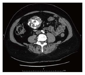
Figure 1 Axial contrast-enhanced computed tomography scan showing a well-defined heterogeneously enhanced gastric mass with coarse and diffuse calcifications.
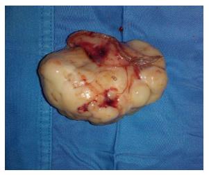
Figure 2 Macroscopic presentation of the resected mass, consisting of an exophytic and yellowish-gray tumor.
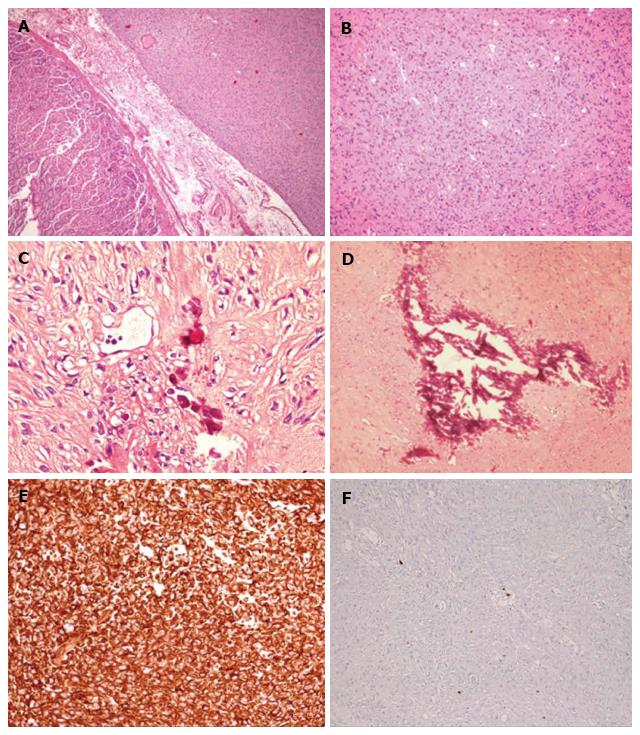
Figure 3 Histologic appearance of the tumor.
A and B: Interlacing bundles of spindle cells with unremarkable mitotic activity (H and E stain; magnification × 10); C and D: Calcified areas (H and E stain; magnification × 20); E: Intense immunoreactivity for DOG1 (Immunohistochemistry stain; magnification × 20); F: Inconspicuous Ki67-MIB1 label index (count rate < 1%; immunohistochemistry stain; magnification × 20).
- Citation: Salati M, Orsi G, Reggiani Bonetti L, Di Benedetto F, Longo G, Cascinu S. Heavily calcified gastrointestinal stromal tumors: Pathophysiology and implications of a rare clinicopathologic entity. World J Gastrointest Oncol 2017; 9(3): 135-141
- URL: https://www.wjgnet.com/1948-5204/full/v9/i3/135.htm
- DOI: https://dx.doi.org/10.4251/wjgo.v9.i3.135


