Copyright
©The Author(s) 2017.
World J Gastrointest Oncol. Mar 15, 2017; 9(3): 129-134
Published online Mar 15, 2017. doi: 10.4251/wjgo.v9.i3.129
Published online Mar 15, 2017. doi: 10.4251/wjgo.v9.i3.129
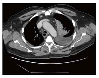
Figure 1 Computed tomography chest showing lung opacity, pleural effusion and lymph nodes.
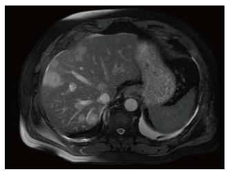
Figure 2 Magnetic resonance imaging showing multiple metastatic liver lesions.
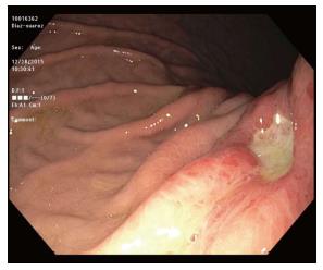
Figure 3 Gastric ulcer in the body.
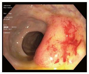
Figure 4 Rectal mass.
Figure 5 Pathology from both rectal mass and gastric ulcer showed metastatic adenocarcinoma, consistent with lung primary.
A: Gastric mucosa with metastatic adenocarcinoma; B: Rectal mass showing submucosa and deep mucosa with metastatic adenocarcinoma.
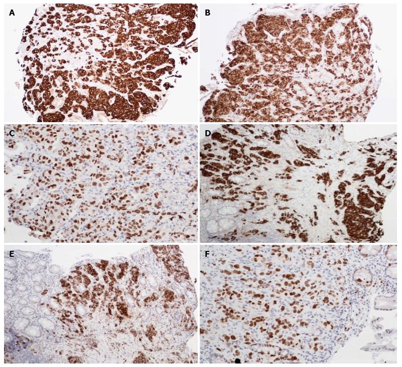
Figure 6 Immunohistochemical staining.
A: Gastric biopsy showing positivity to CK 7; B: Gastric biopsy showing positivity to Napsin-A; C: Gastric biopsy showing positivity to TTF-1; D: Rectal biopsy showing positivity to CK 7; E: Rectal biopsy showing positivity to Napsin-A; F: Rectal biopsy showing positivity to TTF-1. CK 7: Cytokeratin 7; TTF-1: Thyroid transcription factor-1.
- Citation: Badipatla KR, Yadavalli N, Vakde T, Niazi M, Patel HK. Lung cancer metastasis to the gastrointestinal system: An enigmatic occurrence. World J Gastrointest Oncol 2017; 9(3): 129-134
- URL: https://www.wjgnet.com/1948-5204/full/v9/i3/129.htm
- DOI: https://dx.doi.org/10.4251/wjgo.v9.i3.129


