Copyright
©The Author(s) 2017.
World J Gastrointest Oncol. Nov 15, 2017; 9(11): 444-451
Published online Nov 15, 2017. doi: 10.4251/wjgo.v9.i11.444
Published online Nov 15, 2017. doi: 10.4251/wjgo.v9.i11.444
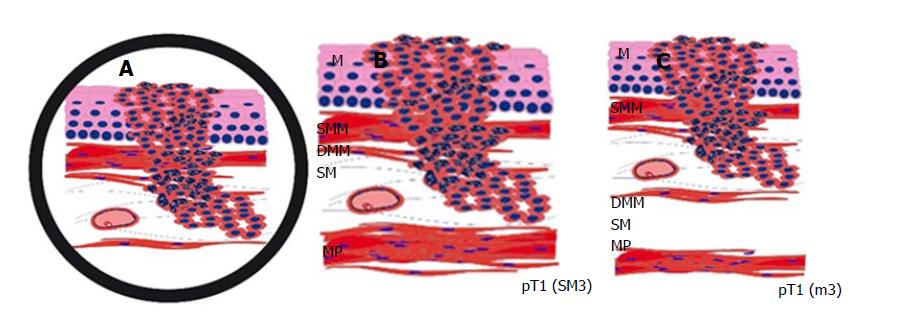
Figure 1 The difficulty in distinguishing between the smooth muscle cells of the muscularis mucosae, which can eventually be splintered in the superficial muscularis mucosae and the deep muscularis mucosae, and those of the muscularis propria.
This schematic drawing shows a section of a Barrett’s adenocarcinoma that may, at first glance appear, relatively straightforward, but determining the type of muscle layer can be challenging. Identifying large vessels (A) may suggest the diagnosis of submucosal invasion (B) and is, therefore, a well-known pitfall, as large vessels can also be found in between the superficial and DMM (C). An intramucosal carcinoma pT1 (m3) can therefore easily be mistaken as a submucosal carcinoma pT1 (sm). M: Mucosa; SMM: Superficial muscularis mucosae; DMM: Deep muscularis mucosae; MP: Muscularis propria.
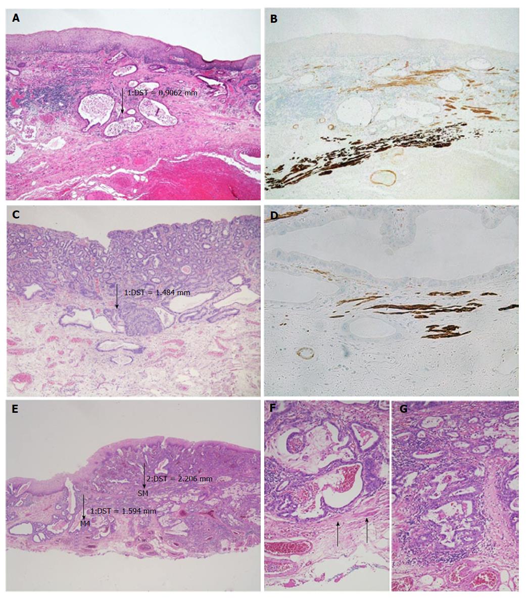
Figure 2 In case of undermining growth beneath the intact epithelium, tumour thickness was measured from the most superficial neoplastic cell layer to the point of deepest invasion.
A: HE 50 ×; ESD specimen with early Barrett’s adenocarcinoma pT1 (m3) with infiltration between the SMM and DMM and a tumour thickness of 900 μm. Tumour thickness was measured from the most superficial tumour cell layer to the deepest point of the invasion; B: Smoothelin IHC 50 ×; Immunohistochemical staining discriminates between the SMM (light brown) and DMM (dark brown). A tumour gland can be seen in-between the two muscle layers; C: HE 16 ×; Neoplastic glands reach the DMM. Smooth muscle fibres are found in the neighbourhood of the glands. The tumour thickness is approximately 1500 μm (m4); D: Smoothelin IHC 200 ×; Immunohistochemical staining confirms the m4 stage. Dark brown fibres of the DMM are found on the same level as the tumour glands; E: HE 16 ×; ESD specimen with an adenocarcinoma of the oesophagus that reaches the submucosa. The left-sided measurement was performed in an area where the tumour was restricted to a m4-stage (tumour thickness approxinately 1600 μm). The right-sided measurement was in an area where the tumour already showed the beginning of an infiltration of the submucosa (tumour thickness approxinately 2200 μm); F: HE 100 ×; Higher magnification of the m4-area of (E). Smooth muscle fibres (arrows) discriminate from sm-stage; G: HE 100 ×; Higher magnification of the sm-area of (E). The lack of muscle fibres indicates the sm-stage. ESD: Endoscopic submucosa dissection; SMM: Superficial muscularis mucosae; DMM: Deep muscularis mucosae; IHC: Immunohistochemistry; HE: Haematoxylin and eosin.
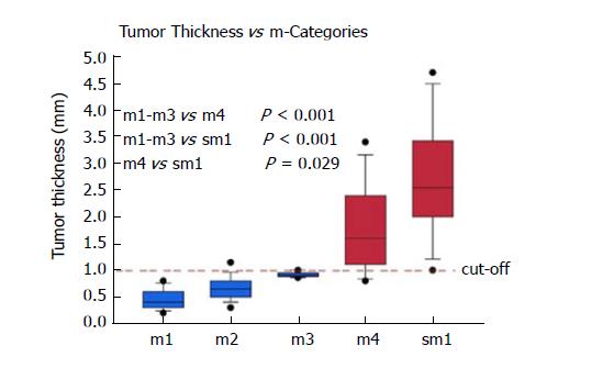
Figure 3 Box plot of the median tumour thickness in the different m-categories.
Circles = 5/95 percentiles. The dashed line indicates the proposed cut-off to discriminate between m1-3 and m4/sm1.
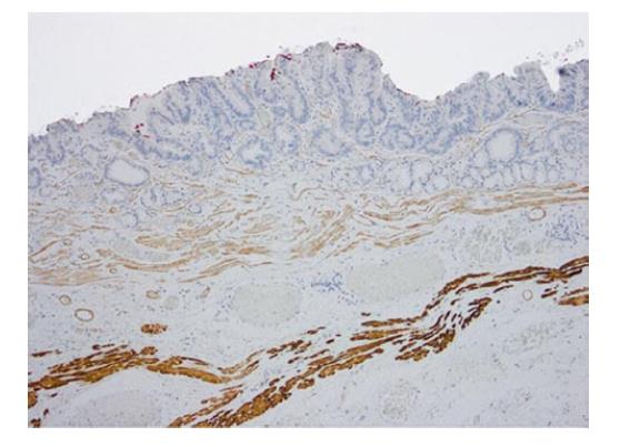
Figure 4 Expression of smoothelin immunohistochemistry demonstrating the different staining intensity in the duplicated muscularis mucosae layers.
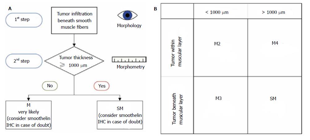
Figure 5 Suggested algorithm for the pT staging in early adenocarcinoma of the oesophagus.
A: In the first step, the relationship between the tumour and the smooth muscle fibres is determined. The second step includes the measurement of the tumour thickness; B: This table illustrates the combinations of the tumour relationships to the smooth muscle fibres/tumour thickness and the corresponding T-sub-stages. M: Mucosa; SM: Superficial muscularis.
- Citation: Endhardt K, Märkl B, Probst A, Schaller T, Aust D. Value of histomorphometric tumour thickness and smoothelin for conventional m-classification in early oesophageal adenocarcinoma. World J Gastrointest Oncol 2017; 9(11): 444-451
- URL: https://www.wjgnet.com/1948-5204/full/v9/i11/444.htm
- DOI: https://dx.doi.org/10.4251/wjgo.v9.i11.444


