©The Author(s) 2015.
World J Gastrointest Oncol. Nov 15, 2015; 7(11): 369-374
Published online Nov 15, 2015. doi: 10.4251/wjgo.v7.i11.369
Published online Nov 15, 2015. doi: 10.4251/wjgo.v7.i11.369
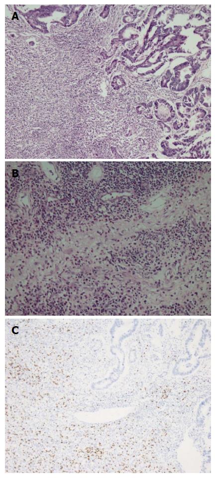
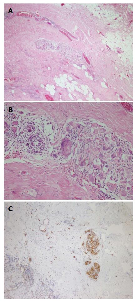
Figure 1 Adenocarcinoma.
A: Adenocarcinoma showing fibrosis and inflammatory cell infiltration in the tumor base (HE × 10); B: Inflammatory cell infiltration consisting of plasma cells, lymphocytes, eosinophils and PNLs was seen in these areas (HE × 20); C: Inflammatory cell infiltration observed in the basis of tumors was rich in CD8 positive T lymphocytes (anti-CD8, × 5).
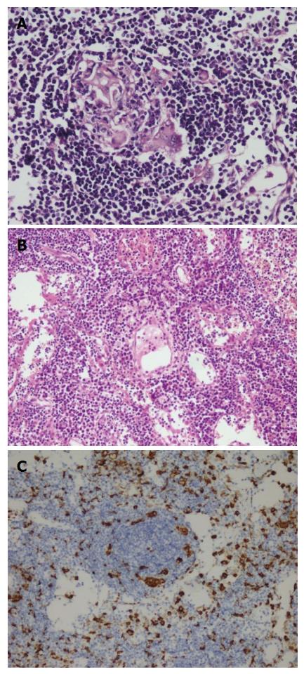
Figure 2 Germinal centers of some lymphoid follicles had giant cells and increased number of plasma cells in inter-follicular areas.
A: Giant cells were detected in germinal centers of some lymph nodes (HE × 20); B: Interfollicular areas of some lymph nodes had increased number of plasma cells (HE × 10); C: Giant cells were stained with CD68 immunohistochemically (anti-CD68 × 10).
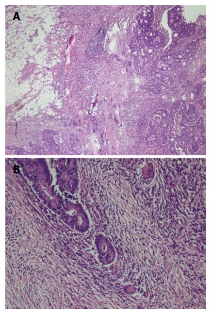
Figure 3 Moderately differentiated adenocarcinoma with mixed inflammatory cell infiltration rich of lymphoplasmocytes, eosinophils and few giant cells (A and B) (HE × 5, HE × 20).
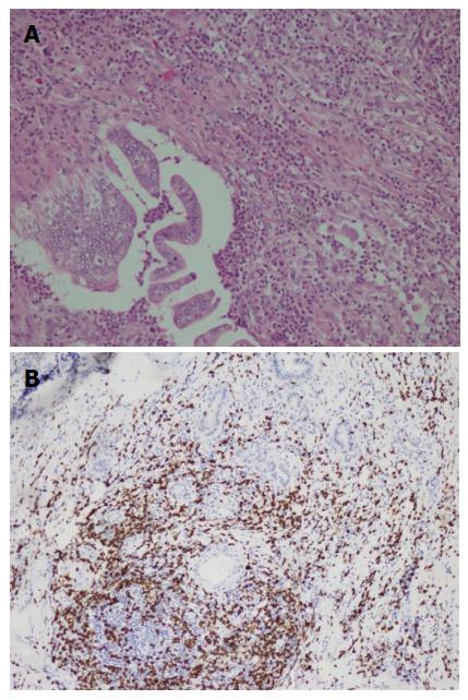
Figure 4 Adenocarcinoma showing mixed inflammatory cell infiltration rich in eosinophils and T - lymphocytes.
A: Moderately differentiated adenocarcinoma showing mixed inflammatory cell infiltration rich in eosinophils and T - lymphocytes (HE × 20); B: The most prominent cellular component on immunohistochemical examination was CD8 positive T - lymphocytes (CD8 × 10).
Figure 5 Giant cells in the areas beneath serosal surface and stained with CD68 immunohistochemically.
A, B: Giant cells were seen in the areas beneath serosal surface (HE × 5, HE × 20); C: CD68 positivity in giant cells (anti -CD68 × 10).
- Citation: Karaarslan S, Cokmert S, Cokmez A. Does St. John’s Wort cause regression in gastrointestinal system adenocarcinomas? World J Gastrointest Oncol 2015; 7(11): 369-374
- URL: https://www.wjgnet.com/1948-5204/full/v7/i11/369.htm
- DOI: https://dx.doi.org/10.4251/wjgo.v7.i11.369


