©2013 Baishideng Publishing Group Co.
World J Gastrointest Oncol. Feb 15, 2013; 5(2): 29-33
Published online Feb 15, 2013. doi: 10.4251/wjgo.v5.i2.29
Published online Feb 15, 2013. doi: 10.4251/wjgo.v5.i2.29
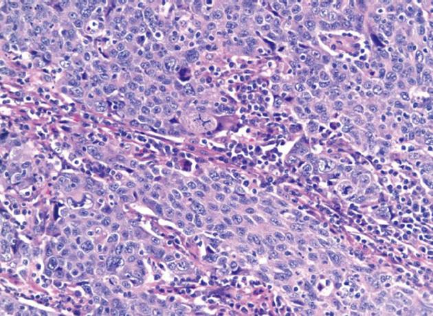
Figure 1 Tumor cells are large with numerous mitotic figures consistent with poorly differentiated carcinoma of the gallbladder.
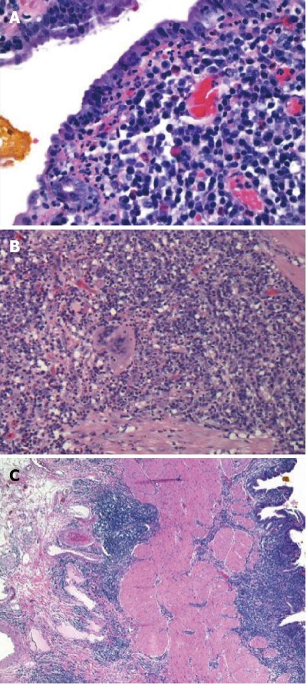
Figure 2 Crohn’s disease involving the gallbladder.
A: Superficial acute inflammation and numerous plasma cells; B: Non necrotizing granuloma in the gallbladder wall; C: Transmural inflammation of the gallbladder wall.
- Citation: Attraplsi S, Shobar RM, Lamzabi I, Abraham R. Gallbladder carcinoma in a pregnant patient with Crohn's disease complicated with gallbladder involvement. World J Gastrointest Oncol 2013; 5(2): 29-33
- URL: https://www.wjgnet.com/1948-5204/full/v5/i2/29.htm
- DOI: https://dx.doi.org/10.4251/wjgo.v5.i2.29


