©2013 Baishideng Publishing Group Co.
World J Gastrointest Oncol. Dec 15, 2013; 5(12): 235-239
Published online Dec 15, 2013. doi: 10.4251/wjgo.v5.i12.235
Published online Dec 15, 2013. doi: 10.4251/wjgo.v5.i12.235
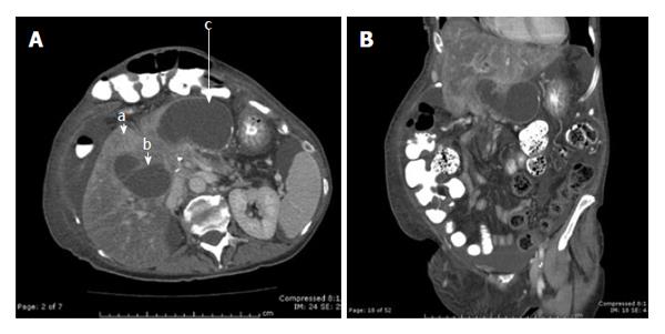
Figure 1 Computed tomography horizontal image.
A: Dilated jejunal Roux limb in the right upper quadrant. No evidence of biliary dilatation was evident of the computed tomography scan. Furthermore, the liver parenchyma showed evidence of hepatic steatosis. There is also the presence of a large incisional hernia in the anterior abdominal wall. Arrow a points to an intrahepatic metastasis from cancer. Arrow b points to the dilated Roux limb prior to the obstruction at the level of the mesentery of the colon. Arrow c points to the proximal portion of the dilated Roux limb that was close to the stomach used for the venting gastrojejunostomy; B: The close proximity of the stomach to the dilated Roux limb.
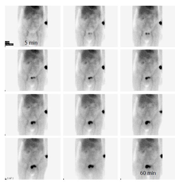
Figure 2 Hepatobiliary iminodiacetic acid scan showing delayed uptake of the liver up to 60 min after administration of radiotracer.
The cause of the delayed uptake likely reflects long term liver dysfunction and likely contributed to the inability of the biliary tree to dilate in a prompt fashion.
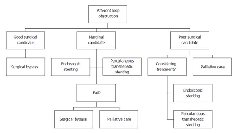
Figure 3 Management algorithm for afferent loop obstruction following pancreaticoduodenectomy.
- Citation: Bakes D, Cain C, King M, Dong XD(. Management of afferent loop obstruction from recurrent metastatic pancreatic cancer using a venting gastrojejunostomy. World J Gastrointest Oncol 2013; 5(12): 235-239
- URL: https://www.wjgnet.com/1948-5204/full/v5/i12/235.htm
- DOI: https://dx.doi.org/10.4251/wjgo.v5.i12.235


