Copyright
©2012 Baishideng Publishing Group Co.
World J Gastrointest Oncol. Feb 15, 2012; 4(2): 22-25
Published online Feb 15, 2012. doi: 10.4251/wjgo.v4.i2.22
Published online Feb 15, 2012. doi: 10.4251/wjgo.v4.i2.22
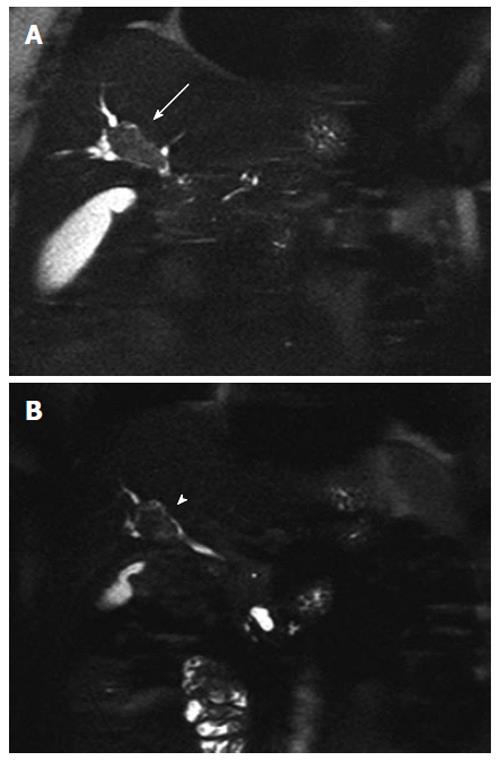
Figure 1 Haste in coronal plane.
A: Biliary lesion ( 40 mm in size) extending to the biliary confluence (arrow); B: A reduction of the biliary lesion (30 mm in size) after radio-chemotherapy with a prevalence of the endoluminal component, in the absence of bile duct dilatation (arrowhead).
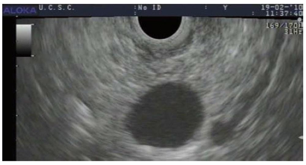
Figure 2 Ultrasound endoscopy image of the pancreatic lesion in the uncinate process showing a 30-mm anechoic lesion without any solid nodule or solid component.
- Citation: Valente R, Capurso G, Pierantognetti P, Iannicelli E, Piciucchi M, Romiti A, Mercantini P, Larghi A, Federici GF, Barucca V, Osti MF, Di Giulio E, Ziparo V, Delle Fave G. Simultaneous intraductal papillary neoplasms of the bile duct and pancreas treated with chemoradiotherapy. World J Gastrointest Oncol 2012; 4(2): 22-25
- URL: https://www.wjgnet.com/1948-5204/full/v4/i2/22.htm
- DOI: https://dx.doi.org/10.4251/wjgo.v4.i2.22


