Copyright
©2011 Baishideng Publishing Group Co.
World J Gastrointest Oncol. Apr 15, 2011; 3(4): 67-70
Published online Apr 15, 2011. doi: 10.4251/wjgo.v3.i4.67
Published online Apr 15, 2011. doi: 10.4251/wjgo.v3.i4.67
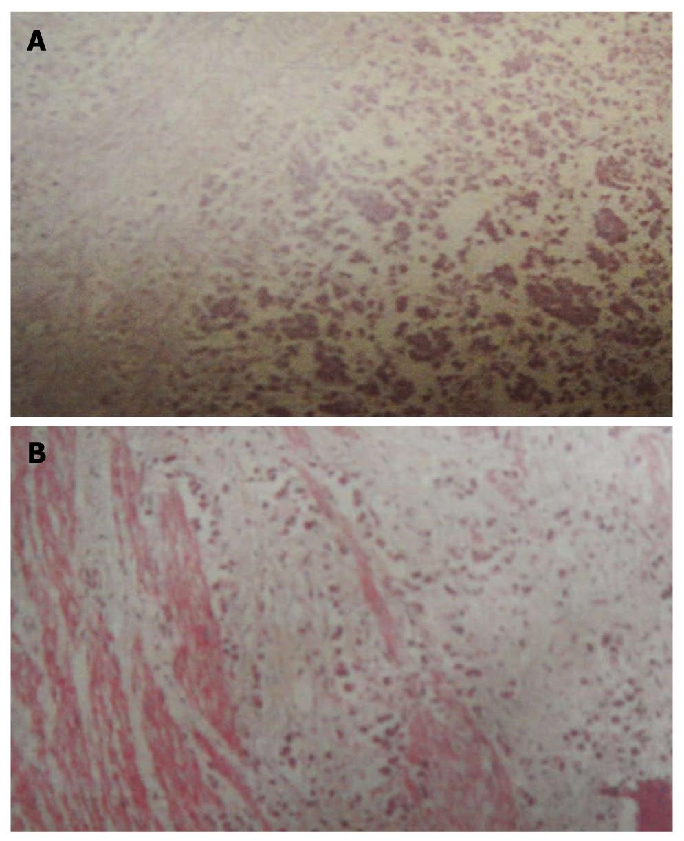
Figure 1 Histopathological findings of the stomach.
A: Neoplastic cells resemble signet-rings since they contain abundant intra-cytoplasmic mucin which pushes the nuclei to the periphery; B: Neoplastic cells resemble signet-rings spread though mucosa, submucosa, muscularis externa and adventitia.
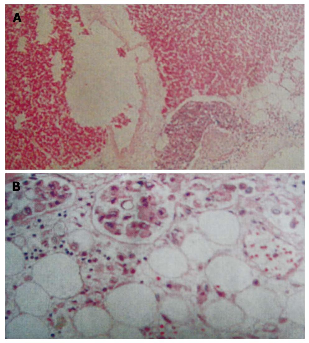
Figure 2 Histopathological findings of the heart.
A: Diffuse metastatic cancer cells and lymphocytes infiltration demonstrating hemorrhage, abnormal glandular development and cellular atypia in left anterior ventricular wall and posterior epicardium; B: Slightly enlarged thickness of coronary artery endothelium with surrounding fatty tissues hemorrhage. The neoplastic cells are seen in small blood vessels and small lymph vessels, resemble variety size nuclei and partly with scanty-size nuclei which pushes the nuclei to the periphery.
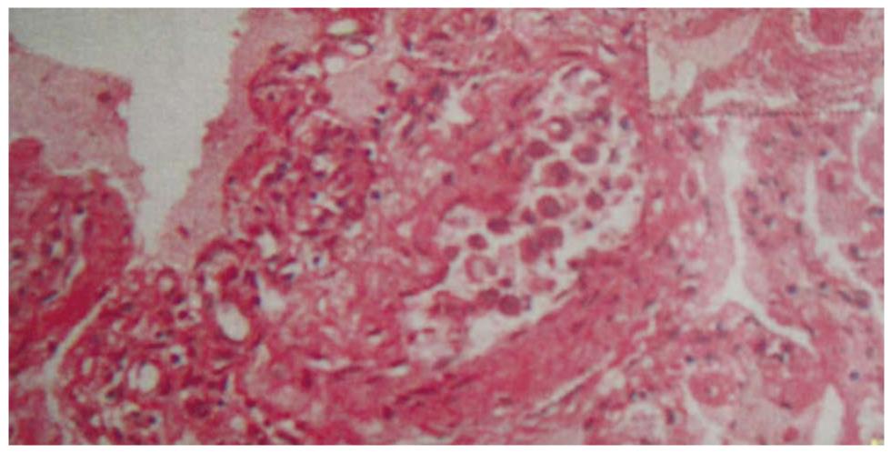
Figure 3 Histopathological findings of the lung.
Blood congestion and edema in the pulmonary area. Partial alveolar walls are thickened, some alveolar walls with an increased amount of small blood vessels and capillaries and neoplastic cells are seen inside some small blood vessels.
- Citation: Huang JY, Jiang HP, Chen D, Tang HL. Primary gastric signet ring cell carcinoma presenting as cardiac tamponade. World J Gastrointest Oncol 2011; 3(4): 67-70
- URL: https://www.wjgnet.com/1948-5204/full/v3/i4/67.htm
- DOI: https://dx.doi.org/10.4251/wjgo.v3.i4.67


