Copyright
©2010 Baishideng.
World J Gastrointest Oncol. Apr 15, 2010; 2(4): 205-208
Published online Apr 15, 2010. doi: 10.4251/wjgo.v2.i4.205
Published online Apr 15, 2010. doi: 10.4251/wjgo.v2.i4.205
Figure 1 Perivascular epithelioid cell neoplasm (PEComa).
A: PEComa in sigmoid colon site (HE, × 12.5); B: PEComa in sigmoid colon showing mucosal ulceration (HE, × 12.5); C: PEComa with individual cell nests invading mucosa (HE, × 100).
Figure 2 PEComa tumor body.
A: PEComa tumor body with intervening delicate capillary network (HE, × 100); B: PEComa, tumor body (HE, × 400).
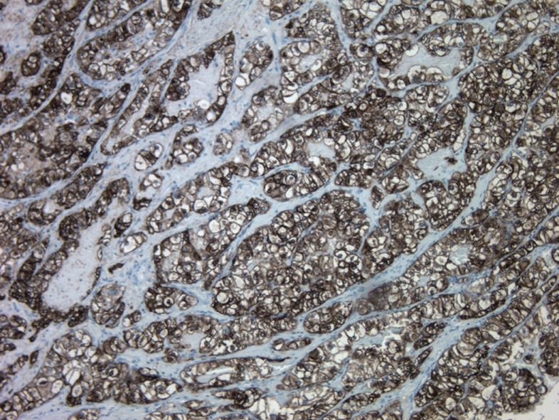
Figure 3 PEComa with HMB-45 antibody strongly stained cytoplasm of neoplastic cells (HMB-45, × 100).
- Citation: Freeman HJ, Webber DL. Perivascular epithelioid cell neoplasm of the colon. World J Gastrointest Oncol 2010; 2(4): 205-208
- URL: https://www.wjgnet.com/1948-5204/full/v2/i4/205.htm
- DOI: https://dx.doi.org/10.4251/wjgo.v2.i4.205


