
















Copyright: ©Author(s) 2026.
World J Gastrointest Oncol. Aug 15, 2026; 18(8): 122077
Published online Aug 15, 2026. doi: 10.4251/wjgo.122077
Published online Aug 15, 2026. doi: 10.4251/wjgo.122077
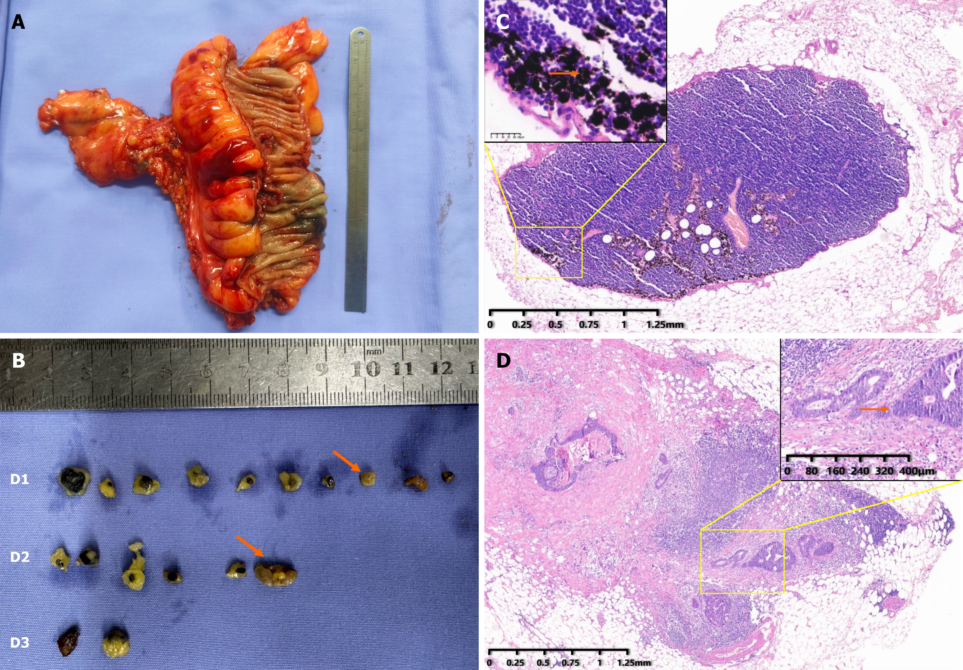
Figure 1 Lymph nodes stained or non-stained with carbon nanoparticles.
A: Representative images tumor specimen stained with carbon nanoparticles (CNPs); B: Representative images of lymph nodes (LNs) from D1 (pericolic/perirectal), D2 (intermediate), and D3 (main) regions. Arrow indicates non-stained LNs; C: Hematoxylin and eosin (HE) staining of a nonmetastatic lymph node stained with CNPs. Arrow indicates CNPs; D: HE staining of a metastatic lymph node non-stained with CNPs. Arrow indicates metastatic adenocarcinoma cells.
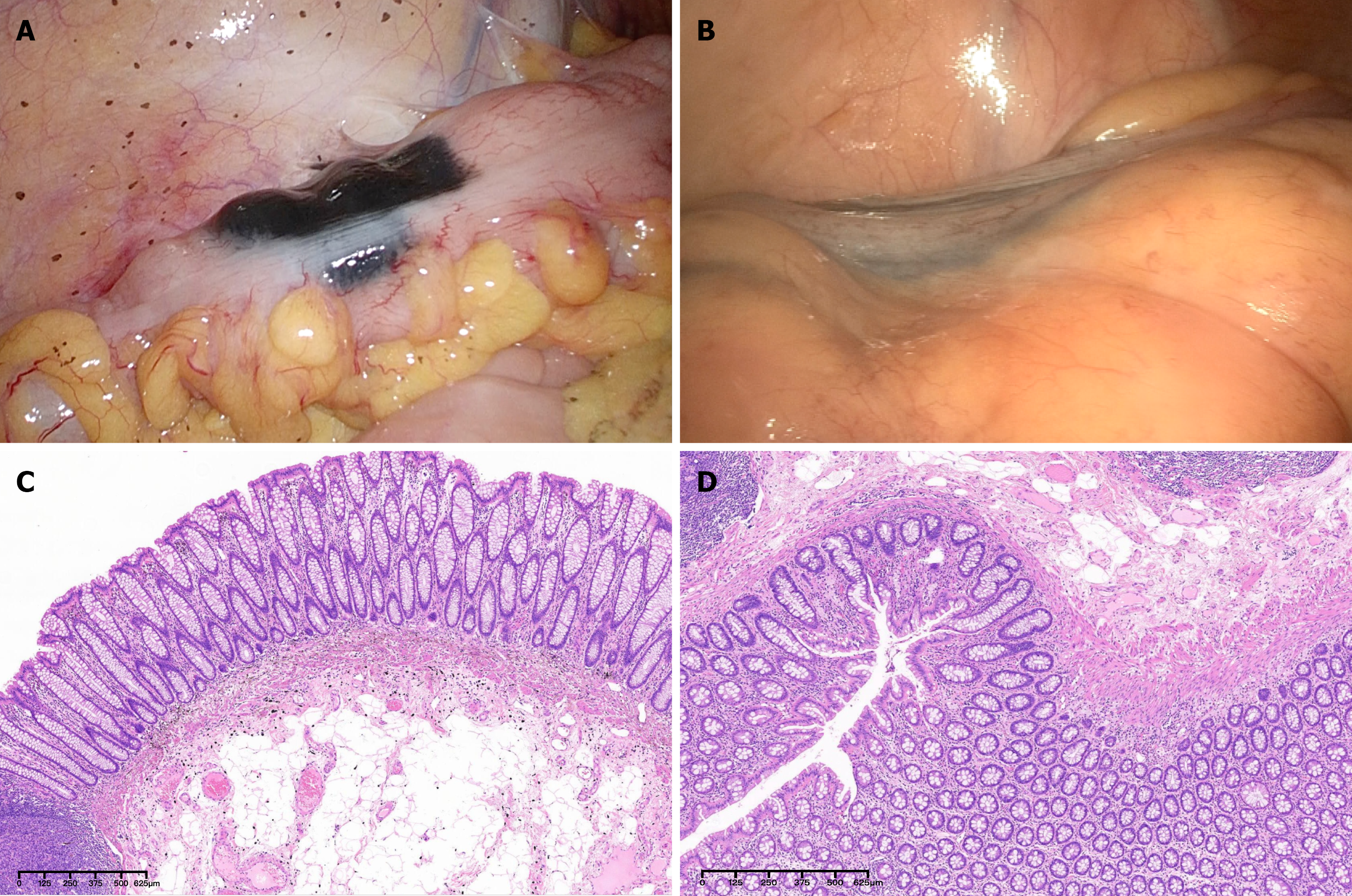
Figure 2 Submucosal injection of carbon nanoparticle suspension.
A: Traditional injection technique: 0.5 mL carbon nanoparticle (CNP) suspension mixed with 4 mL normal saline is endoscopically injected into the submucosal layer at four points. Leakage of CNP into the abdominal cavity is observed; B: “Sandwich” injection technique: 1 mL normal saline, followed by 2.5 mL CNP suspension (0.5 mL CNP mixed with 2 mL normal saline), and another 1 mL normal saline. The abdominal cavity remains clean, with no CNP suspension leakage; C: Hematoxylin and eosin (HE) staining of intestinal wall stained with CNPs; D: HE staining of intestinal wall without CNP staining.
- Citation: Bai BJ, Xu DY, Chen L, Ding LY, Song ZF. Clinical value of carbon nanoparticles in identifying metastatic lymph nodes in colorectal cancer: A single-center observational study. World J Gastrointest Oncol 2026; 18(8): 122077
- URL: https://www.wjgnet.com/1948-5204/full/v18/i8/122077.htm
- DOI: https://dx.doi.org/10.4251/wjgo.122077