
















Copyright: ©Author(s) 2026.
World J Gastrointest Oncol. Aug 15, 2026; 18(8): 121533
Published online Aug 15, 2026. doi: 10.4251/wjgo.v18.i8.121533
Published online Aug 15, 2026. doi: 10.4251/wjgo.v18.i8.121533
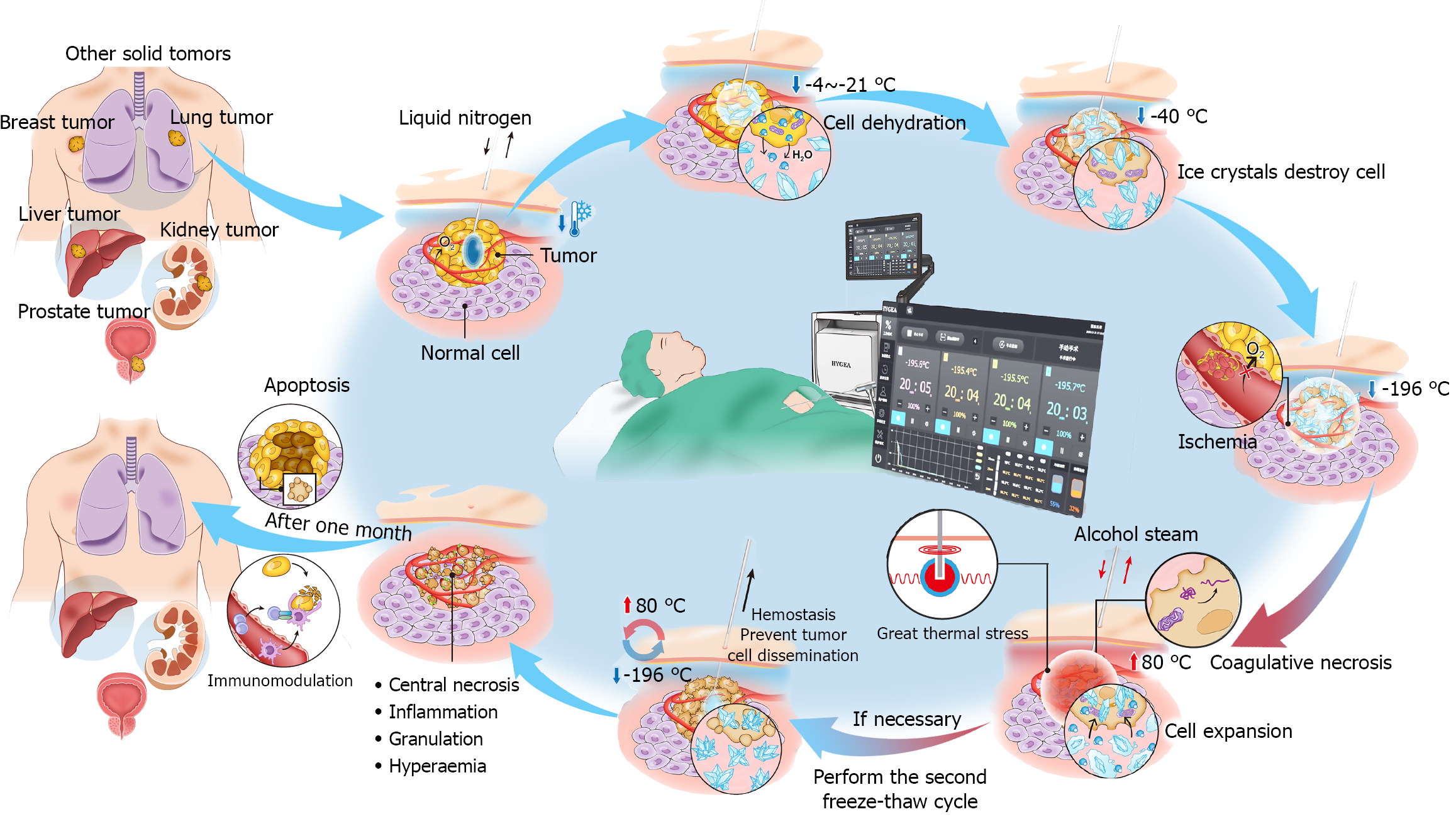
Figure 1 Working mechanism of coablation.
The figure was obtained from Siemens computed tomography. When liquid nitrogen and absolute ethanol are used as the refrigeration and heating media, the achievable temperature extremes are -196 °C and 80 °C, respectively.
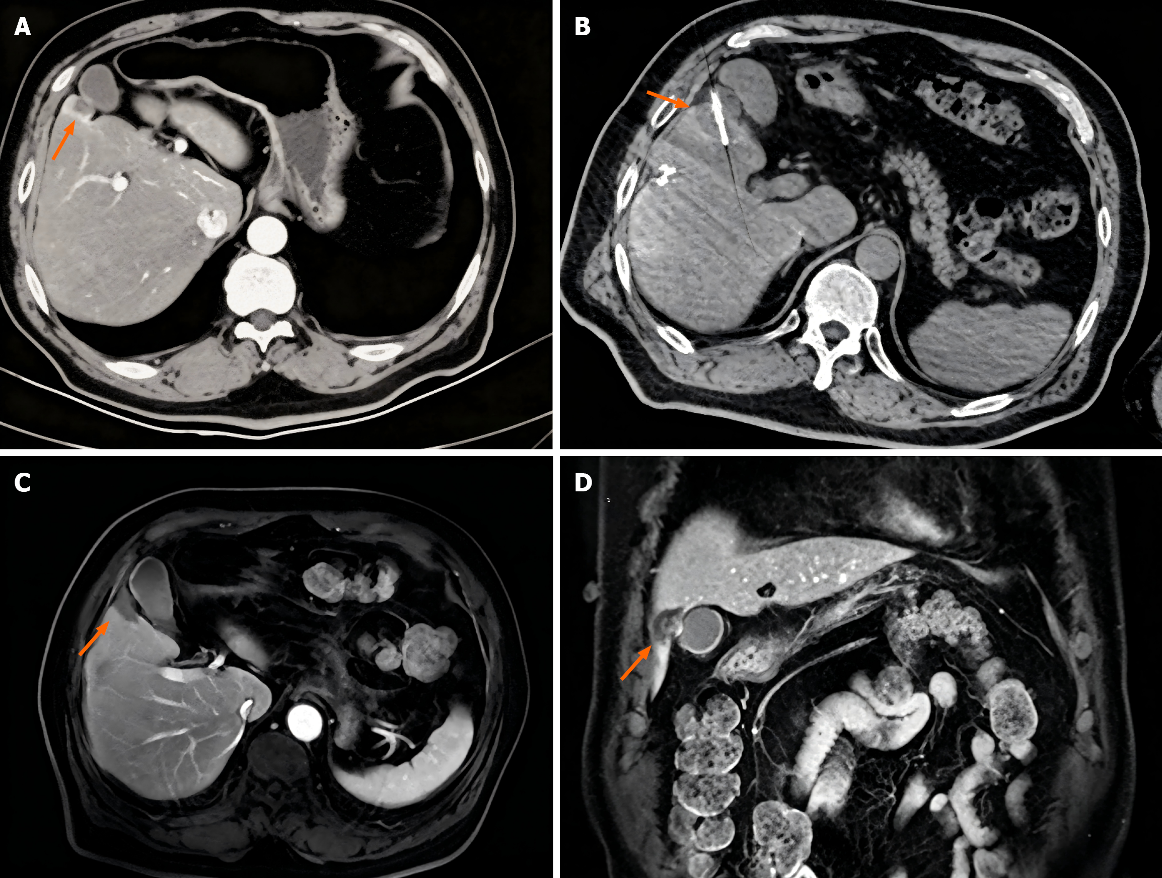
Figure 2 Lesion close to the gallbladder.
Images from a 61-year-old man diagnosed with hepatocellular carcinoma who underwent coablation. A: Computed tomography (CT) image showing a lesion close to the gallbladder before coablation; B: The area of the ice ball was 3.9 cm × 3.5 cm; C and D: Three-month follow-up CT images show the previous ablation zone (orange arrow) and the absence of local recurrence after coablation.
- Citation: Liu BJ, Kou FX, Guo JH, Wu D, Zhang X, Feng AW, Wang XD, Cao G, Xu L, Chen H, Liu P, Xu HF, Gao QZ, Yang RJ, Gao S, Zhu X. Efficacy and safety of computed tomography-guided coablation for hepatic malignancies. World J Gastrointest Oncol 2026; 18(8): 121533
- URL: https://www.wjgnet.com/1948-5204/full/v18/i8/121533.htm
- DOI: https://dx.doi.org/10.4251/wjgo.v18.i8.121533