
















Copyright: ©Author(s) 2026.
World J Gastrointest Oncol. Aug 15, 2026; 18(8): 120982
Published online Aug 15, 2026. doi: 10.4251/wjgo.v18.i8.120982
Published online Aug 15, 2026. doi: 10.4251/wjgo.v18.i8.120982
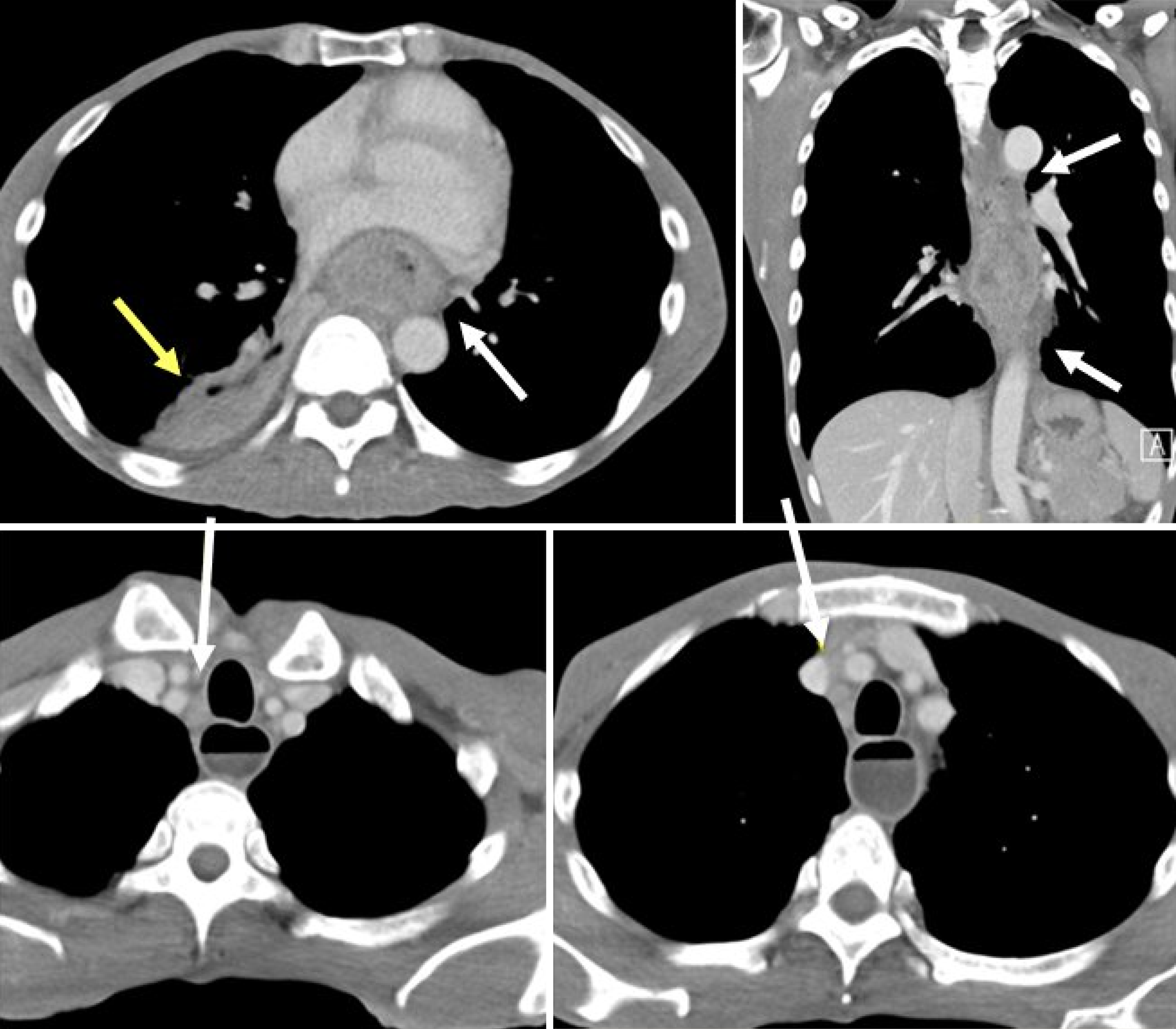
Figure 1 Initial chest computed tomography showing esophageal tumor and pneumonia.
Contrast-enhanced axial computed tomography scan demonstrates a large soft-tissue mass involving the mid- to lower-third esophagus (white arrow), with associated paratracheal and paraesophageal lymphadenopathy. Consolidation and abscess formation suggestive of pneumonia (yellow arrow) with a suspicious tracheoesophageal fistula are also observed.
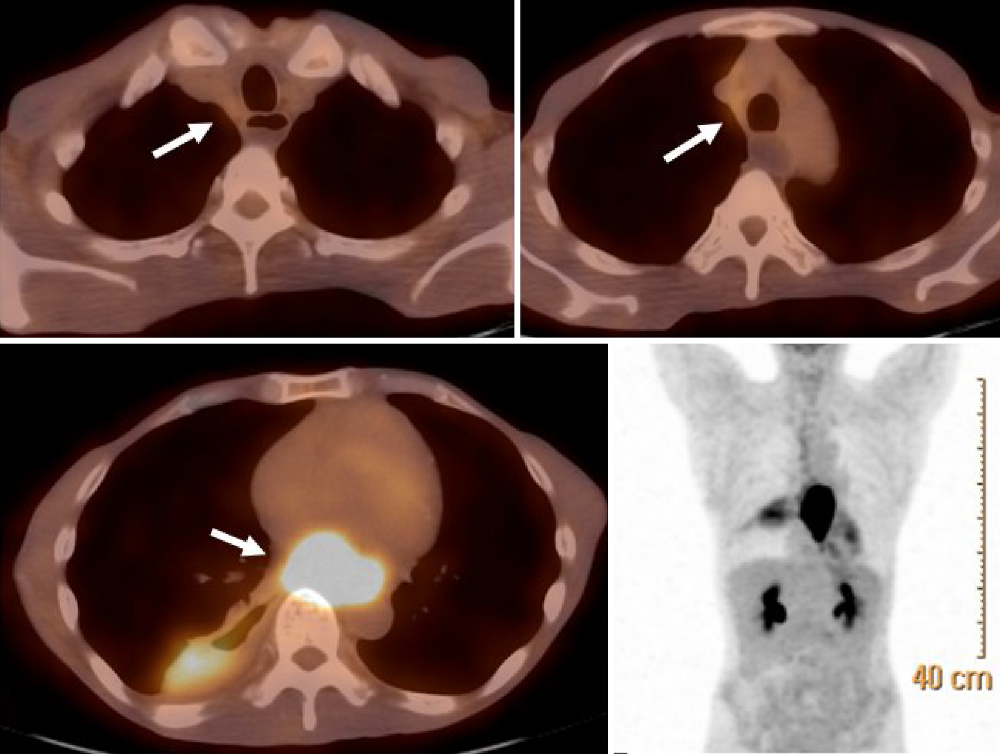
Figure 2 Baseline positron emission tomography/computed tomography confirming locoregional disease without distant metastasis.
Axial fused positron emission tomography/computed tomography image shows hypermetabolic activity at the primary esophageal tumor site and involved mediastinal lymph nodes (arrows), without evidence of distant metastatic disease.
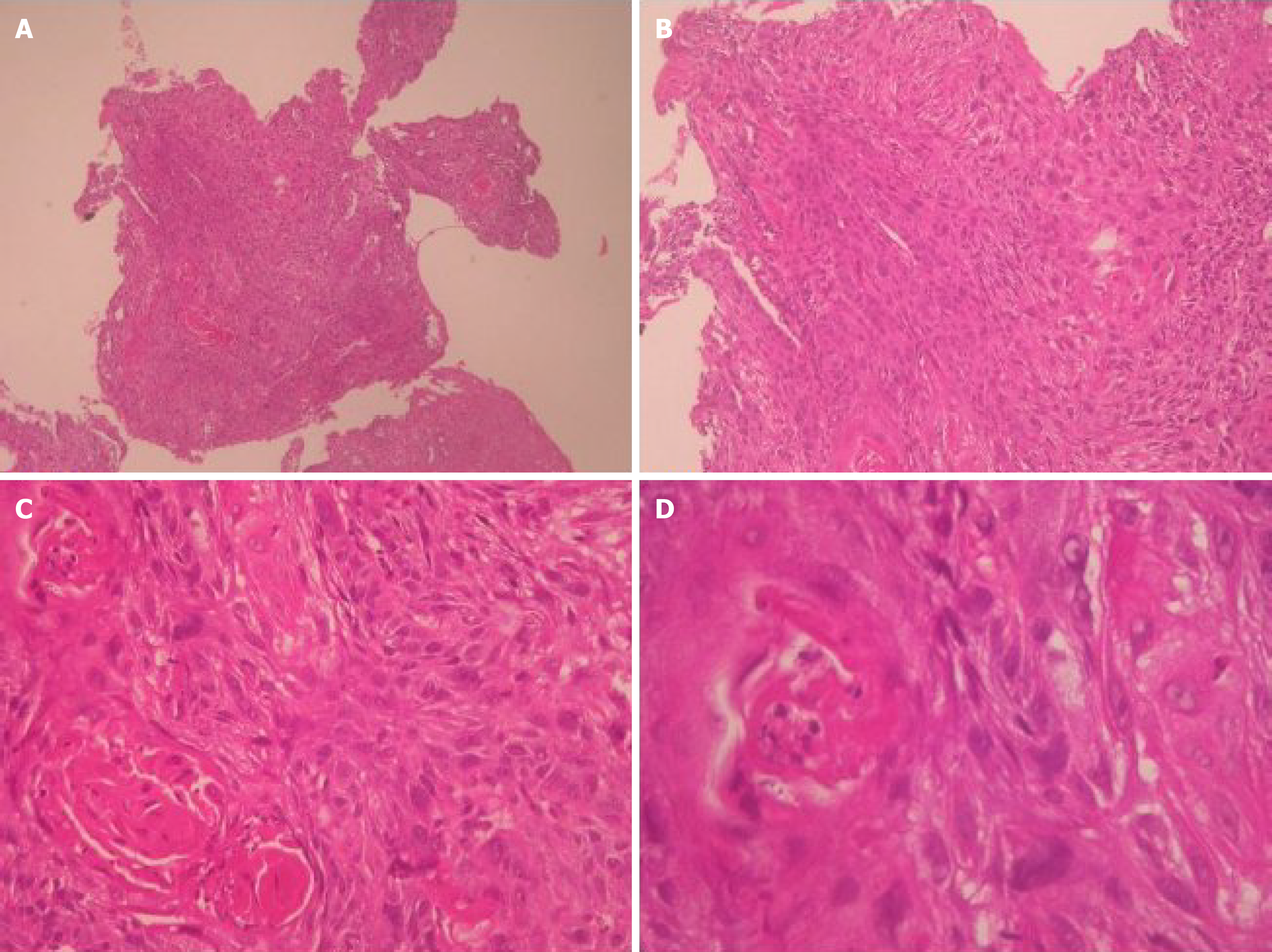
Figure 3 Histopathological features of esophageal squamous cell carcinoma prior to treatment.
Hematoxylin and eosin-stained sections from endoscopic biopsy specimens were examined. A: Low-power view (× 40): Multiple small, fragmented pieces of esophageal mucosa are shown. The limited tissue size (0.3 cm × 0.2 cm × 0.1 cm) constrains evaluation of deeper structures; B: Medium-power view (× 100): The squamous epithelium exhibits complete loss of normal stratification and maturation, with atypical cells occupying the entire layer; C: High-power view (× 200): Neoplastic squamous cells exhibit prominent hyperchromatic nuclei and pleomorphism, involving the full thickness of the epithelium. These features are diagnostic of at least high-grade squamous intraepithelial neoplasia (squamous cell carcinoma in situ); D: High-power view (× 400): Focal areas show keratin pearl formation, supporting squamous differentiation. While subepithelial stromal invasion is suspected, a definitive diagnosis of invasive carcinoma is precluded by the limited depth of the specimen. These findings are consistent with squamous cell carcinoma in situ with features suspicious for early invasion.
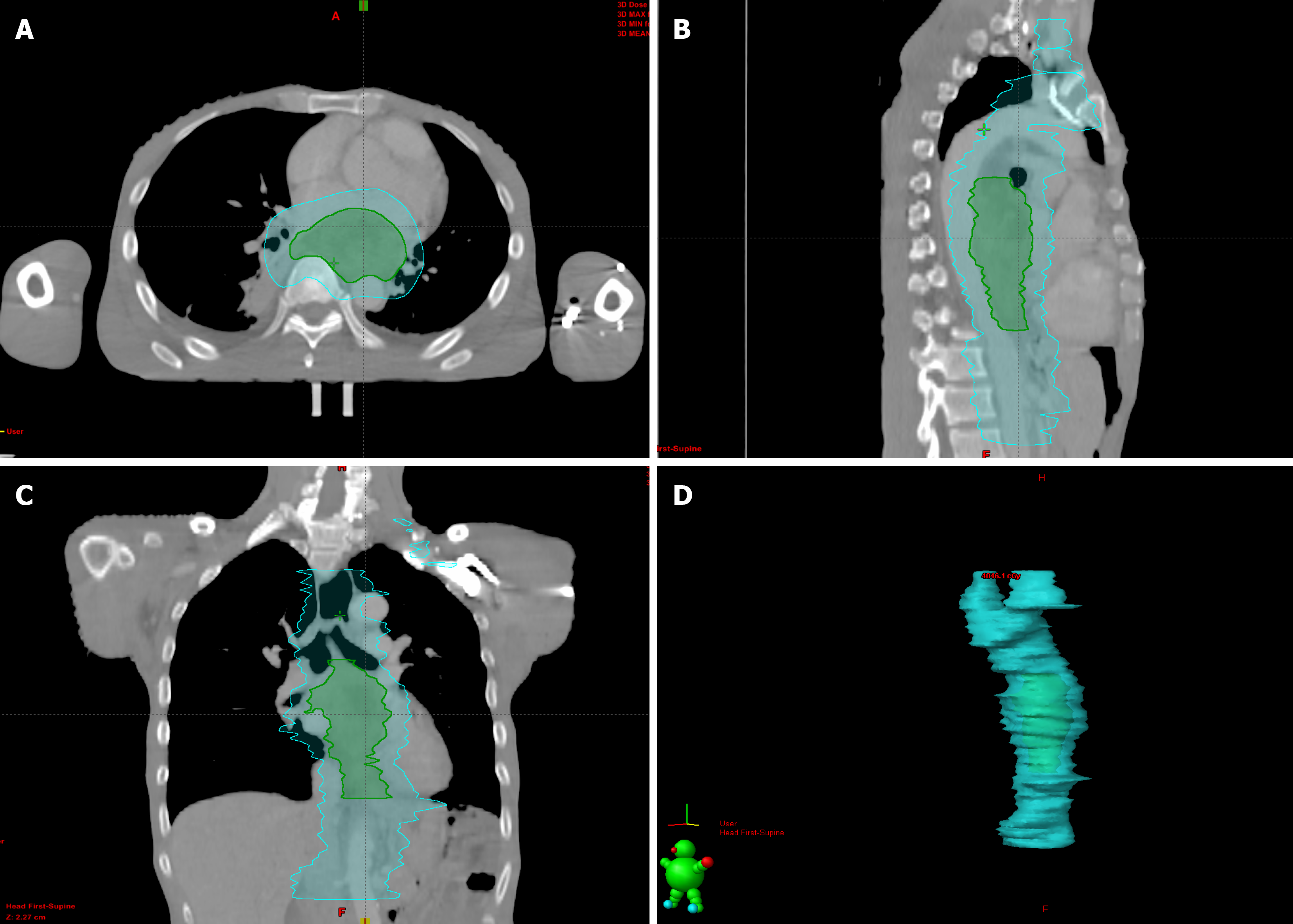
Figure 4 Radiotherapy planning images demonstrating gross tumor and planning target volumes.
A-C: Representative axial (A), sagittal (B), and coronal (C) views from volumetric modulated arc therapy radiotherapy treatment planning are shown. The gross tumor volume and involved lymphadenopathy regions were delineated for a sequential boost irradiation (green line), delivering a total dose of 59.4 Gy in 33 fractions, while maintaining dose constraints for surrounding normal organs. The cyan line represents the planning target volume; D: Three-dimensional reconstruction of the target volumes.
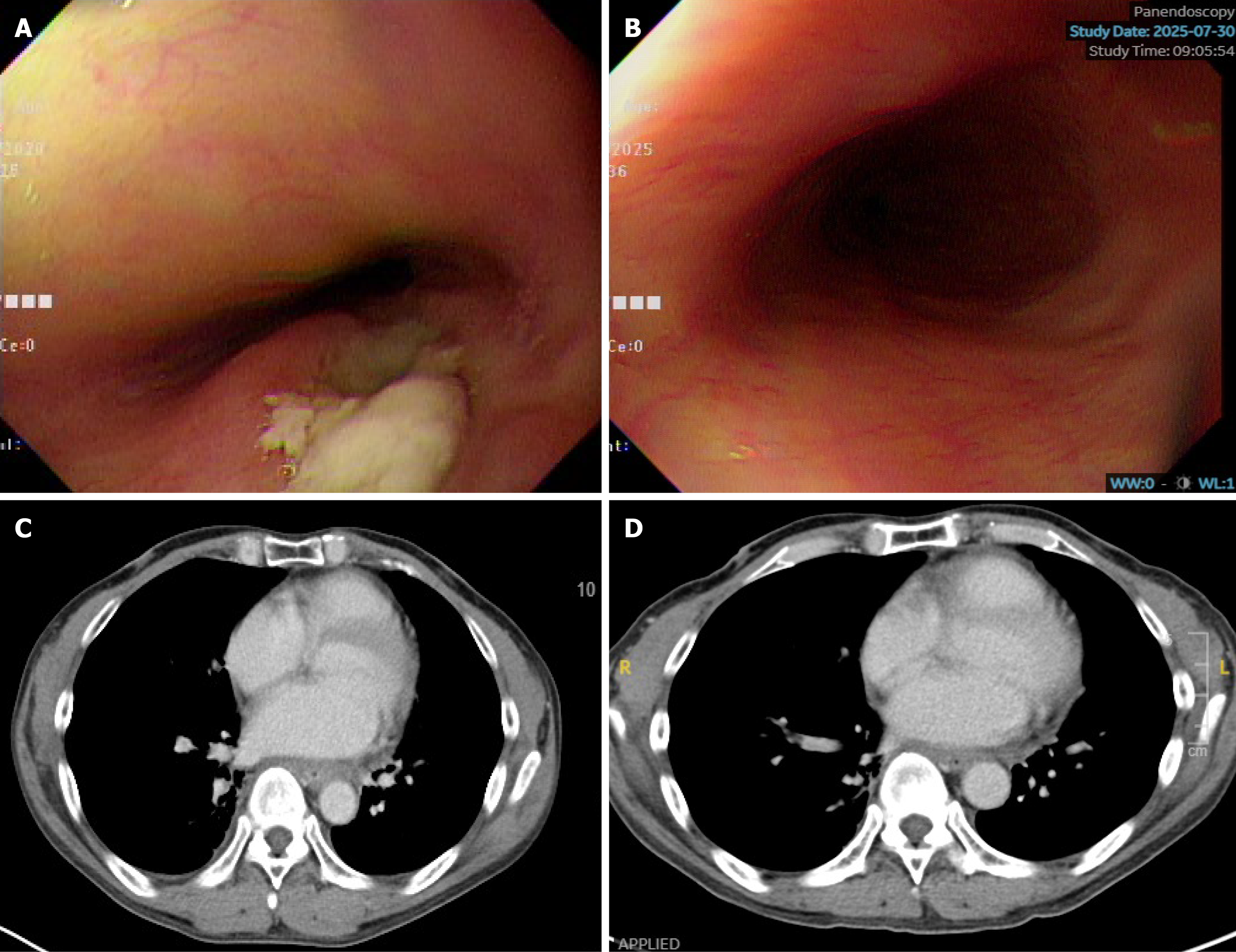
Figure 5 Follow-up endoscopy and imaging after treatment completion.
Endoscopic and radiological follow-up after multimodal treatment. A: Endoscopic view at approximately 2.2 months post-chemoradiotherapy showed a healing ulcer without evidence of residual tumor; B: Follow-up endoscopy at 58 months post-chemoradiotherapy revealed no signs of local recurrence; C: Follow-up computed tomography scan at approximately 2.5 months post-chemoradiotherapy, showing significant tumor regression and resolution of pneumonia; D: Follow-up computed tomography scan at 60 months (5 years) post-chemoradiotherapy, confirming a sustained complete response and progression-free status.
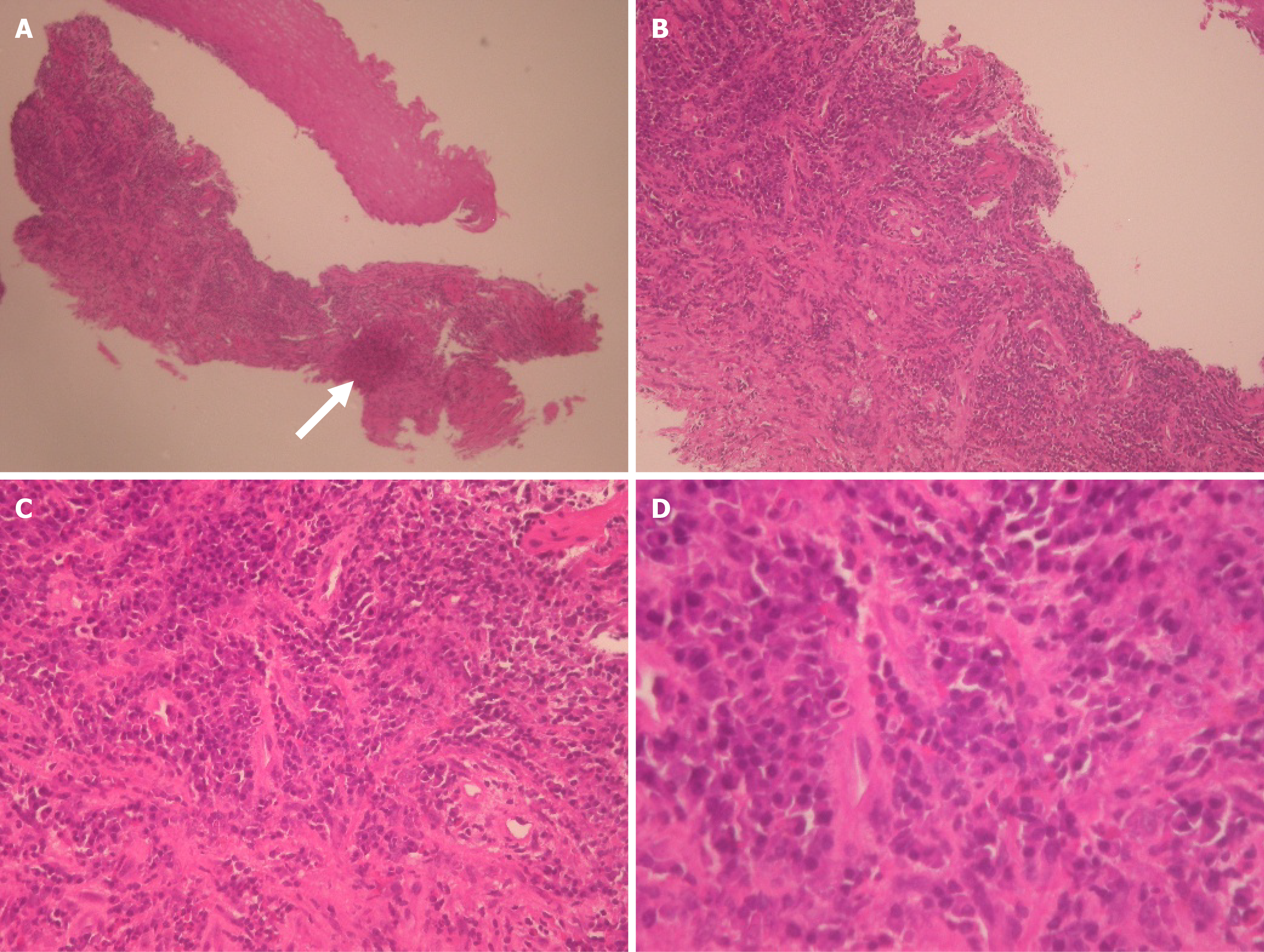
Figure 6 Histopathological findings after treatment.
Hematoxylin and eosin-stained sections from post-treatment endoscopic biopsy specimens were evaluated. A: Low-power view (× 40): Post-treatment biopsy showing preserved architecture of the esophageal mucosa with evidence of chronic inflammatory cell infiltration in the stroma. The arrow indicates a representative area for detailed evaluation; B: Medium-power view (× 100): Magnified view of the arrow-indicated area in (A). The squamous epithelium demonstrates regenerative changes with restoration of normal stratification and maturation. No evidence of expansile epithelial proliferation or tumor nests is observed; C: High-power view (× 200): Further magnification of the same region. The epithelial cells lack cytologic atypia, with normal nuclear-to-cytoplasmic ratios. No dysplastic or malignant features are identified; D: High-power view (× 400): Highest magnification of the arrow-indicated area. Post-treatment endoscopic biopsy specimen demonstrates subepithelial connective tissue with prominent inflammatory infiltrates composed predominantly of neutrophils and lymphocytes. No cytologic atypia or malignant squamous cells are identified in the examined field. These findings support the presence of a pathological complete response at the primary tumor site.
- Citation: Cheng CW, Li WT, Lee YT, Kam KH, Tsang YW. Long-term complete response in stage IVa esophageal squamous cell carcinoma treated with chemoradiotherapy and modulated electro-hyperthermia: A case report. World J Gastrointest Oncol 2026; 18(8): 120982
- URL: https://www.wjgnet.com/1948-5204/full/v18/i8/120982.htm
- DOI: https://dx.doi.org/10.4251/wjgo.v18.i8.120982