
















Copyright: ©Author(s) 2026.
World J Gastrointest Oncol. Aug 15, 2026; 18(8): 120889
Published online Aug 15, 2026. doi: 10.4251/wjgo.v18.i8.120889
Published online Aug 15, 2026. doi: 10.4251/wjgo.v18.i8.120889
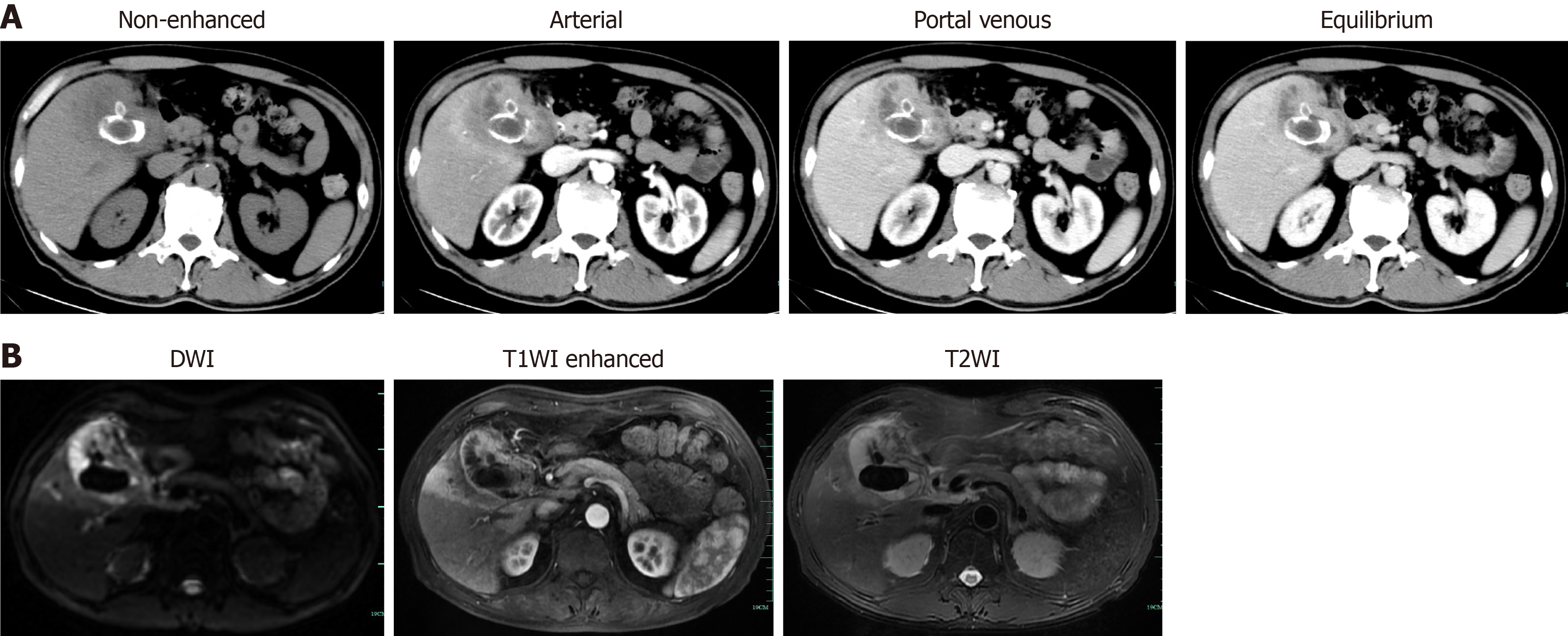
Figure 1 Preoperative imaging examinations of the first case.
The imaging examinations did not exclude gallbladder cancer. A: Contrast-enhanced computed tomography showed diffused inhomogeneous thickening of the gallbladder wall. Cholecystitis with abscess was considered initially; B: Magnetic resonance imaging suggested gallbladder cancer with liver involvement. DWI: Diffusion weighted imaging; T1WI: T1 Weighted imaging; T2WI: T2 Weighted imaging.
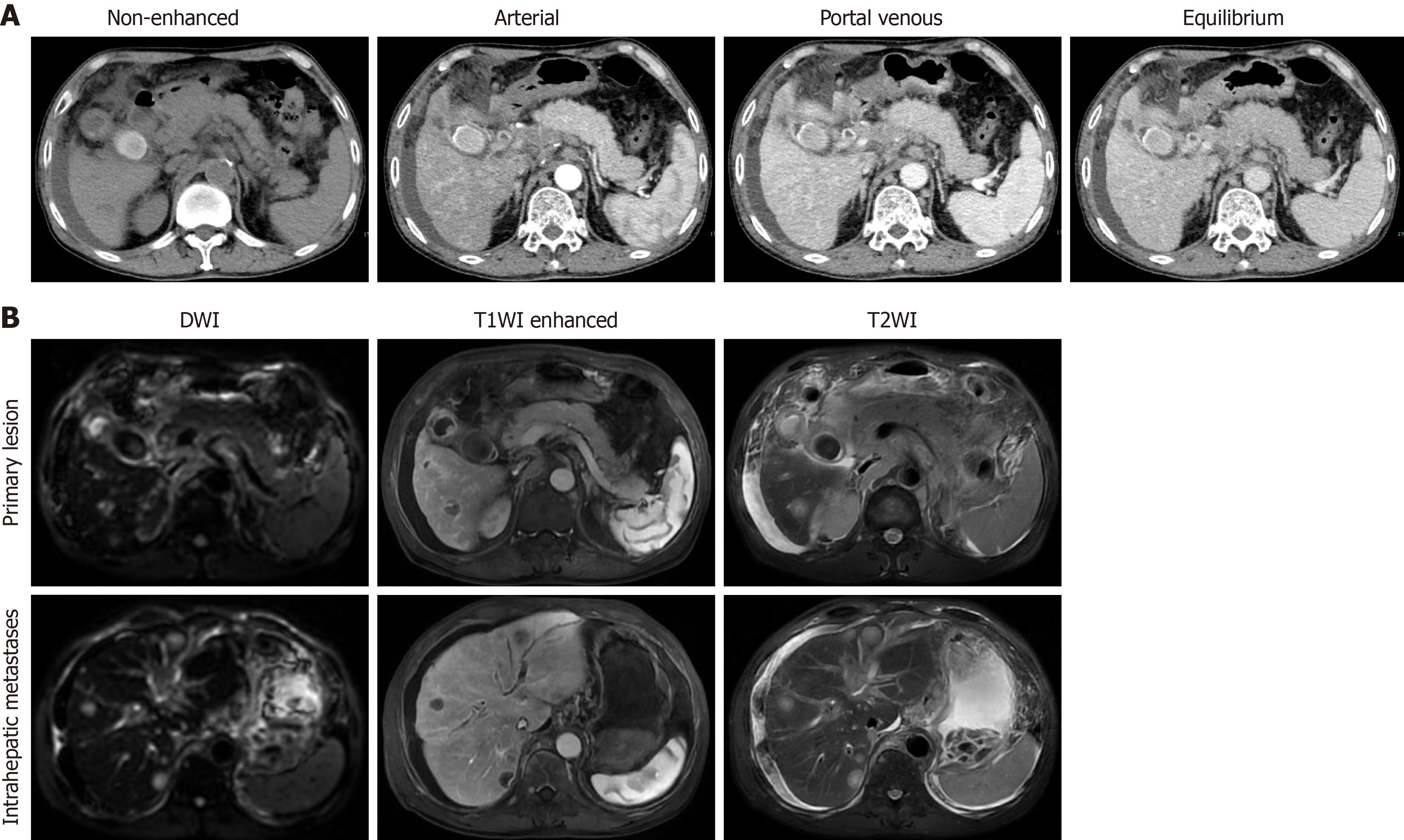
Figure 2 Preoperative imaging examinations of the second case.
A: Contrast-enhanced computed tomography; B: Magnetic resonance imaging of the second case suggested gallbladder cancer or hilar cholangiocarcinoma, with involvement of all extrahepatic bile ducts and the portal vein and its branches. Multiple intrahepatic metastases, multiple portal and retroperitoneal lymph node metastases, and cholecystolithiasis were observed. DWI: Diffusion weighted imaging; T1WI: T1 Weighted imaging; T2WI: T2 Weighted imaging.
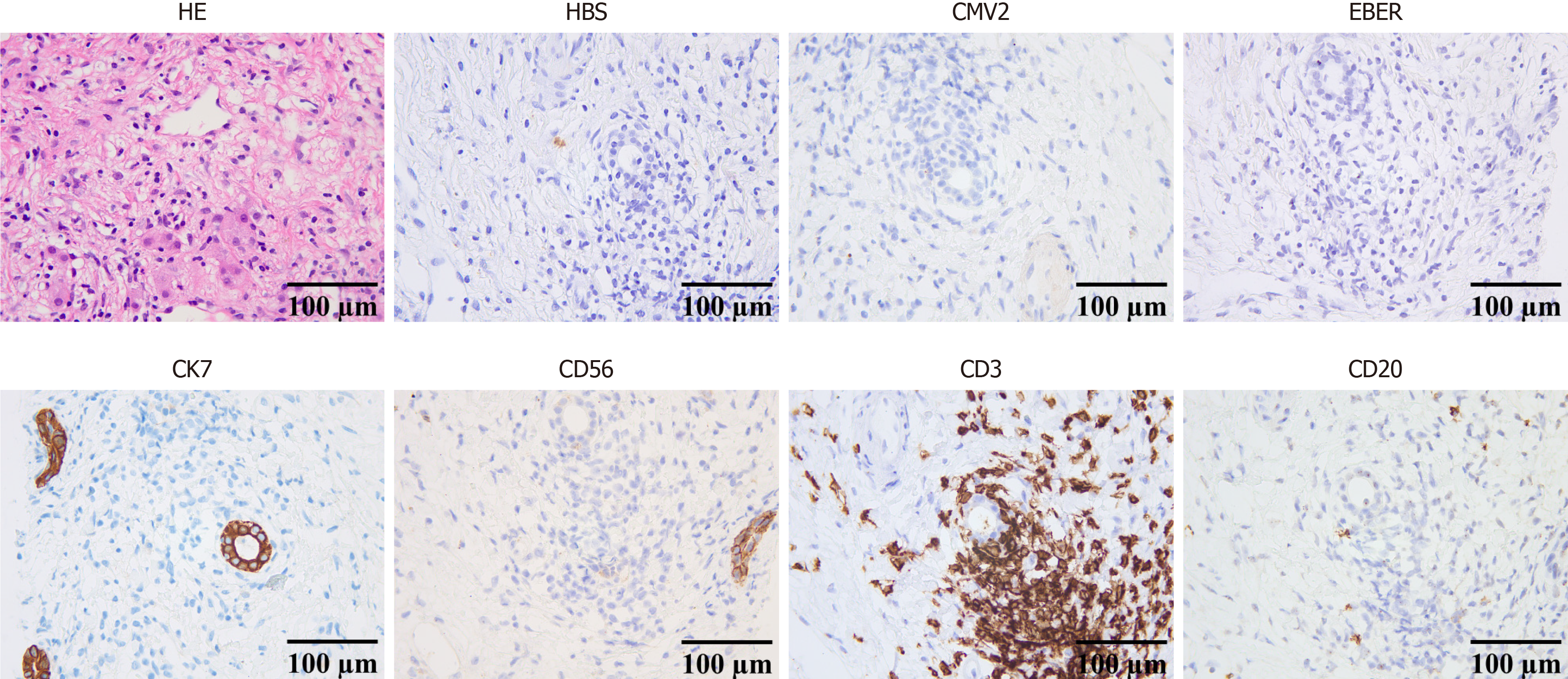
Figure 3 Biopsy pathology of the first case showing no evidence of tumor.
Hematoxylin and eosin staining showed necrosis of liver tissue, with chronic inflammatory cell infiltration and proliferation of fibrous tissue, in accordance with the positive cluster of differentiation 3 (CD3) and CD20 staining. Other immunohistochemistry assays suggested positive cytokeratin 7 and CD56 staining, and negative staining for hepatitis B surface antigen, cytomegalovirus type 2, and Epstein-Barr virus-encoded small RNAs. HE: Hematoxylin and eosin; HBS: Hepatitis B surface antigen; CMV2: Cytomegalovirus type 2; EBER: Epstein-Barr virus-encoded small RNAs; CK: Cytokeratin; CD: Cluster of differentiation.
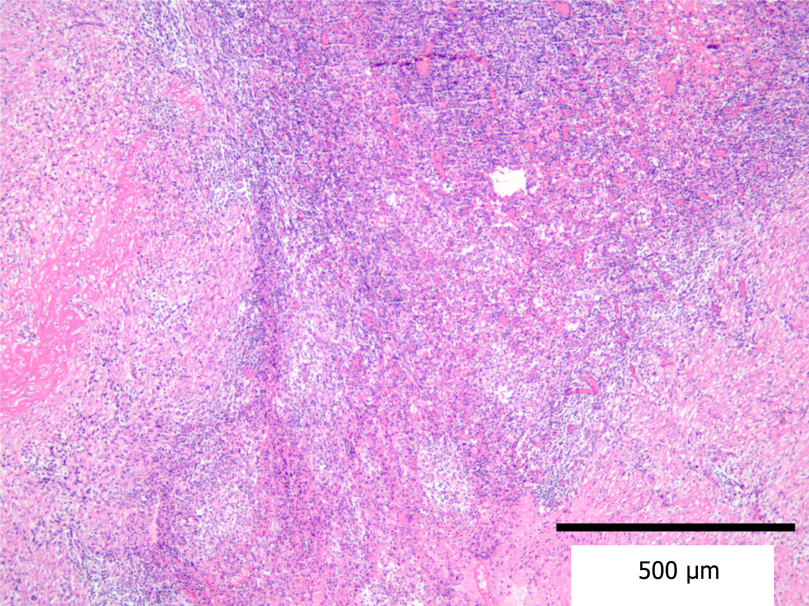
Figure 4 Postoperative routine pathology of the first case.
Routine pathology revealed focal gallbladder wall mucosal shedding, infiltration by a large number of acute and chronic inflammatory cells, hemorrhage and necrosis, and a multinucleated giant cell reaction. All of these findings are in accordance with xanthogranulomatous cholecystitis.
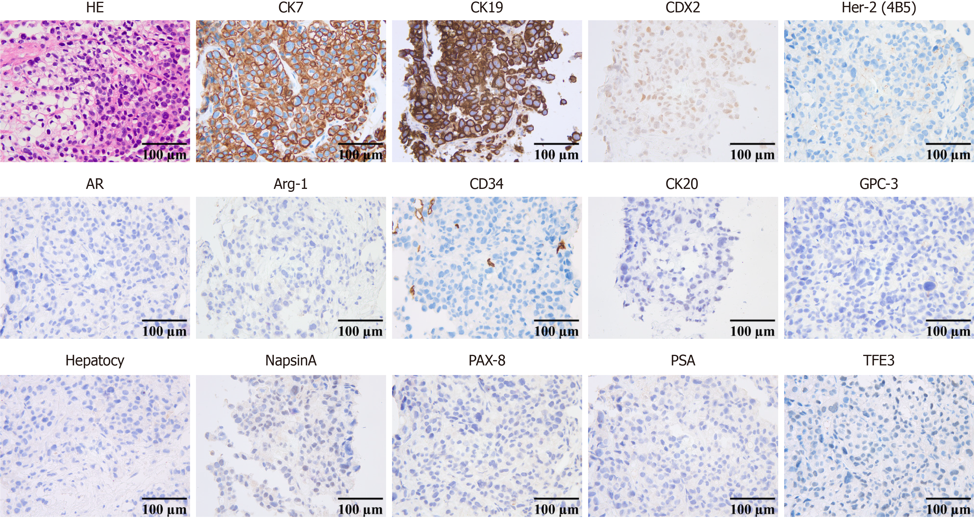
Figure 5 Biopsy pathology of the second case.
The biopsy pathology of the second case revealed poorly differentiated carcinoma, and immu
- Citation: Zhang J, Xi B, Li J, Wen L, Deng Y, Wang CW, Zhang Y. Prophylactic cholecystectomy for asymptomatic gallstone disease: Two case reports and review of literature. World J Gastrointest Oncol 2026; 18(8): 120889
- URL: https://www.wjgnet.com/1948-5204/full/v18/i8/120889.htm
- DOI: https://dx.doi.org/10.4251/wjgo.v18.i8.120889