
















Copyright: ©Author(s) 2026.
World J Gastrointest Oncol. Aug 15, 2026; 18(8): 120791
Published online Aug 15, 2026. doi: 10.4251/wjgo.v18.i8.120791
Published online Aug 15, 2026. doi: 10.4251/wjgo.v18.i8.120791
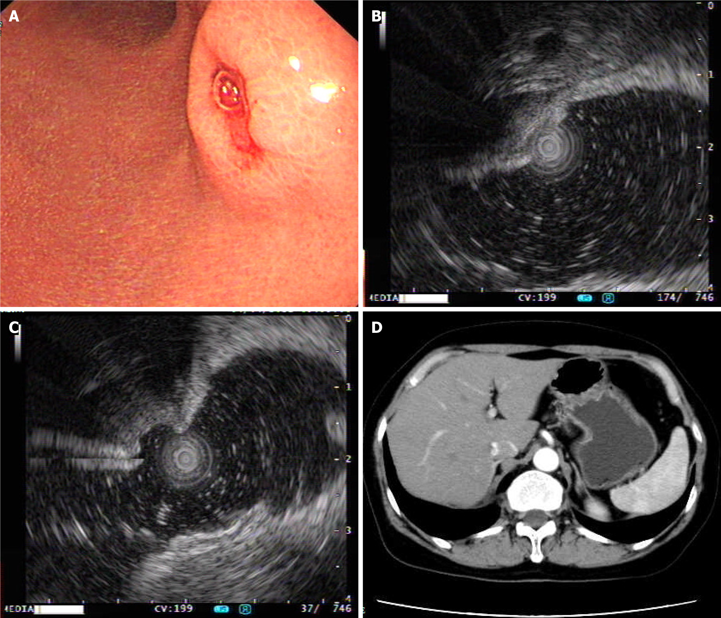
Figure 1 Endoscopic and computed tomography-related images.
A: Gastroscopy showing a hemispherical mucosal elevation on the posterior wall at the junction of the gastric body and antrum; B: Endoscopic ultrasound showing a submucosal elevation with ulceration at the apex on the posterior wall at the junction of the gastric body and antrum; C: Intraluminal water-injection-assisted endoscopic ultrasound imaging showing mucosal layer loss at the elevation site; D: Abdominal contrast-enhanced computed tomography showing gastric wall thickening on the posterior wall near the junction of the gastric body and antrum.
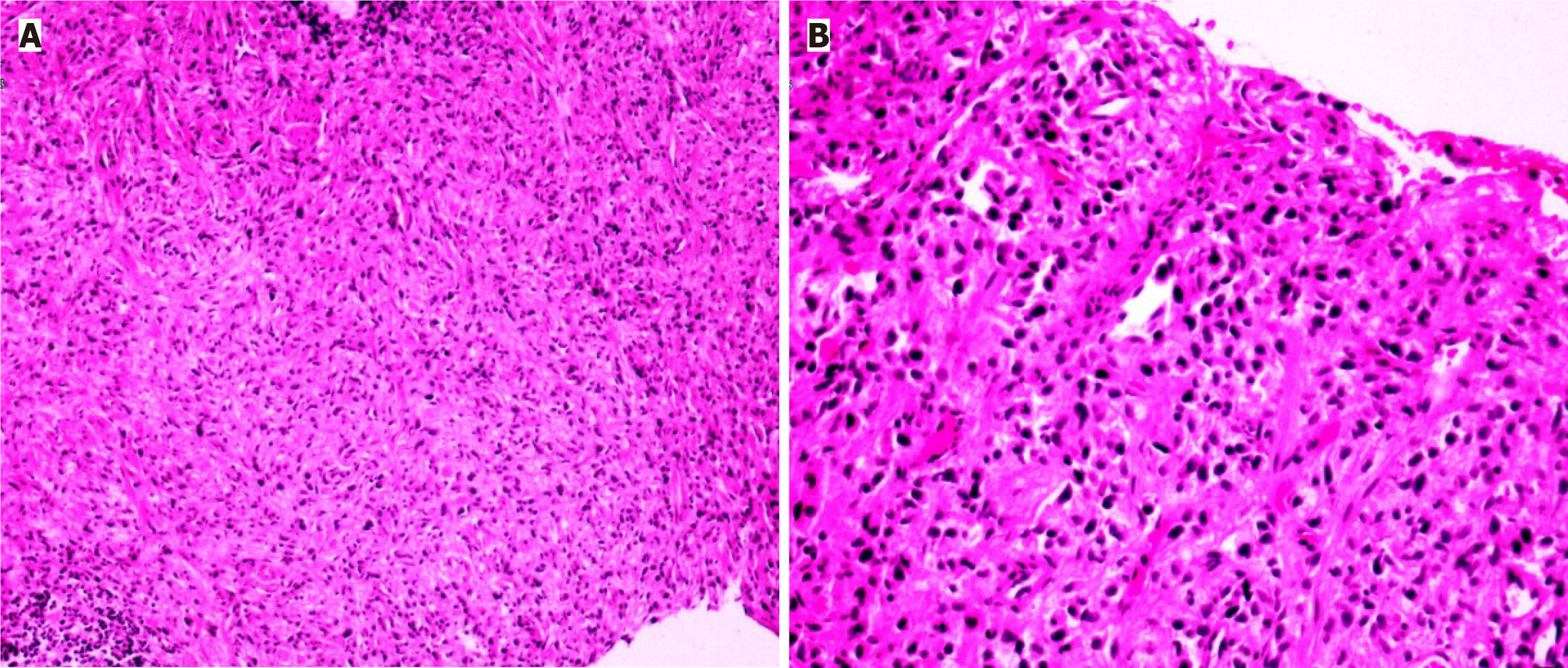
Figure 2 Diagnostic endoscopic submucosal dissection biopsy pathology.
A: Hematoxylin and eosin staining of the biopsy specimen from the posterior wall at the junction of the gastric body and antrum, showing a spindle cell-rich lesion (× 100); B: Higher-power hematoxylin and eosin view showing spindle cell-like morphology with elongated tumor cells (× 200).
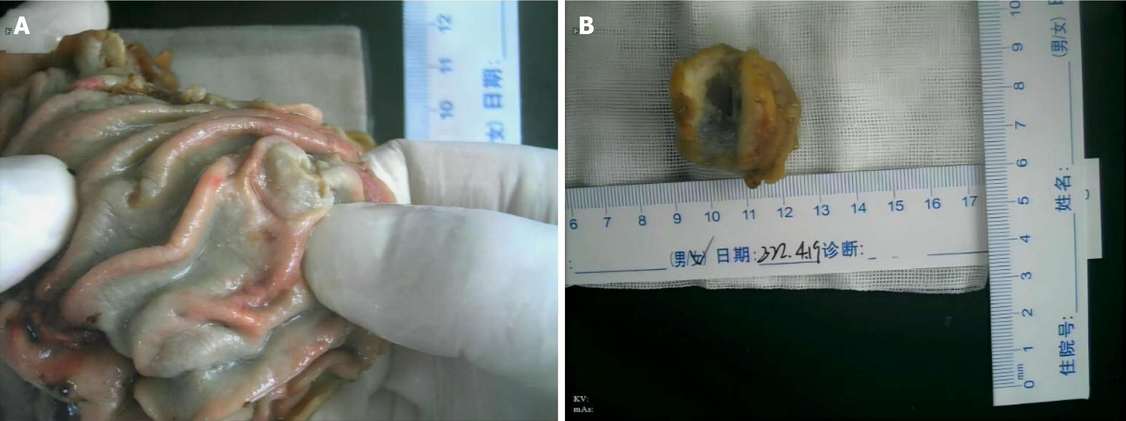
Figure 3 Surgical specimens.
A: Gross view of the resected stomach showing a lesion measuring approximately 2.5 cm × 1.2 cm × 1 cm; B: Opened gastrectomy specimen showing an ulcerative carcinoma located on the posterior wall at the junction of the gastric body and antrum, with clear anatomical orientation.
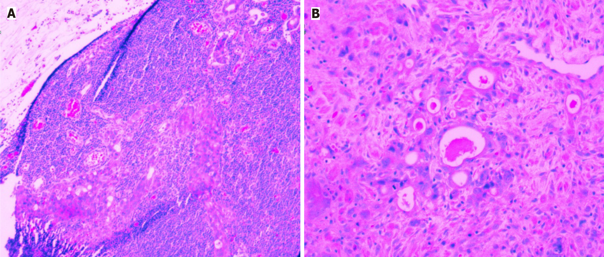
Figure 4 Surgical histopathology.
A: Periodic acid-Schiff (PAS) staining showing infiltrative tumor growth in the gastric wall (× 100); B: Higher-power PAS staining showing PAS-positive mucin in tumor cells and glandular lumina (× 200).
- Citation: Xu LY, Lin YQ, Yao RY, Yang ML, Shen HF, Zhang GS. Poorly differentiated adenocarcinoma misdiagnosed as gastrointestinal stromal tumor: A case report. World J Gastrointest Oncol 2026; 18(8): 120791
- URL: https://www.wjgnet.com/1948-5204/full/v18/i8/120791.htm
- DOI: https://dx.doi.org/10.4251/wjgo.v18.i8.120791